Virions Collection (#7)

Virions, the tiny particles that cause infections, come in various forms and can be seen under a transmission electron microscope (TEM

Background imageVirions Collection: Influenza virus particles

Influenza virus particles. Computer artwork of influenza (flu) virus particles. Each virus consists of a core of RNA (ribonucleic acid) genetic material surrounded by a protein coat

Background imageVirions Collection: Flu virus particles

Flu virus particles
Influenza virus particles, computer artwork. The particles comprise a core (central sphere) surrounded by membranes. The outer membrane bears the proteins neuraminidase and haemagglutinin (spikes)

Background imageVirions Collection: Avian influenza virus, TEM

Avian influenza virus, TEM
Avian influenza virus, type A strain H5N1, coloured transmission electron micrograph (TEM). This virus was isolated in Vietnam, during the avian flu outbreak in early 2004

Background imageVirions Collection: Influenza viruses

Influenza viruses. Coloured transmission electron micrograph (TEM) of influenza (flu) viruses. The flu virus belongs to the orthomyxovirus group of viruses which have an affinity for mucus

Background imageVirions Collection: Adenoviruses

Adenoviruses

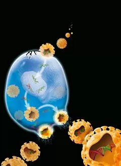
Background imageVirions Collection: Virus replication cycle, artwork

Virus replication cycle, artwork. The cycle starts at upper centre, as virus particles (orange) enter a cell (blue). Once in the cell the viral genetic material, deoxyribonucleic acid (DNA, green)

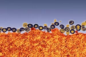
Background imageVirions Collection: Budding HIV particles, TEM

Budding HIV particles, TEM
Budding HIV particles. Coloured transmission electron micrograph (TEM) of HIV particles (blue and yellow) budding from the membrane of the host cell (orange)

Background imageVirions Collection: Budding HIV particle, computer artwork

Budding HIV particle, computer artwork
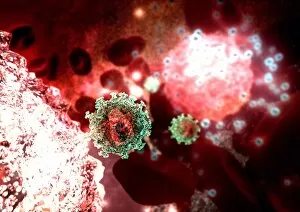
Budding HIV particle. Computer artwork of an HIV (human immunodeficiency virus) particle (green) exiting a T-lymphocyte (a type of white blood cell, lower left)

Background imageVirions Collection: HIV particles

HIV particles, computer artwork. HIV (human immunodeficiency virus) causes AIDS (acquired immune deficiency syndrome). The virus consists of an RNA genome

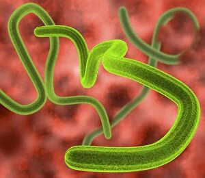
Background imageVirions Collection: Ebola virus particles

Ebola virus particles, computer artwork. This thread-like RNA (ribonucleic acid) virus is the cause of ebola haemorrhagic fever in humans

Background imageVirions Collection: Rhinovirus particles

Rhinovirus particles, computer artwork. This virus consists of a protein capsid (multicoloured), enclosing an RNA (ribonucleic acid) genome

Background imageVirions Collection: AIDS viruses budding from a cell, TEM

AIDS viruses budding from a cell, TEM
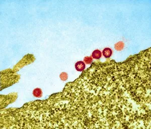
AIDS virus particles budding from the membrane of their host cell, coloured transmission electron micrograph (TEM). AIDS is a disease caused by the human immunodeficiency virus (HIV, red)

Background imageVirions Collection: Viruses

Viruses, computer artwork

Background imageVirions Collection: Simian AIDS viruses, TEM

Simian AIDS viruses, TEM
Simian AIDS virus particles, coloured transmission electron micrograph (TEM). These virus particles (small, round) are bursting out of a cell (across bottom) after using the cell to replicate

Background imageVirions Collection: West Nile virus, TEM

West Nile virus, TEM
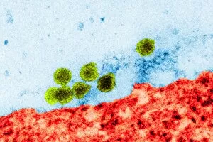
West Nile virus particles. Coloured transmission electron micrograph (TEM) of West Nile virus (WNV) particles (green) next to a host cell (red)

Background imageVirions Collection: Vaccinia viruses, TEM

Vaccinia viruses, TEM
Vaccinia virus particles. Coloured transmission electron micrograph (TEM) of sectioned vaccinia virus particles. The genetic cores (red) are covered by membrane layers (green)

Background imageVirions Collection: Polio viruses, TEM

Polio viruses, TEM
Polio virus. Coloured transmission electron micrograph (TEM) of a section through a para- crystalline array of polio virus particles

Background imageVirions Collection: Herpes simplex virus infection, TEM

Herpes simplex virus infection, TEM
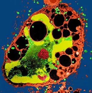
Herpes simplex virus infection. Coloured transmission electron micrograph (TEM) of a section through a cell being destroyed by herpes simplex virus infection

Background imageVirions Collection: Adeno-associated virus, TEM

Adeno-associated virus, TEM
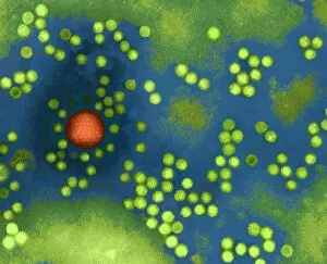
Adeno-associated virus. Coloured transmission electron micrograph (TEM) of particles of an unidentified adeno-associated virus (aV, green). A helper adenovirus (orange) is also seen

Background imageVirions Collection: Yellow fever virus particles, TEM

Yellow fever virus particles, TEM
Yellow fever virus particles. Coloured transmission electron micrograph (TEM) of yellow fever virus particles (white) growing in culture

Background imageVirions Collection: Crimean-Congo haemorrhagic fever virus

Crimean-Congo haemorrhagic fever viruses. Coloured transmission electron micrograph (TEM) of Crimean- Congo haemorrhagic fever (CCHF) viruses (brown)

Background imageVirions Collection: Virus, conceptual image

Virus, conceptual image

Background imageVirions Collection: Ebola viruses, artwork

Ebola viruses, artwork
Ebola viruses, computer artwork

Background imageVirions Collection: Viral research, conceptual artwork

Viral research, conceptual artwork
Viral research, conceptual computer artwork


For sale as Licensed Images

Choose your image, Select your licence and Download the media

Virions, the tiny particles that cause infections, come in various forms and can be seen under a transmission electron microscope (TEM). Norovirus particles, responsible for stomach bugs often spread through sneezing or contaminated surfaces, are captured in artwork C013 / 5949. Similarly, coronavirus particles can also be observed using TEM and have recently gained attention due to their role in the ongoing pandemic. Influenza virus particles, another common cause of respiratory illnesses, appear distinct under TEM as well. Hepatitis C viruses and Rift Valley fever virus are other examples that have been visualized using this powerful microscopy technique. Herpes virus particles are beautifully depicted through computer artwork, showcasing their intricate structure. Paramyxovirus particles and herpes simplex viruses are two more types that have been studied extensively with TEM. The images reveal their unique shapes and features. One striking image shows AIDS viruses budding from a cell under TEM observation. This highlights the devastating impact of these particular virions on human health. Lastly, dengue fever virus particles can also be observed using TEM technology. These tiny entities play a significant role in spreading dengue fever through mosquito bites.