Virions Collection (#9)

Virions, the tiny particles that cause infections, come in various forms and can be seen under a transmission electron microscope (TEM

Background imageVirions Collection: Flu infection, conceptual artwork

Flu infection, conceptual artwork

Background imageVirions Collection: Flu virus spreading, artwork

Flu virus spreading, artwork
Flu virus spreading, conceptual computer artwork. Virus particles (virions, spheres) around a human silhouette

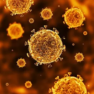
Background imageVirions Collection: HIV virus particles, artwork

HIV virus particles, artwork
HIV virus particles, computer artwork. Each particle (virion) consists of RNA (ribonucleic acid) contained within a capsid and an envelope covered in glycoprotein spikes

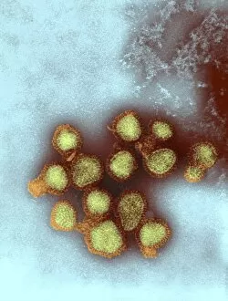
Background imageVirions Collection: Influenza viruses, TEM

Influenza viruses, TEM
Influenza viruses. Coloured transmission electron micrograph (TEM) of influenza (flu) viruses (yellow) budding from a host cell. The host cell nucleus is black

Background imageVirions Collection: Cell infected with HIV, electronic image

Cell infected with HIV, electronic image
Experts of the Ivanovsky Virusology Institute [USSR Academy of Medical Sciences] have made this electronic image of a cell infected with the AIDS virus

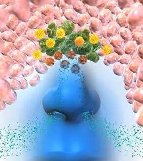
Background imageVirions Collection: Respiratory virus infection treatment

Respiratory virus infection treatment. Computer artwork showing virus particles (spherical) inside a persons nose and how they might be treated by inhaling a drug in the form of a nasal spray

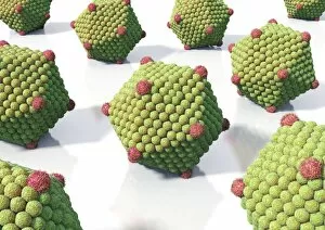
Background imageVirions Collection: Icosahedral virus particles, artwork

Icosahedral virus particles, artwork
Icosahedral virus particles, computer artwork. Icosahedral viruses have a 20-sided symmetry of their capsid (protein coat)

Background imageVirions Collection: Viruses, artwork

Viruses, artwork
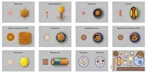
Viruses. Computer artwork showing three typical virus shapes: helical (blue), bacteriophage (red), and icosahedral (green)

Background imageVirions Collection: Bacteriophages, artwork

Bacteriophages, artwork
Bacteriophages, computer artwork. A bacteriophage, or phage, is a virus that infects bacteria. It consists of an icosahedral (20-sided) head (top), which contains the genetic material

Background imageVirions Collection: Helical virus particles, artwork

Helical virus particles, artwork. These are viruses with their capsids (protein coats) arranged in a helical structure. Viruses contain a core of genetic material

Background imageVirions Collection: Viral transfer of bacterial DNA, artwork

Viral transfer of bacterial DNA, artwork
Viral transfer of bacterial DNA. Computer artwork showing the process of transduction, whereby genetic information is transferred from one bacterium (right column) to another (left column)

Background imageVirions Collection: Virus types, artwork

Virus types, artwork

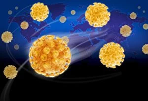
Background imageVirions Collection: Virus pandemic, conceptual image

Virus pandemic, conceptual image. Computer artwork of virus particles superimposed on a map of the world, representing the worldwide spread of an infectious viral disease

Background imageVirions Collection: Viral infection

Viral infection

Background imageVirions Collection: H1N1 swine flu virus, SEM

H1N1 swine flu virus, SEM
H1N1 swine flu virus. Coloured scanning electron micrograph (SEM) of swine influenza (flu) virus particles (virions, yellow)

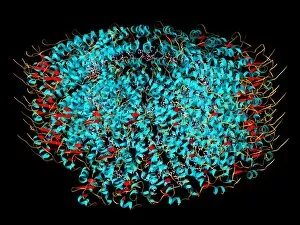
Background imageVirions Collection: West Nile virus particle, molecular model

West Nile virus particle, molecular model. This virus consists of an RNA (ribonucleic acid) core of genetic material, surrounded by a nucleocapsid (red, smooth)

Background imageVirions Collection: Virus structures, artwork

Virus structures, artwork
Virus structures. Computer artwork showing the structure of three different types of virus: bacteriophage (upper right), enveloped icosahedral (lower left) and naked icosahedral (lower right)

Background imageVirions Collection: Icosahedral viruses, artwork

Icosahedral viruses, artwork
Icosahedral viruses. Computer artwork of icosahedral virus particles (virions). Viruses contain a core of genetic material, either DNA (deoxyribonucleic acid) or RNA (ribonucleic acid)

Background imageVirions Collection: Tobacco mosaic virus, molecular model

Tobacco mosaic virus, molecular model
Tobacco mosaic virus. Computer model showing the molecular structure of a tobacco mosaic virus (TMV) particle (virion). This plant virus is a tobamovirus, and has an RNA (ribonucleic acid) genome

Background imageVirions Collection: Influenza C virus, TEM

Influenza C virus, TEM
Influenza C virus. Coloured transmission electron micrograph (TEM) of the influenza C virus, which is one of the causes of influenza (flu) in humans and pigs

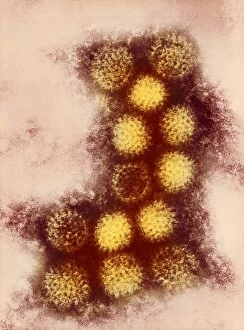
Background imageVirions Collection: Adenovirus particles, TEM

Adenovirus particles, TEM
Adenovirus particles. Coloured transmission electron micrograph (TEM) of adenovirus particles. Each particle consists of an icosahedral protein coat (capsid)

Background imageVirions Collection: H5N3 influenza A virus particles, TEM

H5N3 influenza A virus particles, TEM
H5N3 influenza A virus particles. Coloured transmission electron micrograph (TEM) of the H5N3 strain of influenza virus type A. All five H5 strains (H5N1, H5N2, H5N3, H5N8 and H5N9) cause bird flu

Background imageVirions Collection: Human coronavirus, TEM

Human coronavirus, TEM
Human coronavirus. Coloured transmission electron micrograph (TEM) of Human coronavirus particles. Coronaviruses primarily infect the upper respiratory and gastrointestinal tract

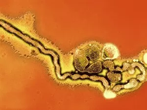
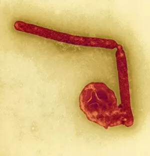
Background imageVirions Collection: Marburg virus, TEM

Marburg virus, TEM
Marburg virus, coloured transmission electron micrograph (TEM). This tubular RNA (ribonucleic acid) virus causes Marburg haemorrhagic fever in humans and non-human primates

Background imageVirions Collection: La Crosse encephalitis virus, TEM

La Crosse encephalitis virus, TEM
La Crosse encephalitis virus. Coloured transmission electron micrograph (TEM) of La Cross (LAC) encephalitis virus particles (virions)

Background imageVirions Collection: California encephalitis virus, TEM

California encephalitis virus, TEM
California encephalitis virus. Coloured transmission electron micrograph of California encephalitis virus particles (virions)

Background imageVirions Collection: Changuinola virus, TEM

Changuinola virus, TEM
Changuinola virus. Coloured transmission electron micrograph of Changuinola virus particles (virions). Each particle consists of a protein coat (capsid)

Background imageVirions Collection: Avian influenza virus particles, TEM

Avian influenza virus particles, TEM
Avian influenza virus particles. Coloured transmission electron micrograph (TEM) of a H5 strain of influenza virus type A. All five H5 strains (H5N1, H5N2, H5N3, H5N8 and H5N9) cause bird flu


For sale as Licensed Images

Choose your image, Select your licence and Download the media

Virions, the tiny particles that cause infections, come in various forms and can be seen under a transmission electron microscope (TEM). Norovirus particles, responsible for stomach bugs often spread through sneezing or contaminated surfaces, are captured in artwork C013 / 5949. Similarly, coronavirus particles can also be observed using TEM and have recently gained attention due to their role in the ongoing pandemic. Influenza virus particles, another common cause of respiratory illnesses, appear distinct under TEM as well. Hepatitis C viruses and Rift Valley fever virus are other examples that have been visualized using this powerful microscopy technique. Herpes virus particles are beautifully depicted through computer artwork, showcasing their intricate structure. Paramyxovirus particles and herpes simplex viruses are two more types that have been studied extensively with TEM. The images reveal their unique shapes and features. One striking image shows AIDS viruses budding from a cell under TEM observation. This highlights the devastating impact of these particular virions on human health. Lastly, dengue fever virus particles can also be observed using TEM technology. These tiny entities play a significant role in spreading dengue fever through mosquito bites.