
Virions Collection (#3)
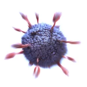
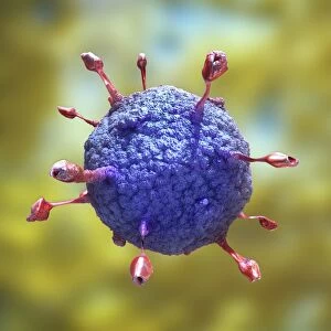
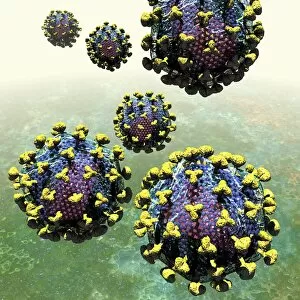
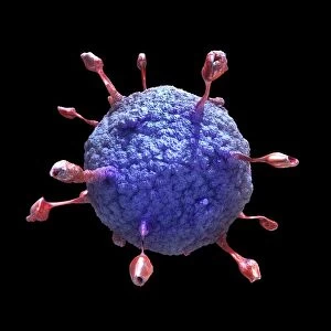
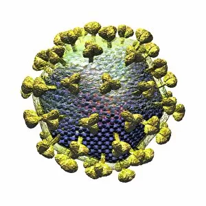
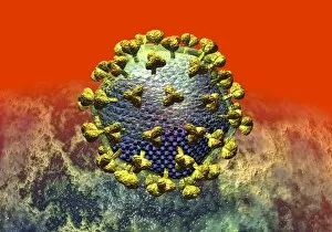
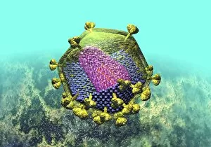
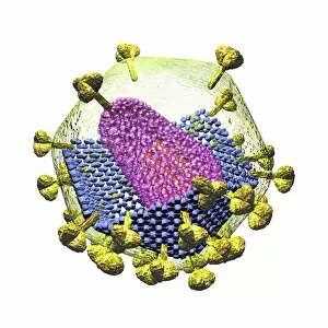
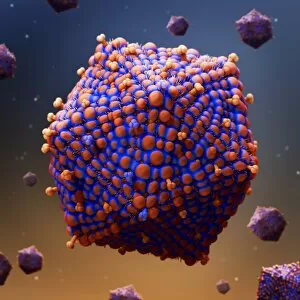
Virions, the tiny particles that cause infections, come in various forms and can be seen under a transmission electron microscope (TEM


For sale as Licensed Images
Choose your image, Select your licence and Download the media
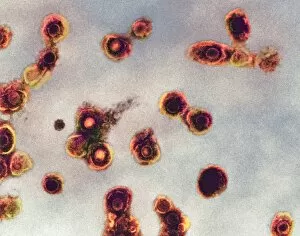
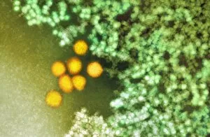
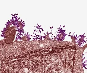
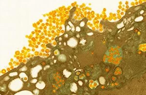
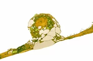
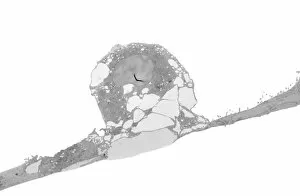
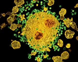
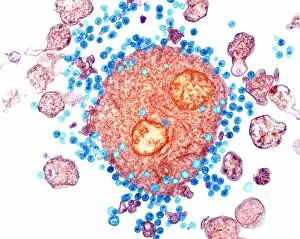
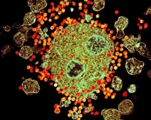
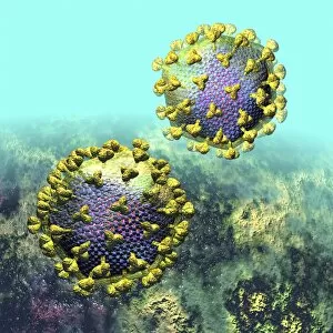
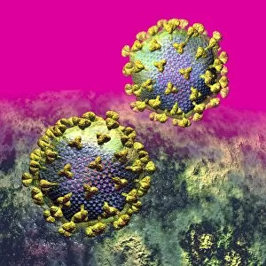
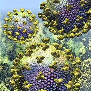
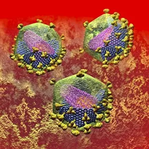
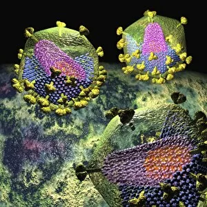
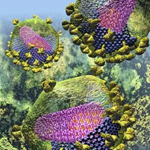
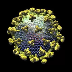
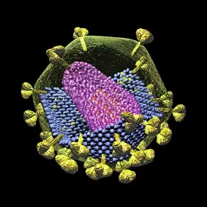
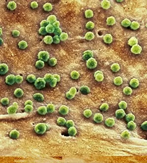
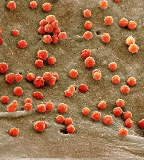
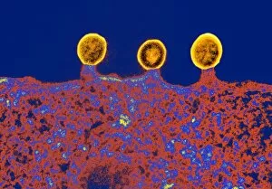
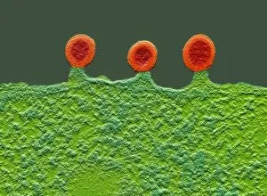
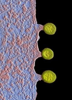
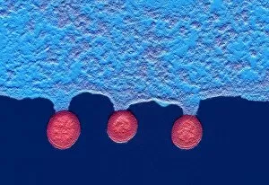
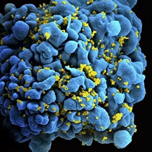
Virions, the tiny particles that cause infections, come in various forms and can be seen under a transmission electron microscope (TEM). Norovirus particles, responsible for stomach bugs often spread through sneezing or contaminated surfaces, are captured in artwork C013 / 5949. Similarly, coronavirus particles can also be observed using TEM and have recently gained attention due to their role in the ongoing pandemic. Influenza virus particles, another common cause of respiratory illnesses, appear distinct under TEM as well. Hepatitis C viruses and Rift Valley fever virus are other examples that have been visualized using this powerful microscopy technique. Herpes virus particles are beautifully depicted through computer artwork, showcasing their intricate structure. Paramyxovirus particles and herpes simplex viruses are two more types that have been studied extensively with TEM. The images reveal their unique shapes and features. One striking image shows AIDS viruses budding from a cell under TEM observation. This highlights the devastating impact of these particular virions on human health. Lastly, dengue fever virus particles can also be observed using TEM technology. These tiny entities play a significant role in spreading dengue fever through mosquito bites.