Xylem Collection (#2)

Xylem, the intricate network of plant tissues responsible for water transport and structural support, is a fascinating subject to explore under various microscopes

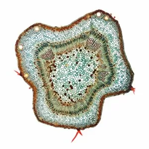
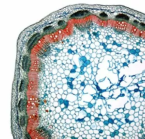
Background imageXylem Collection: Cotton stem, light micrograph

Cotton stem, light micrograph
Cotton stem. Light micrograph of a cross-section through a cotton (Gossypium sp.) stem. There is a large area of central pith surrounded by secondary xylem (light green)

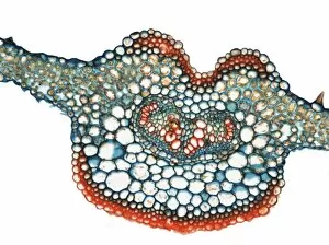
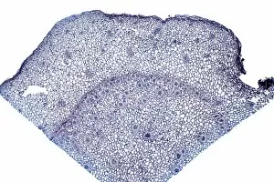
Background imageXylem Collection: Fern leaf, light micrograph

Fern leaf, light micrograph
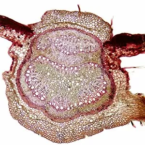
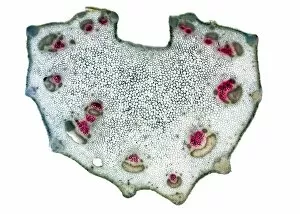
Fern leaf. Light micrograph of a transverse section through the midrib of a fern leaf. A vascular bundle is at centre. Magnification: x40 when printed at 10 centimetres wide

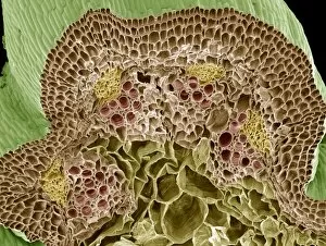
Background imageXylem Collection: Nasturtium stem, SEM

Nasturtium stem, SEM
Nasturtium stem. Coloured scanning electron micrograph (SEM) of a freeze-fractured Nasturtium (Tropaeolum sp.) stem, showing numerous vascular bundles (such as at upper centre)

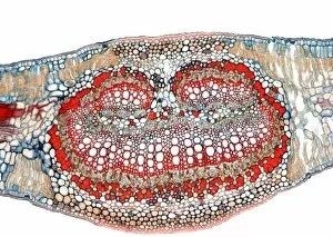
Background imageXylem Collection: Olive leaf, light micrograph

Olive leaf, light micrograph
Olive leaf. Light micrograph of a section through the mid rib of an olive leaf (Olea sativa). It has a thickened epidermis for water retention

Background imageXylem Collection: Peanut stem, light micrograph

Peanut stem, light micrograph
Peanut stem. Light micrograph of a section through a peanut stem (Arachis hypogaea). The central pith is blue. Xylem is red and phloem is black. Magnification: x60 when printed at 10 centimetres wide

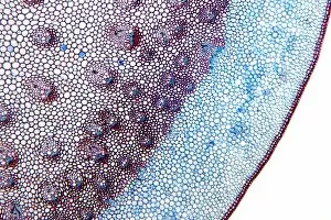
Background imageXylem Collection: Grape stem, light micrograph

Grape stem, light micrograph
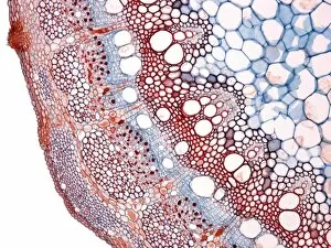
Grape stem. Light micrograph of a section through a grape stem. At top right is the pith (blue), surround that is the xylem (purple) and then the phloem (red)

Background imageXylem Collection: Sago palm leaf, light micrograph

Sago palm leaf, light micrograph
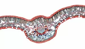
Sago palm leaf. Light micrograph of a transverse section through a sago palm (Cycas revoluta) leaf. It has a thick epidermis (outer red layer) typical of drought resistant plants

Background imageXylem Collection: Water lily stem, SEM

Water lily stem, SEM
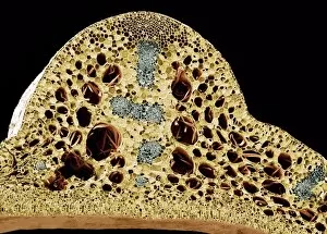
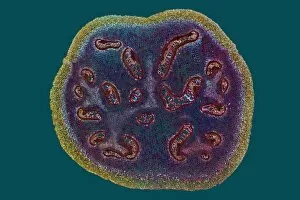
Water lily stem. Coloured scanning electron micrograph (SEM) of a freeze-fractured water lily stem showing numerous vascular bundles (grey) and large intercellular air spaces (holes)

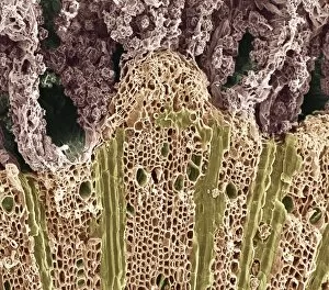
Background imageXylem Collection: Wood, SEM

Wood, SEM
Wood. Coloured scanning electron micrograph (SEM) of a section through wood showing the phloem vessels (dark green holes) and xylem tissue (lower frame)

Background imageXylem Collection: Acorus calamus rhizome, light micrograph

Acorus calamus rhizome, light micrograph
Acorus calamus rhizome. Light micrograph of a section through a rhizome from a sweet flag (Acorus calamus) plant. The rhizome has an outer layer with scattered collateral vascular bundles

Background imageXylem Collection: Dracaena draco stem, light micrograph

Dracaena draco stem, light micrograph
Dracaena draco stem. Light micrograph of a section through the stem of a young dragon tree (Dracaena draco). Shown here is an outer ring of vascular bundles, containing phloem (blue) and xylem (red)

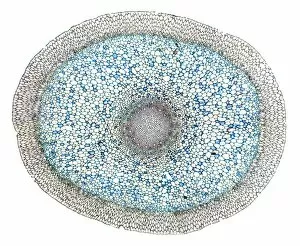
Background imageXylem Collection: Dendrobium orchid root, light micrograph

Dendrobium orchid root, light micrograph
Dendrobium orchid root. Light micrograph of a section through an aerial root from a Dendrobium sp. orchid. The outer tissue (velamen radicum, grey) is composed of hexagonal cells

Background imageXylem Collection: Sharp rush stem, light micrograph

Sharp rush stem, light micrograph
Sharp rush stem. Light micrograph of a section through the stem of a sharp rush (Juncus acutus) plant. This arid-adapted plant (xerophyte) has scattered vascular bundles

Background imageXylem Collection: Beech tree leaf, light micrograph

Beech tree leaf, light micrograph
Beech tree leaf. Light micrograph of a section through the leaf of a common beech tree (Fagus sylvatica), showing the midrib

Background imageXylem Collection: Marrow stem, light micrograph

Marrow stem, light micrograph
Marrow stem. Light micrograph of a section through the stem of a marrow (Curcurbita sp.), showing the sieve plates in the phloem. A single collateral vascular bundle can be seen

Background imageXylem Collection: Sycamore leaf vein, light micrograph

Sycamore leaf vein, light micrograph
Sycamore leaf vein. Light micrograph of a section through the midrib (vein) of a leaf from a sycamore (Acer pseudoplatanus) tree

Background imageXylem Collection: Sage stem, light micrograph

Sage stem, light micrograph
Sage stem. Light micrograph of a section through a primary stem of a scarlet sage (Salivia splendens) plant. The outer stem is covered with a thin epidermis (green) that contains stomata

Background imageXylem Collection: Rhubarb root, light micrograph

Rhubarb root, light micrograph
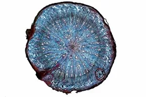
Rhubarb root. Light micrograph of a section through the root of a rhubarb (Rheum sp.) plant. The primary cortex has been shed by the formation of a circular meristem, the periderm

Background imageXylem Collection: Oak root, light micrograph

Oak root, light micrograph
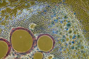
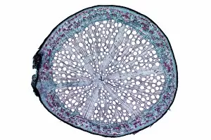
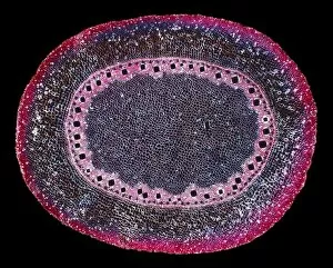
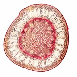
Oak root. Light micrograph of a section through a secondary root from an oak (Quercus sp.) tree. The primary cortex (outer layer) has been shed by the formation of a circular meristem, the periderm

Background imageXylem Collection: White bryony stem, light micrograph

White bryony stem, light micrograph
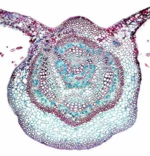
White bryony stem. Light micrograph of a transverse section through the stem of a white bryony (Bryonia alba) plant, showing a single collateral vascular bundle

Background imageXylem Collection: Beech tree leaves, light micrograph

Beech tree leaves, light micrograph
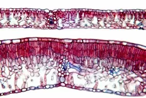
Beech tree leaves. Light micrograph of a section through two leaves from different parts of a common beech tree (Fagus sylvatica)

Background imageXylem Collection: Horse-tail stem, light micrograph

Horse-tail stem, light micrograph
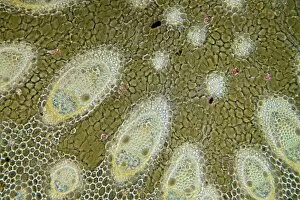
Rice grass stem. Light micrograph of a section through a rice grass stem (Oryza sativa). Vascular bundles (small oval structures) can be seen containing xylem (larger openings)

Background imageXylem Collection: Plant root development, artwork

Plant root development, artwork
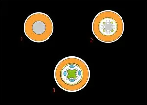
Plant root development. Artwork showing the first three stages in the development of a plant root. The first of these three stages (upper left)

Background imageXylem Collection: Plant root development, diagram

Plant root development, diagram
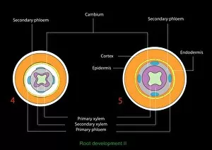
Plant root development. Diagram showing the the fourth (left) and fifth (right) stages in a series showing the development of a plant root

Background imageXylem Collection: Grape vine stem, light micrograph

Grape vine stem, light micrograph
Grape vine stem. Polarised light micrograph of a cross-section through a young stem from the grape vine (Vitis vinifera). Most of the stem is a large central pith made up of parenchyma tissue (blue)

Background imageXylem Collection: Tree growth rings, light micrograph

Tree growth rings, light micrograph
Tree growth rings. Polarised light micrograph of a cross-section through a woody stem, showing a portion of an annual ring of xylem tissue with periods of seasonal growth

Background imageXylem Collection: Virginia creeper stem, light micrograph

Virginia creeper stem, light micrograph
Virginia creeper stem. Polarised light micrograph of a cross-section through a stem from a Virginia creeper (Parthenocissus tricuspidatus)

Background imageXylem Collection: Celery stalk, light micrograph

Celery stalk, light micrograph
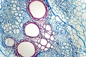
Celery stalk. Light micrograph of a cross-section through a stalk from a celery plant (Apium graveolens). Red eosin dye has been used to stain the xylem tissues

Background imageXylem Collection: Tree-of-heaven stem, light micrograph

Tree-of-heaven stem, light micrograph
Tree-of-heaven stem. Polarised light micrograph of a cross-section through the stem of the tree-of-heaven (Ailanthus glandulosa). Below the outer layers (red) is a ring of vascular bundles

Background imageXylem Collection: Aralia leaf stalk, light micrograph

Aralia leaf stalk, light micrograph
Aralia leaf stalk. Light micrograph of a cross-section through a leaf stalk (petiole) from an Araliatree. The outer layer directly below the epidermis is a supporting collenchyma ring (red)

Background imageXylem Collection: Bracken fern rhizome, light micrograph

Bracken fern rhizome, light micrograph
Bracken fern rhizome. Polarised light micrograph of a cross-section through a rhizome from a bracken fern (Pteris aquilinum). The outer layer (yellow-green) consists of supporting fibres

Background imageXylem Collection: Burdock stem, light micrograph

Burdock stem, light micrograph
Burdock stem. Polarised light micrograph of a cross-section through the stem of a burdock plant (Arctium lappa). The outer layers below the epidermis are the collenchyma (red patches)

Background imageXylem Collection: Corn (Zea mays) root, light micrograph

Corn (Zea mays) root, light micrograph
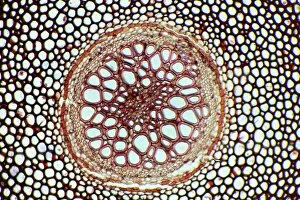
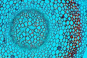
Maize root. Polarised light micrograph of a section through the root of a maize plant (Zea mays) showing a vascular cylinder

Background imageXylem Collection: Fern stem, light micrograph

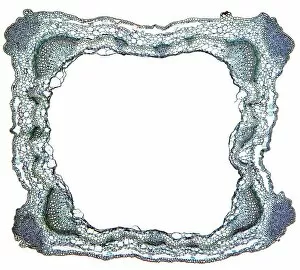
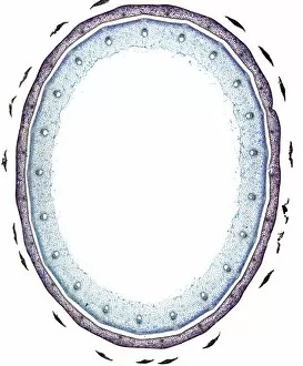
Fern stem, light micrograph. Transverse section through a rachis (stem) of the bracken fern (Pteridium aquilinum). Under the outer epidermis (black) is a thin cortex (deep red)
Background imageXylem Collection: Fern rhizome, light micrograph

Fern rhizome, light micrograph. Transverse section through the center of a rhizome from the Killarney fern (Trichomanes speciosum)
Background imageXylem Collection: Clubmoss stem, light micrograph

Clubmoss stem, light micrograph
Clubmoss stem, polarised light micrograph. Transverse section through the stem of the clubmoss Lycopodium clavatum. This is the central portion of the stem consisting of the inner cortex (red)
Background imageXylem Collection: Butchers broom stem, light micrograph

Butchers broom stem, light micrograph
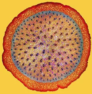
Butchers broom stem. Polarised light micrograph of a section through a butchers broom stem (Ruscus aculeatis) showing many vascular bundles (dark clusters) within the plant cortex (orange)

Background imageXylem Collection: Umbrella grass stem, light micrograph

Umbrella grass stem, light micrograph
Umbrella grass stem. Polarised light micrograph of a section through a umbrella grass stem (Cyperus alternifolius). Vascular bundles (brown) can be seen containing xylem (larger openings)

Background imageXylem Collection: Flax plant stem, light micrograph

Flax plant stem, light micrograph
Flax plant stem. Light micrograph of a transverse section through a stem of the flax plant (Linum usitatissimum). The layers from outer to inner (some very thin) are the epidermis (bottom)

Background imageXylem Collection: Purple moor grass stem, light micrograph

Purple moor grass stem, light micrograph
Purple moor grass stem. Polarised light micrograph of a section through a purple moor grass stem (Molinia caerula). Vascular bundles (round orange structures)


For sale as Licensed Images

Choose your image, Select your licence and Download the media

Xylem, the intricate network of plant tissues responsible for water transport and structural support, is a fascinating subject to explore under various microscopes. From scanning electron microscopy (SEM) images capturing the detailed vascular bundles to light micrographs showcasing different plant parts, xylem reveals its beauty and functionality. In a castor oil stem captured by a light micrograph, we witness the complexity tissue as it forms an interconnected system of vessels that efficiently transports water from roots to leaves. Similarly, in a maize root image taken with a light microscope, we can observe the organized arrangement cells that enable efficient uptake of water and nutrients. Moving on to leaf structures, a captivating light micrograph showcases the delicate veins within a water lily leaf. These intricate networks are composed mainly tissue which ensures proper hydration throughout the leaf's surface area. Another stunning SEM image captures silver birch twig's xylem vessels in detail - their elongated shapes perfectly adapted for fluid transportation. Exploring further into plants' underground systems, we encounter an enchanting light micrograph displaying the rhizome structure of a water fern. Xylem tissues play an essential role here too by providing support and facilitating nutrient absorption from surrounding soil particles. Light micrographs also capture xylem wonders in stems like those found in pine trees or pondweeds. Pine tree stems exhibit dense clusters of tracheids – specialized cells within xylem – while pondweed stems showcase long strands forming hollow tubes for efficient water movement. Even at smaller scales observed through SEM imaging techniques, such as examining isolated samples of pure xylem tissue or wood itself under high magnification power; one can appreciate its intricate cellular arrangements that allow for optimal fluid flow and mechanical stability. Lastly, exploring below ground once again brings us to oak roots depicted through another mesmerizing light micrograph.