Metal Print > Arts > Artists > P > Francis Place
Metal Print : Eye anatomy, artwork


![]()
Metal Prints from Science Photo Library
Eye anatomy, artwork
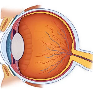
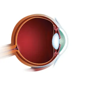
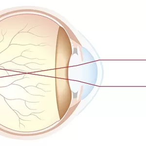
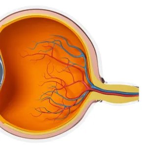
Eye anatomy, computer artwork. At the front of the eye is the cornea, a transparent coating. Behind this is the lens, which is partly covered by the iris. The lens focuses light on the retina at the back of the eye. Light sensitive cells in the retina transmit impulses to the brain via the optic nerve. Behind the retina is the choroid, which contains blood vessels that nourish the back of the eye. The outer layer is the sclera, the white of the eye. The eyeball is filled with a gelatinous substance called vitreous humor. Several muscles hold the eyeball in place and allow it to rotate
Science Photo Library features Science and Medical images including photos and illustrations
Media ID 6326713
© FRANCIS LEROY, BIOCOSMOS/SCIENCE PHOTO LIBRARY
Choroid Cornea Interior Iris Label Labeled Labelled Labels Lens Muscles Ocular Retina Sclera Sense Sight Text Vision Visual Sectioned
16"x20" (51x41cm) Metal Print
Discover the intricacies of the human eye with our captivating Metal Prints from Media Storehouse, featuring Science Photo Library's stunning artwork. This mesmerizing image showcases the anatomy of the eye, highlighting the transparency of the cornea at the forefront and the partially covered lens behind it, with the iris adding a dynamic touch. Bring this scientific masterpiece into your home or office to ignite curiosity and inspire conversation. Our high-quality Metal Prints are known for their vibrant colors, sharp details, and long-lasting durability. Order yours today and embark on a visual journey into the marvels of the human body.
Made with durable metal and luxurious printing techniques, our metal photo prints go beyond traditional canvases, adding a cool, modern touch to your space. Wall mount on back. Eco-friendly 100% post-consumer recycled ChromaLuxe aluminum surface. The thickness of the print is 0.045". Featuring a Scratch-resistant surface and Rounded corners. Backing hangers are attached to the back of the print and float the print 1/2-inch off the wall when hung, the choice of hanger may vary depending on size and International orders will come with Float Mount hangers only. Finished with a brilliant white high gloss surface for unsurpassed detail and vibrance. Printed using Dye-Sublimation and for best care we recommend a non-ammonia glass cleaner, water, or isopropyl (rubbing) alcohol to prevent harming the print surface. We recommend using a clean, lint-free cloth to wipe off the print. The ultra-hard surface is scratch-resistant, waterproof and weatherproof. Avoid direct sunlight exposure.
Made with durable metal and luxurious printing techniques, metal prints bring images to life and add a modern touch to any space
Estimated Image Size (if not cropped) is 50.8cm x 35.7cm (20" x 14.1")
Estimated Product Size is 51.4cm x 41.2cm (20.2" x 16.2")
These are individually made so all sizes are approximate
Artwork printed orientated as per the preview above, with landscape (horizontal) orientation to match the source image.
FEATURES IN THESE COLLECTIONS
> Arts
> Artists
> P
> Francis Place
EDITORS COMMENTS
This print showcases the intricate anatomy of the human eye, beautifully depicted through computer artwork. The image highlights various components that contribute to our sense of sight. At the forefront is the transparent cornea, acting as a protective coating for the eye. Just behind it lies the lens, partially covered by the colorful iris, which plays a crucial role in adjusting light intake. The lens diligently focuses incoming light onto the retina positioned at the back of the eye. Within this delicate layer reside light-sensitive cells that transmit visual impulses to our brain via the optic nerve. Behind this vital network lies another essential structure called choroid, housing blood vessels responsible for nourishing and maintaining optimal functioning of our eyes. The outermost layer known as sclera forms an elegant white covering around our eyeball, providing structural support and protection. Filling up most of its interior is a gelatinous substance called vitreous humor, ensuring proper shape and stability. To facilitate movement and rotation, several muscles work in harmony to hold and position our eyeball accurately within its socket. This comprehensive illustration serves as an invaluable tool for understanding normal eye anatomy with labeled details aiding comprehension. Immerse yourself in this visually stunning piece that not only celebrates biology but also invites contemplation on how we perceive and interpret our surroundings through this remarkable organ - our eyes!
MADE IN THE USA
Safe Shipping with 30 Day Money Back Guarantee
FREE PERSONALISATION*
We are proud to offer a range of customisation features including Personalised Captions, Color Filters and Picture Zoom Tools
SECURE PAYMENTS
We happily accept a wide range of payment options so you can pay for the things you need in the way that is most convenient for you
* Options may vary by product and licensing agreement. Zoomed Pictures can be adjusted in the Cart.