
Canvas Print > Science > SEM
Canvas Print : Blood clot, SEM


![]()
Canvas Prints from Science Photo Library
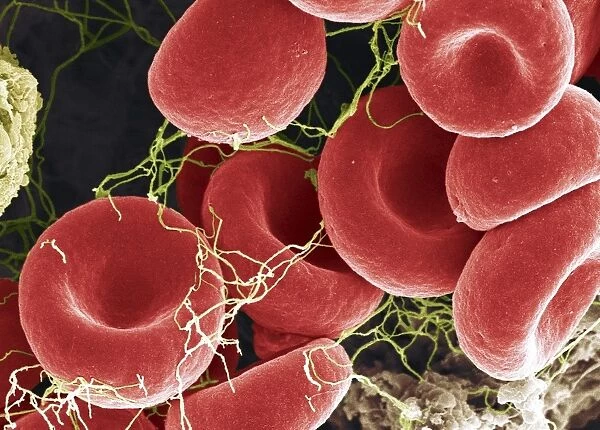
Blood clot, SEM
Blood clot, coloured scanning electron micrograph (SEM). Red blood cells are red and fibrin protein strands are green. Platelets are at bottom right. Magnification: x4000 when printed at 10 centimetres wide
Science Photo Library features Science and Medical images including photos and illustrations
Media ID 6367575
© STEVE GSCHMEISSNER/SCIENCE PHOTO LIBRARY
Blood Blood Cell Blood Clot Clotting Coagulated Coagulating Coagulation Erythrocyte Erythrocytes False Colour Fibrin Haematology Hematology Platelet Platelets Rbcs Red Blood Cell Red Blood Cells Strand Strands Thrombocyte Vascular False Coloured Protein
20"x16" (51x41cm) Canvas Print
Discover the intricacy of life with Media Storehouse's Canvas Prints featuring this captivating Scanning Electron Micrograph (SEM) image from Science Photo Library. Witness the complex structure of a blood clot as red blood cells intertwine with green fibrin protein strands and platelets at the bottom right. Bring this mesmerizing scientific discovery into your home or office as a unique conversation starter and a testament to the beauty of science. Our high-quality canvas prints are designed to bring vibrant colors and stunning detail to your space, making this an unforgettable addition to your decor.
Delivered stretched and ready to hang our premium quality canvas prints are made from a polyester/cotton blend canvas and stretched over a 1.25" (32mm) kiln dried knot free wood stretcher bar. Packaged in a plastic bag and secured to a cardboard insert for safe transit.
Canvas Prints add colour, depth and texture to any space. Professionally Stretched Canvas over a hidden Wooden Box Frame and Ready to Hang
Estimated Product Size is 50.8cm x 40.6cm (20" x 16")
These are individually made so all sizes are approximate
Artwork printed orientated as per the preview above, with landscape (horizontal) orientation to match the source image.
EDITORS COMMENTS
A captivating coloured scanning electron micrograph (SEM) of a blood clot reveals the intricate structure of this vital biological process. The image showcases a complex network of red blood cells (erythrocytes, shown in red) and fibrin protein strands (green) that come together to form a clot and prevent excessive blood loss. The platelets (thrombocytes, located at the bottom right) play a crucial role in the clotting process by releasing chemicals that initiate the coagulation cascade. Magnified at 4000x when printed at 10 centimetres wide, this false-coloured image offers a mesmerizing glimpse into the world of haematology. The delicate strands of fibrin, which act as a scaffold for the clot, can be seen intertwining and binding the red blood cells together. The platelets, which are essential for the clotting process, can be observed at the bottom right of the image, clustered together and appearing translucent in this micrograph. The intricacy of the blood clotting process is a testament to the marvels of biology. This image, taken using a scanning electron microscope, provides a unique perspective on the complex interplay of various blood components in the formation of a clot. The vibrant colours and high magnification offer a striking visual representation of the coagulation process that is essential for maintaining vascular health and preventing excessive blood loss.
MADE IN THE USA
Safe Shipping with 30 Day Money Back Guarantee
FREE PERSONALISATION*
We are proud to offer a range of customisation features including Personalised Captions, Color Filters and Picture Zoom Tools
SECURE PAYMENTS
We happily accept a wide range of payment options so you can pay for the things you need in the way that is most convenient for you
* Options may vary by product and licensing agreement. Zoomed Pictures can be adjusted in the Cart.