Framed Print : Cell nucleus, SEM


![]()
Framed Photos from Science Photo Library
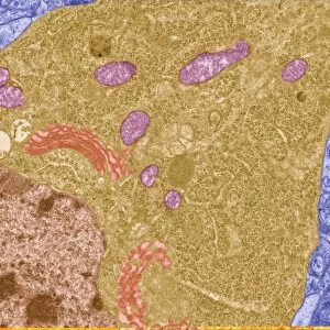
Cell nucleus, SEM
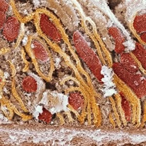
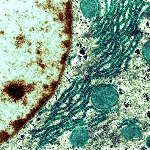
Cell nucleus. Coloured scanning electron micrograph (SEM) of a section through the nucleus (centre right) of a kidney cell. The cell nucleus contains the cells genetic information in the form of DNA (deoxyribonucleic acid). Here the DNA is complexed with proteins to form chromatin (light brown structures). Surrounding the nucleus are mitochondria (ovoids), which supply the cell with energy
Science Photo Library features Science and Medical images including photos and illustrations
Media ID 6302919
© DR DAVID FURNESS, KEELE UNIVERSITY/SCIENCE PHOTO LIBRARY
Cell Biology Chromatin Cross Section Cytology Cytoplasm Energy False Colour Genetic Information Kidney Mitochondrion Nucleus Organelle Renal Structures Deoxyribonucleic Acid False Coloured Section Sectioned
12"x10" Modern Frame
Bring the wonders of the microscopic world into your home or office with our Framed Prints from Media Storehouse. This stunning coloured Scanning Electron Micrograph (SEM) of a kidney cell's nucleus, captured by Science Photo Library, offers a captivating glimpse into the intricacies of cellular biology. Each print is meticulously framed in a sleek, modern design, ensuring your new addition becomes a conversation starter and a source of inspiration. Experience the beauty of science in high definition, right at your fingertips.
10x8 Print in an MDF Wooden Frame with 180 gsm Satin Finish Paper. Glazed using shatter proof thin plexi glass. Frame thickness is 1 inch and depth 0.75 inch. Fluted cardboard backing held with clips. Supplied ready to hang with sawtooth hanger and rubber bumpers. Spot clean with a damp cloth. Packaged foam wrapped in a card.
Contemporary Framed and Mounted Prints - Professionally Made and Ready to Hang
Estimated Image Size (if not cropped) is 25.4cm x 25.4cm (10" x 10")
Estimated Product Size is 30.5cm x 25.4cm (12" x 10")
These are individually made so all sizes are approximate
Artwork printed orientated as per the preview above, with landscape (horizontal) or portrait (vertical) orientation to match the source image.
EDITORS COMMENTS
This print showcases the intricate beauty of a cell nucleus, captured through a scanning electron microscope (SEM). At the center-right of the image, we are drawn to the nucleus of a kidney cell. This vital organelle houses the genetic information that defines and directs cellular functions - DNA or deoxyribonucleic acid. The DNA within this nucleus is not alone; it forms complex structures with proteins known as chromatin, which appear as light brown formations. These chromatin structures play a crucial role in regulating gene expression and maintaining genomic stability. Surrounding the nucleus, we observe mitochondria – ovoid-shaped powerhouses responsible for supplying energy to the cell. Their presence emphasizes their significance in sustaining cellular activities and ensuring proper functioning. Through this false-colored SEM image, we gain insight into both structure and function at an incredibly detailed level. It serves as a reminder of how biology unravels its secrets through microscopic exploration. This mesmerizing snapshot delves into various aspects of cellular biology: from cross-sections to genetic information storage; from energy supply mechanisms to intricate protein-DNA complexes. Science Photo Library has once again provided us with an awe-inspiring glimpse into nature's wonders at its most fundamental level.
MADE IN THE USA
Safe Shipping with 30 Day Money Back Guarantee
FREE PERSONALISATION*
We are proud to offer a range of customisation features including Personalised Captions, Color Filters and Picture Zoom Tools
SECURE PAYMENTS
We happily accept a wide range of payment options so you can pay for the things you need in the way that is most convenient for you
* Options may vary by product and licensing agreement. Zoomed Pictures can be adjusted in the Cart.