
Anatomie Collection
"Exploring the Intricacies of Anatomie: From Ligaments to Artwork" Delve into the fascinating world of anatomie, where the human body and art intertwine

For sale as Licensed Images
Choose your image, Select your licence and Download the media
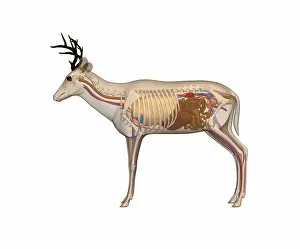
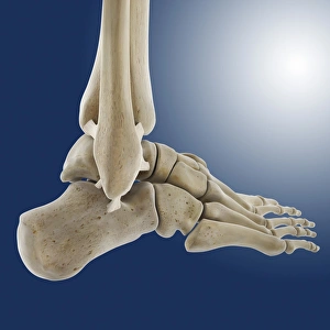
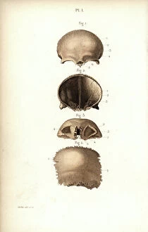
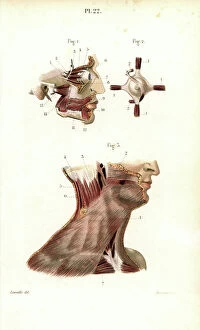
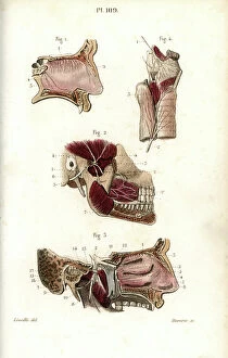
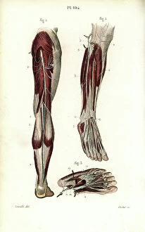
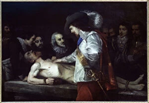
"Exploring the Intricacies of Anatomie: From Ligaments to Artwork" Delve into the fascinating world of anatomie, where the human body and art intertwine. Journey through a diverse collection of captivating images and historical references that shed light on our understanding of anatomy. Discover the delicate outer ankle ligaments in artwork C013/4452, beautifully depicted with meticulous detail. Witness how artists skillfully capture not only the physicality but also the essence of these intricate structures. Step into a different realm as dog anatomy takes center stage in another mesmerizing artwork. Uncover the intricacies of canine physiology, marveling at how their bodies are both similar and distinct from ours. Travel back in time to Uncle Tom's Cabin, as George Cruikshank's illustration brings Harriet Beecher Stowe's powerful novel to life. Explore how literature can provide insight into societal perceptions and challenges faced by individuals during that era. Max Ernst's painting "The Couple" offers an intriguing glimpse into human relationships and emotions. Delicate brushstrokes convey a depth of feeling that transcends time, inviting us to contemplate love, connection, and vulnerability. Witness Doctor Alfred Velleau conducting an autopsy amidst his attentive students in Francois Feyen Perrin's 19th-century masterpiece. This thought-provoking image captures both scientific curiosity and reverence for knowledge as medical professionals strive to unravel mysteries hidden within our bodies. Jean-Baptiste Marc Bourgery's colored engraving showcases amputations and prosthetics—a testament to humanity's resilience even in times of war-induced adversity. These artificial limbs symbolize hope for soldiers who have endured unimaginable hardships on the battlefield. "The Anatomy Lesson" series by Drs Nicolaes Tulp and Willem van der Meer transports us back centuries ago when dissection was a groundbreaking practice. These oil paintings immortalize moments where science meets artistry while unraveling secrets concealed beneath our skin.