
Binding Collection (#8)
"Binding: From Ancient Techniques to Modern Science" Step into the world of binding, where artistry and science intertwine


For sale as Licensed Images
Choose your image, Select your licence and Download the media
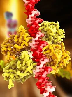
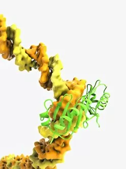
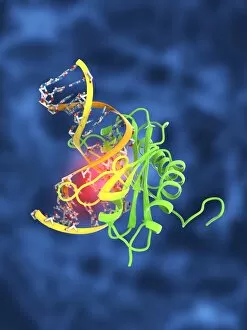
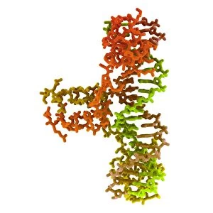
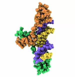
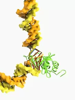
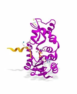
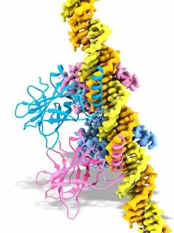
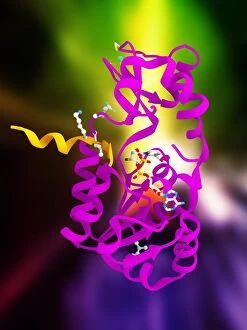
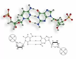
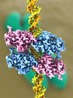
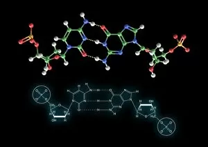
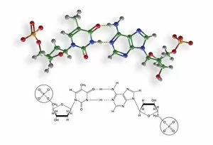
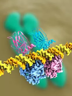
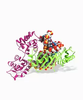
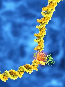
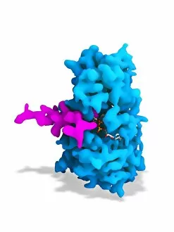
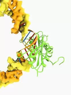
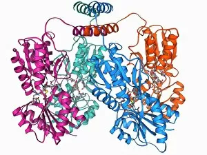
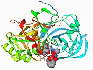
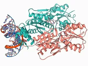
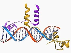
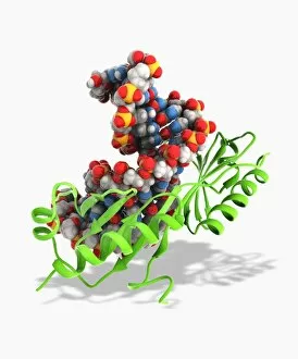
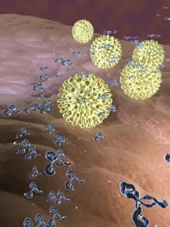
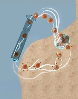
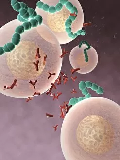
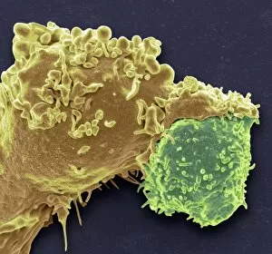
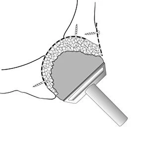
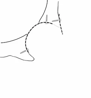
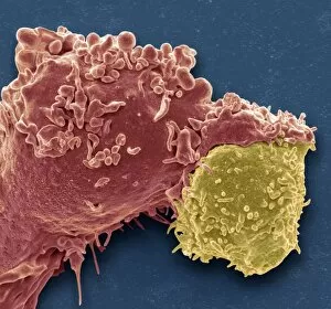
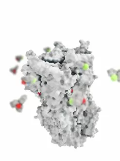
"Binding: From Ancient Techniques to Modern Science" Step into the world of binding, where artistry and science intertwine. Explore the delicate craft of bookbinding, as Bookbinding Tools from 1875 take center stage in preserving knowledge through time. Travel back in time with a Greek vase painting depicting Achilles and Patroclus, showcasing their unbreakable bond. Witness how even in ancient times, binding went beyond physical objects. Discover the microscopic realm where T lymphocytes bind to cancer cells, captured beautifully under SEM C001 / 1679. Marvel at the intricate dance between life and disease on a cellular level. Uncover the secrets behind anesthesia's power to inhibit ion channels with Anaesthetic inhibiting an ion channel C015 / 6718. See how this binding effect brings relief and comfort during medical procedures. Enter a bustling Bookbinding Workshop filled with artisans meticulously stitching pages together, breathing life into stories yet untold. Witness their dedication to preserving history one book at a time. Delve into Japanese culture as you admire Netsuke in the form of a demon queller and small demon from the Meiji Period (1868-1912). These tiny sculptures embody both beauty and protection through their bound forms. Marvel at EDTA crystals under a light microscope; these enchanting structures showcase nature's ability to create mesmerizing patterns when elements bind together harmoniously. Witness Achilles' unwavering loyalty as he binds Patroclus' wound on an ancient battlefield - an act that transcends mere friendship and symbolizes sacrifice for those we hold dear. Immerse yourself in artwork depicting cannabinoid receptor binding - a visual representation of how substances can interact with our bodies on molecular levels, unlocking new possibilities for medicine and wellness. Explore official wording within Apprenticeship Indenture documents that bind individuals to learn valuable skills from experienced masters - highlighting the importance of passing down knowledge through generations. Contemplate Chinese Foot-Binding, a practice that bound women's feet for centuries.