Binding Collection (#9)

"Binding: From Ancient Techniques to Modern Science" Step into the world of binding, where artistry and science intertwine

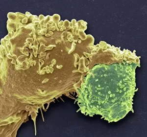
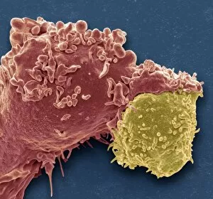
Background imageBinding Collection: White blood cell antigen presentation C016 / 9058

White blood cell antigen presentation C016 / 9058
White blood cell antigen presentation. Coloured scanning electron micrograph (SEM) showing the interaction between a macrophage (yellow) and a T helper lymphocyte (Th cell, green)

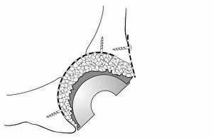
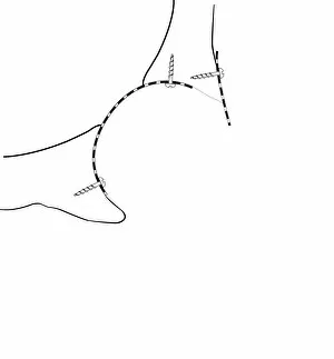
Background imageBinding Collection: Hip socket bone grafting, diagram C016 / 6786

Hip socket bone grafting, diagram C016 / 6786
Hip socket bone grafting. Diagram showing completed hip socket bone grafting as part of a total hip replacement operation

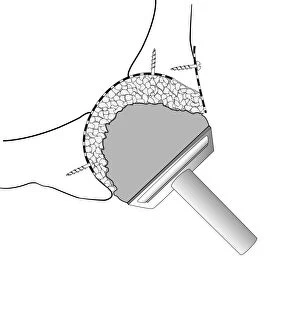
Background imageBinding Collection: Hip socket bone grafting, diagram C016 / 6784

Hip socket bone grafting, diagram C016 / 6784
Hip socket bone grafting. Diagram showing the second stage in hip socket bone grafting as part of a total hip replacement operation

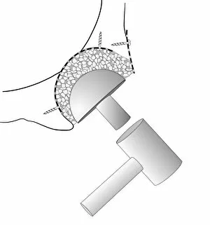
Background imageBinding Collection: Hip socket bone grafting, diagram C016 / 6785

Hip socket bone grafting, diagram C016 / 6785
Hip socket bone grafting. Diagram showing the third stage in hip socket bone grafting as part of a total hip replacement operation

Background imageBinding Collection: Hip socket bone grafting, diagram C016 / 6783

Hip socket bone grafting, diagram C016 / 6783
Hip socket bone grafting. Diagram showing the first stage in hip socket bone grafting as part of a total hip replacement operation

Background imageBinding Collection: White blood cell antigen presentation C016 / 9057

White blood cell antigen presentation C016 / 9057
White blood cell antigen presentation. Coloured scanning electron micrograph (SEM) showing the interaction between a macrophage (red) and a T helper lymphocyte (Th cell, yellow)

Background imageBinding Collection: Nerve growth factor bound to receptor

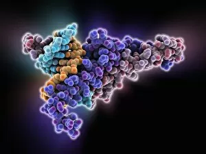
Nerve growth factor bound to receptor, molecular model. Nerve growth factor (NGF) complexed with the TrkA receptor. NGF is a neurotrophin that acts on the development and function of nerves

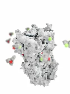
Background imageBinding Collection: Ketamine drug binding to ion channel

Ketamine drug binding to ion channel, molecular model. Several molecules of the drug ketamine binding to a pentameric ligand-gated ion channel (pLGIC)

Background imageBinding Collection: MyoD muscle protein-DNA complex

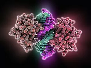
MyoD muscle protein-DNA complex. Molecular model of the MyoD muscle protein (green and blue) bound to a strand of DNA (deoxyribonucleic acid, orange and green)

Background imageBinding Collection: Tumour suppressor protein and DNA C016 / 6264

Tumour suppressor protein and DNA C016 / 6264
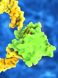
Tumour suppressor protein and DNA. Computer artwork showing a molecule of the tumour suppressor protein p53 (blue and green) bound to a strand of DNA (deoxyribonucleic acid, grey)

Background imageBinding Collection: Interferon-DNA transcription complex C015 / 8251

Interferon-DNA transcription complex C015 / 8251
Interferon-DNA transcription complex, molecular model. Bound to the DNA (deoxyribonucleic acid, green and yellow) is transcription factor p65, interferon regulatory factor 7

Background imageBinding Collection: Interferon-DNA transcription complex C015 / 8252

Interferon-DNA transcription complex C015 / 8252
Interferon-DNA transcription complex, molecular model. Bound to the DNA (deoxyribonucleic acid, pink and white) is transcription factor p65, interferon regulatory factor 7, interferon fusion protein

Background imageBinding Collection: Vitamin B12 import proteins C015 / 9942

Vitamin B12 import proteins C015 / 9942
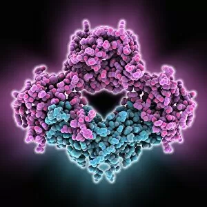
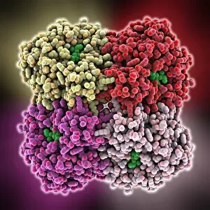
Vitamin B12 import proteins, molecular model. This complex is the import proteins btuC, btuD, and btuF. The first two together form BtuCD

Background imageBinding Collection: Vitamin B12 import proteins C015 / 9943

Vitamin B12 import proteins C015 / 9943
Vitamin B12 import proteins, molecular model. This complex is the import proteins btuC, btuD, and btuF. The first two together form BtuCD

Background imageBinding Collection: Tumour suppressor protein and DNA C017 / 3643

Tumour suppressor protein and DNA C017 / 3643
Tumour suppressor protein and DNA. Computer artwork showing a molecule of the tumour suppressor protein p53 (blue and pink) bound to a molecule of DNA (deoxyribonucleic acid, yellow and orange)

Background imageBinding Collection: Homeodomain from Ubx and Exd protein C017 / 7006

Homeodomain from Ubx and Exd protein C017 / 7006
Structure of a DNA-bound Ultrabithorax (Ubx) and Extradenticle (Exd) homeodomain complex bound to DNA, showing how one of the helical regions fits into a major groove on the doulbe-helical DNA

Background imageBinding Collection: Thymine-adenine interaction, artwork C017 / 7368

Thymine-adenine interaction, artwork C017 / 7368
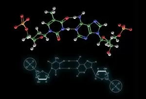
Thymine-adenine interaction. Computer artwork showing the structure of bound thymine and adenine molecules. Atoms are shown as colour-coded spheres: carbon (green), hydrogen (white)
Background imageBinding Collection: TATA box-binding protein complex C017 / 7087

TATA box-binding protein complex C017 / 7087
TATA box-binding protein complex. Molecular model showing a TATA box-binding protein (TBP, green) complexed with a strand of DNA (deoxyribonucleic acid, yellow) and transcription factor IIB

Background imageBinding Collection: E. coli DNA mismatch repair complex

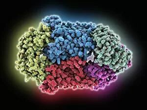
E. coli DNA mismatch repair complex. Computer model showing the structure of a MutS (Mutator S) protein from Escherichia coli (E)

Background imageBinding Collection: Bone morphogenetic protein complex

Bone morphogenetic protein complex, molecular model. Bone Morphogenetic Protein-7 (BMP-7, blue) in complex with the secreted antagonist Noggin (pink)

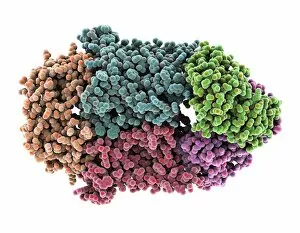
Background imageBinding Collection: FP2 malaria protease enzyme complex

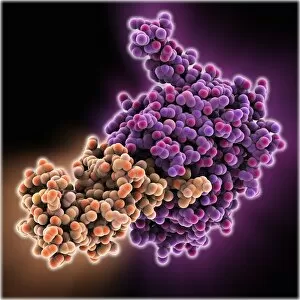
FP2 malaria protease enzyme complex, molecular model. This complex consists of the falcipain-2 (FP2) protease enzyme (purple, right) bound to a cystatin (orange, left), a form of protease inhibitor

Background imageBinding Collection: Oestrogen related receptor-DNA complex

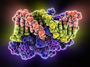
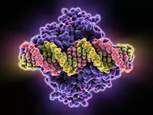
Oestrogen related receptor-DNA complex. Molecular model of human estrogen related receptor-2 (heRR-2, purple) binding to a strand of DNA (deoxyribonucleic acid, red and yellow-green)

Background imageBinding Collection: Lambda repressor-operator complex

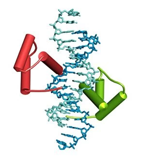
Lambda repressor-operator complex. Molecular model of the lambda repressor protein (red and green) binding to a region of DNA (deoxyribonucleic acid, orange and blue) known as the lambda operator

Background imageBinding Collection: Repair protein and DNA, molecular model

Repair protein and DNA, molecular model
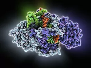
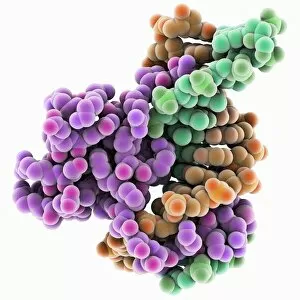
Repair protein and DNA. Molecular model of the Ku heterodimer (grey, blue and purple) bound to a strand of DNA (deoxyribonucleic acid, orange and green) as part of the repair process

Background imageBinding Collection: Max transcription factor-DNA complex

Max transcription factor-DNA complex. Molecular model of the Max transcription factor (purple and red) bound to a strand of DNA (deoxyribonucleic acid, light blue and orange)

Background imageBinding Collection: Antibody fragment-lysozyme complex

Antibody fragment-lysozyme complex
fab d1.3, , chicken egg white lysozyme, protein, biomolecule, macromolecule, lysozyme, enzyme, fab, antigen, antibody, biochemistry, biology, molecular biology, proteomics, artwork, illustration

Background imageBinding Collection: Bacterial regulator-DNA complex

Bacterial regulator-DNA complex. Molecular model of a complex formed between a bacterial regulator called SarA (orange and brown) and a fragment of DNA (pink and yellow-green strands)

Background imageBinding Collection: Bacterial protein-chaperone complex

Bacterial protein-chaperone complex. Molecular model of a bacterial effector protein binding to a chaperone protein that helps prevent keep the bacterial protein in an unfolded or partially folded

Background imageBinding Collection: Hin recombinase-DNA complex

Hin recombinase-DNA complex. Molecular model of the Hin recombinase protein (pink) bound to a double helix (green and orange) strand of DNA (deoxyribonucleic acid)

Background imageBinding Collection: Tumour suppressor protein molecular model C016 / 2065

Tumour suppressor protein molecular model C016 / 2065
Tumour suppressor protein. Molecular model of the tumour suppressor protein p53 (left and right) bound to a molecule of DNA (deoxyribonucleic acid, down centre) at the p53 response element

Background imageBinding Collection: DNA mismatch repair complex

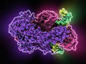
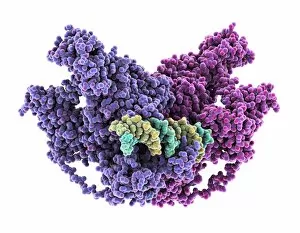
DNA mismatch repair complex. Computer model showing the structure of a MutS (Mutator S) protein binding to DNA (deoxyribonucleic acid) during DNA mismatch repair

Background imageBinding Collection: Aspirin in action

Aspirin in action. Computer artwork showing how aspirin has its effect. Aspirin (acetylsalicylic acid) is converted to salicylic acid and acetic acid in the body

Background imageBinding Collection: Methyltransferase complexed with DNA C016 / 2033

Methyltransferase complexed with DNA C016 / 2033
Methyltransferase complexed with DNA, molecular model. The strand of DNA (deoxyribonucleic acid, green and blue) is enclosed by DNA methyltransferase 1 (DNMT-1, green and pink)

Background imageBinding Collection: Methyltransferase complexed with DNA C016 / 2032

Methyltransferase complexed with DNA C016 / 2032
Methyltransferase complexed with DNA, molecular model. The strand of DNA (deoxyribonucleic acid, green and yellow) is enclosed by DNA methyltransferase 1 (DNMT-1, purple and pink)

Background imageBinding Collection: Notch transcription, molecular model

Notch transcription, molecular model
Notch transcription. Molecular model showing a strand of DNA (deoxyribonucleic acid) being acted upon by various proteins and other molecules

Background imageBinding Collection: Beta secretase inhibitor, molecular model

Beta secretase inhibitor, molecular model. The inhibitor molecule (centre, also shown in C015/1977) is bound to the beta secretase enzyme (partially seen)

Background imageBinding Collection: Anaesthetic inhibiting an ion channel C015 / 6723

Anaesthetic inhibiting an ion channel C015 / 6723
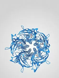
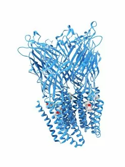
Anaesthetic inhibiting an ion channel. Computer model showing the structure of propofol anaesthetic drug molecules (spheres) bound to a pentameric ligand-gated ion channel (pLGIC, blue ribbons)

Background imageBinding Collection: Anaesthetic inhibiting an ion channel C015 / 6722

Anaesthetic inhibiting an ion channel C015 / 6722
Anaesthetic inhibiting an ion channel. Computer model showing the structure of propofol anaesthetic drug molecules (spheres) bound to a pentameric ligand-gated ion channel (pLGIC, blue ribbons)

Background imageBinding Collection: Anaesthetic inhibiting an ion channel C015 / 6720

Anaesthetic inhibiting an ion channel C015 / 6720
Anaesthetic inhibiting an ion channel. Computer model showing the structure of propofol anaesthetic drug molecules (lower left and right) bound to a pentameric ligand-gated ion channel (pLGIC, grey)

Background imageBinding Collection: Anaesthetic inhibiting an ion channel C015 / 6721

Anaesthetic inhibiting an ion channel C015 / 6721
Anaesthetic inhibiting an ion channel. Computer model showing the structure of propofol anaesthetic drug molecules (spheres)

Background imageBinding Collection: Anaesthetic inhibiting an ion channel C015 / 6719

Anaesthetic inhibiting an ion channel C015 / 6719
Anaesthetic inhibiting an ion channel. Computer model showing the structure of propofol anaesthetic drug molecules (spheres)

Background imageBinding Collection: Flu virus surface protein and drug

Flu virus surface protein and drug. Molecular model of the neuraminidase glycoprotein enzyme found on the surface of the influenza (flu) virus, in complex with the drug oseltamivir

Background imageBinding Collection: Roman library, Late Roman Empire C015 / 6073

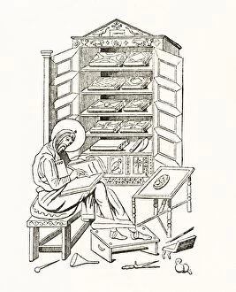
Roman library, Late Roman Empire C015 / 6073
Roman library. 19th-century artwork of a library of the Late Roman Empire, after Garruca. This period in the history of the Roman Empire lasted from around 250-450 AD

Background imageBinding Collection: Transcription repressor protein and DNA C015 / 5810

Transcription repressor protein and DNA C015 / 5810
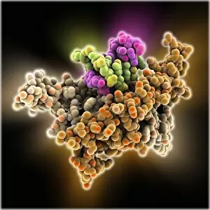
Transcription repressor protein and DNA, molecular model. The repressor protein (green) is binding to a strand of DNA (deoxyribonucleic acid, pink and purple)

Background imageBinding Collection: SMAD4 protein domain bound to DNA C015 / 6552

SMAD4 protein domain bound to DNA C015 / 6552
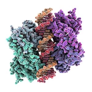
SMAD4 protein domain bound to DNA, molecular model. This strand of DNA (deoxyribonucleic acid, green and pink) is surrounded by MH1 domains of the SMAD4 (Mothers against decapentaplegic homolog 4)

Background imageBinding Collection: SMAD4 protein domain bound to DNA C015 / 6551

SMAD4 protein domain bound to DNA C015 / 6551
SMAD4 protein domain bound to DNA, molecular model. This strand of DNA (deoxyribonucleic acid, red and blue) is surrounded by MH1 domains of the SMAD4 (Mothers against decapentaplegic homolog 4)

Background imageBinding Collection: Transcription repressor protein and DNA C015 / 5811

Transcription repressor protein and DNA C015 / 5811
Transcription repressor protein and DNA, molecular model. The repressor protein (purple) is binding to a strand of DNA (deoxyribonucleic acid, yellow and pink)

Background imageBinding Collection: Beta secretase enzyme, molecular model C015 / 5277

Beta secretase enzyme, molecular model C015 / 5277
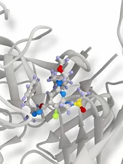
Beta secretase enzyme. Molecular model of the enzyme beta secretase bound to an inhibitor molecule. Beta secretase is a membrane-associated aspartic protease


For sale as Licensed Images

Choose your image, Select your licence and Download the media

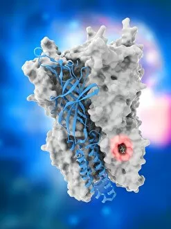
"Binding: From Ancient Techniques to Modern Science" Step into the world of binding, where artistry and science intertwine. Explore the delicate craft of bookbinding, as Bookbinding Tools from 1875 take center stage in preserving knowledge through time. Travel back in time with a Greek vase painting depicting Achilles and Patroclus, showcasing their unbreakable bond. Witness how even in ancient times, binding went beyond physical objects. Discover the microscopic realm where T lymphocytes bind to cancer cells, captured beautifully under SEM C001 / 1679. Marvel at the intricate dance between life and disease on a cellular level. Uncover the secrets behind anesthesia's power to inhibit ion channels with Anaesthetic inhibiting an ion channel C015 / 6718. See how this binding effect brings relief and comfort during medical procedures. Enter a bustling Bookbinding Workshop filled with artisans meticulously stitching pages together, breathing life into stories yet untold. Witness their dedication to preserving history one book at a time. Delve into Japanese culture as you admire Netsuke in the form of a demon queller and small demon from the Meiji Period (1868-1912). These tiny sculptures embody both beauty and protection through their bound forms. Marvel at EDTA crystals under a light microscope; these enchanting structures showcase nature's ability to create mesmerizing patterns when elements bind together harmoniously. Witness Achilles' unwavering loyalty as he binds Patroclus' wound on an ancient battlefield - an act that transcends mere friendship and symbolizes sacrifice for those we hold dear. Immerse yourself in artwork depicting cannabinoid receptor binding - a visual representation of how substances can interact with our bodies on molecular levels, unlocking new possibilities for medicine and wellness. Explore official wording within Apprenticeship Indenture documents that bind individuals to learn valuable skills from experienced masters - highlighting the importance of passing down knowledge through generations. Contemplate Chinese Foot-Binding, a practice that bound women's feet for centuries.