
Bound Collection (page 31)
"Bound: A Captivating Journey Through Art, Nature, and History" "The Thief - Antonio Maria Fabres y Costa's masterpiece captures the essence of boundless desire


For sale as Licensed Images
Choose your image, Select your licence and Download the media
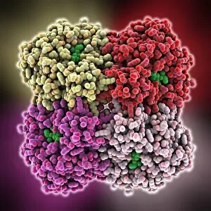
"Bound: A Captivating Journey Through Art, Nature, and History" "The Thief - Antonio Maria Fabres y Costa's masterpiece captures the essence of boundless desire. " "In 'The Young Martyr, ' we witness the unbreakable spirit that remains bound to one's beliefs. " "Emigrant Ship Leaving Belfast: The bittersweet farewell as dreams set sail, forever bound to new horizons. " "T lymphocytes and cancer cells locked in a battle for life – an intricate dance of binding forces. " "Anaesthetic inhibiting an ion channel: Unveiling the delicate balance between numbness and pain-bound consciousness. " "Suffragettes - Christmas Dinner in Holloway: Bound by solidarity, these brave women fought for equality with every bite. " "Shooters Hill stands tall, silently witnessing the ever-changing world below – steadfastly bound to its roots. " "Angerona Goddess guards her secrets within a timeless embrace – eternally bound by ancient mysteries. Siberian Tiger / Amur Tiger - in winter snow: Majestic beauty roams freely yet remains intricately tied to its snowy domain. Netsuke demon queller and small demon unite in a captivating bond from Japan's Meiji Period. Agnus Dei whispers serenity through Caravaggio's brushstrokes – forever bound to divine grace. Lockheed (P2V-5) Neptune MR1 soars above clouds, dutifully serving while tethered by loyalty to the Royal Air Force. Embark on this extraordinary journey where artistry meets nature’s wonders and history intertwines with human emotions; discover how our lives are shaped by what binds us together or holds us captive.