Cells Collection (#3)

"Exploring the Intricacies of Cells: From Cerebellum Tissue to Geranium Anther" Delving into the complexity of cells

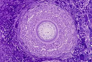
Background imageCells Collection: Ovarian follicle, light micrograph C016 / 0519

Ovarian follicle, light micrograph C016 / 0519
Ovarian follicle. Light micrograph of a section through a secondary ovarian follicle showing signs of maturation into the antral follicle class

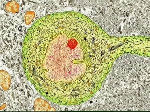
Background imageCells Collection: Purkinje nerve cell, TEM C014 / 0583

Purkinje nerve cell, TEM C014 / 0583
Purkinje nerve cell. Transmission electron micrograph (TEM) of a purkinje nerve cell (green) from the cerebellum of the brain, showing the cell body (centre) and its primary dendrite (cell process)

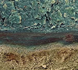
Background imageCells Collection: Skin layers, SEM

Skin layers, SEM

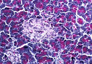
Background imageCells Collection: Pancreatic islet of Langerhans

Pancreatic islet of Langerhans
Islet of Langerhans. Light micrograph of a section through an islet of Langerhans (pale, centre) in pancreas tissue. This clump of secretory cells forms part of the endocrine system of the body

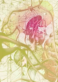
Background imageCells Collection: Brain anatomy

Brain anatomy. Computer artwork of strands of nerve cells (neurons) in front of an exploded view of the brain seen from behind

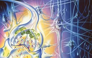
Background imageCells Collection: Nerve synapse

Nerve synapse
Synapse. Illustration of a synaptic knob (at left), the junction between two nerve cells. When an electrical impulse reaches the synapse, vesicles (round blue shapes)

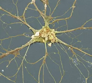
Background imageCells Collection: Nerve cell, SEM

Nerve cell, SEM
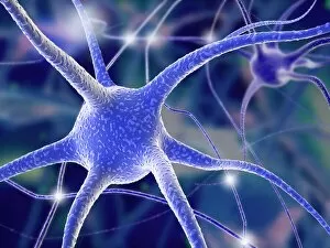
Nerve cell. Coloured scanning electron micrograph (SEM) of a neuron (nerve cell). The cell body is the central structure with neurites (long and thin structures) radiating outwards from it

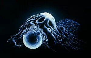
Background imageCells Collection: Macrophage engulfing bead

Macrophage engulfing bead
Illustration based on a scanning electron micrograph of a human macrophage phagocytosing (engulfing) a latex bead. Macrophages are monocytes from circulating blood that have migrated to tissues

Background imageCells Collection: Red blood cells, computer artwork

Red blood cells, computer artwork
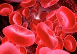
Red blood cells. Computer artwork of human red blood cells (erythrocytes) in a blood vessel. Red blood cells are biconcave, giving them a large surface area for gas exchange, and highly elastic

Background imageCells Collection: Red blood cells

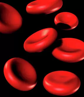
Red blood cells. Computer artwork of human red blood cells (erythrocytes). Red blood cells are biconcave, giving them a large surface area for gas exchange, and highly elastic

Background imageCells Collection: Rift Valley fever virus, TEM

Rift Valley fever virus, TEM

Background imageCells Collection: Stem cells, SEM

Stem cells, SEM
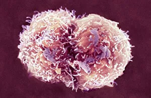
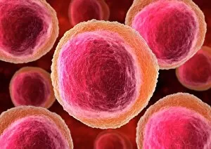
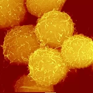
Stem cells, coloured scanning electron micrograph (SEM). Stem cells can differentiate into any other cell type. There are three main types of mammalian stem cell: embryonic stem cells

Background imageCells Collection: Nerve cells, computer artwork

Nerve cells, computer artwork
Nerve cells. Computer artwork of nerve cells, or neurons

Background imageCells Collection: Lymphocyte white blood cells, artwork

Lymphocyte white blood cells, artwork
Lymphocyte white blood cells. Computer artwork of lymphocyte white blood cells, showing their nuclei (red, centre). Lymphocytes, like all white blood cells, are part of the immune system

Background imageCells Collection: Frozen sperm, conceptual image

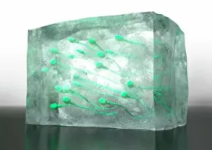
Frozen sperm, conceptual image. Computer artwork of sperm cells frozen inside a block of ice, representing the long term preservation of donated sperm to be used in fertility treatments

Background imageCells Collection: Skin surface, SEM

Skin surface, SEM
Skin surface. Coloured scanning electron micrograph (SEM) of squamous cells on the surface of the skin. These are flat, keratinised

Background imageCells Collection: Tomato leaf, light micrograph

Tomato leaf, light micrograph
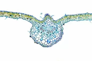
Tomato leaf. Light micrograph of a transverse section through the midrib of a tomato (Lycopersicum esculentum) leaf. The upper and lower epidermis on the surfaces of the leaf are blue

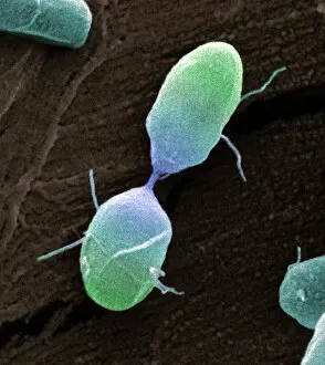
Background imageCells Collection: Salmonella bacterium dividing, SEM

Salmonella bacterium dividing, SEM
Salmonella bacterium dividing. Coloured scanning electron micrograph (SEM) of of a Salmonella bacterium dividing. The two new daughter cells are seen at upper right and lower left

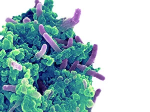
Background imageCells Collection: Bacteria infecting a macrophage, SEM

Bacteria infecting a macrophage, SEM
Bacteria infecting a macrophage. Coloured scanning electron micrograph (SEM) of Mycobacterium tuberculosis bacteria (purple) infecting a macrophage white blood cell

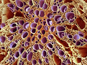
Background imageCells Collection: Xylem tissue, SEM

Xylem tissue, SEM
Xylem tissue. Coloured scanning electron micrograph (SEM) of a section through xylem tissue from a dicotyledon rootlet. Xylem vessels (purple)

Background imageCells Collection: Pollination, SEM

Pollination, SEM
Pollination. Coloured scanning electron micrograph (SEM) of part of a flowers pistil with pollen grains attached. The pistil is the female reproductive part of a flower

Background imageCells Collection: French lavender leaf pore, SEM

French lavender leaf pore, SEM
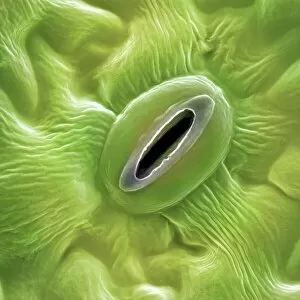
French lavender leaf pore. Coloured scanning electron micrograph (SEM) of an open stoma (centre, black). Stomata are pores that open and close in order to regulate gas exchange in a plant

Background imageCells Collection: Marrow and forget-me-not pollen, SEM

Marrow and forget-me-not pollen, SEM
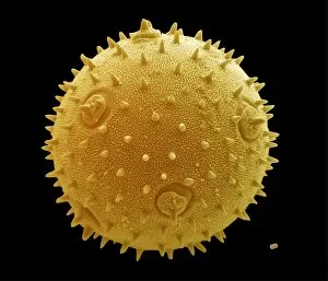
Pollen grains. Coloured scanning electron micrograph (SEM) of a pollen grain from a marrow plant (Cucurbita sp. left) and a forget-me-not plant (Myosotis sp. lower right)

Background imageCells Collection: Haematopoietic stem cells, SEM C013 / 5009

Haematopoietic stem cells, SEM C013 / 5009
Haematopoietic stem cells, coloured scanning electron micrograph (SEM). Stem cells can differentiate into any other cell type

Background imageCells Collection: The microscopic structure of cells

The microscopic structure of cells
2626843 The microscopic structure of cells; (add.info.: Rudolph Virchow (1821-1902) is regarded as perhaps the greatest medical scientist of the nineteenth century

Background imageCells Collection: Human finger 1, magnified 2, epidermis 3, magnified 4, scales 5, magnified skin 6

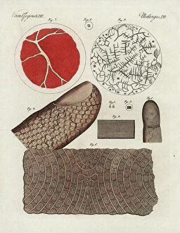
Human finger 1, magnified 2, epidermis 3, magnified 4, scales 5, magnified skin 6, and human blood 7, serum 8
FLO4574028 Human finger 1, magnified 2, epidermis 3, magnified 4, scales 5, magnified skin 6, and human blood 7, serum 8, and salt crystals in blood 9 under the microscope

Background imageCells Collection: John Howard (1726-1790) English prison reformer, entering Savoy military prison, London

John Howard (1726-1790) English prison reformer, entering Savoy military prison, London, after mutiny
530188 John Howard (1726-1790) English prison reformer, entering Savoy military prison, London, after mutiny. Restored peace and persuaded prisoners to return to cells

Background imageCells Collection: Black&White

Black&White
Alexey Pedan

Background imageCells Collection: The Space In Between

The Space In Between
Thoth Adan

Background imageCells Collection: Abacus

Abacus
Thoth Adan

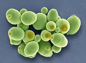
Background imageCells Collection: Yeast cells, SEM

Yeast cells, SEM
Yeast cells. Coloured scanning electron micrograph (SEM) of cells of brewer s, or baker s, yeast (Saccharomyces cerevisiae). This fungus consists of single vegetative cells

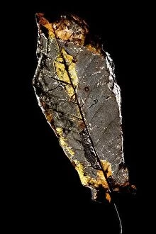
Background imageCells Collection: Desiccated leaf skeleton isolated against black background - North Carolina, USA

Desiccated leaf skeleton isolated against black background - North Carolina, USA

Background imageCells Collection: SINGAPORE - SEPTEMBER 5, 2015: Supertrees at Gardens by the Bay

SINGAPORE - SEPTEMBER 5, 2015: Supertrees at Gardens by the Bay. The tree-like structures are fitted with environmental technologies that mimic the ec SINGAPORE - SEPTEMBER 5

Background imageCells Collection: Colony of Haemophilus influenzae, 1906 (litho)

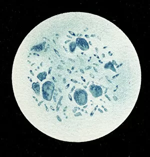
Colony of Haemophilus influenzae, 1906 (litho)
3501914 Colony of Haemophilus influenzae, 1906 (litho) by German School, (20th century); Private Collection; (add.info.: Bacteria constitute a large domain of prokaryotic microorganisms)

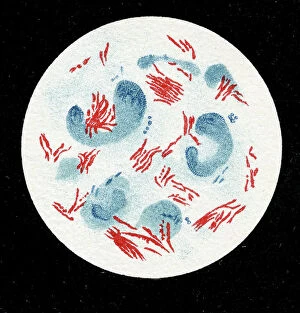
Background imageCells Collection: Colony of Mycobacterium leprae, 1906 (litho)

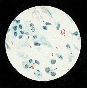
Colony of Mycobacterium leprae, 1906 (litho)
3501912 Colony of Mycobacterium leprae, 1906 (litho) by German School, (20th century); Private Collection; (add.info.: Bacteria constitute a large domain of prokaryotic microorganisms)

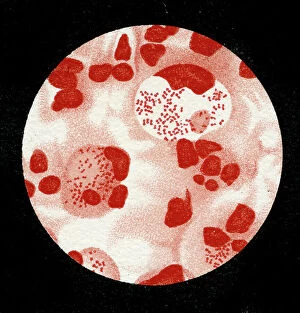
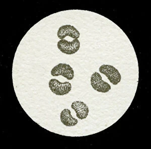
Background imageCells Collection: Colony of Micrococcus Gonorrhoea, 1906 (litho)

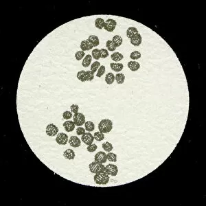
Colony of Micrococcus Gonorrhoea, 1906 (litho)
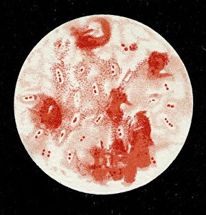
3501903 Colony of Micrococcus Gonorrhoea, 1906 (litho) by German School, (20th century); Private Collection; (add.info.: Bacteria constitute a large domain of prokaryotic microorganisms)

Background imageCells Collection: Colony of Streptococcus pneumoniae with and without bubble capsule, 1906 (litho)

Colony of Streptococcus pneumoniae with and without bubble capsule, 1906 (litho)
3501899 Colony of Streptococcus pneumoniae with and without bubble capsule, 1906 (litho) by German School, (20th century); Private Collection; (add.info)

Background imageCells Collection: Colony of Streptococcus Pneumoniae, 1906 (litho)

Colony of Streptococcus Pneumoniae, 1906 (litho)
3501898 Colony of Streptococcus Pneumoniae, 1906 (litho) by German School, (20th century); Private Collection; (add.info.: Bacteria constitute a large domain of prokaryotic microorganisms)

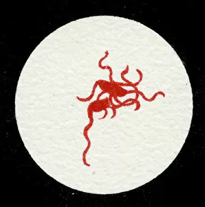
Background imageCells Collection: Spirillum of the Lyme disease causing bacteria, Spirochaetes Borrelia Recurrentis

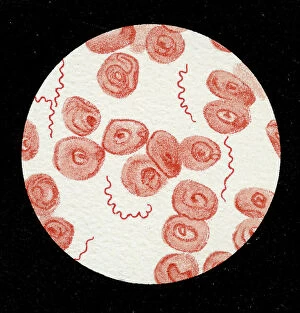
Spirillum of the Lyme disease causing bacteria, Spirochaetes Borrelia Recurrentis, in blood sample, 1906 (litho)
3501919 Spirillum of the Lyme disease causing bacteria, Spirochaetes Borrelia Recurrentis, in blood sample, 1906 (litho) by German School

Background imageCells Collection: Colony of Vibrio cholerae, 1906 (litho)

Colony of Vibrio cholerae, 1906 (litho)
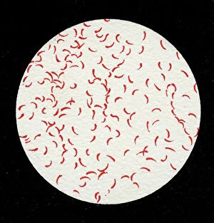
3501918 Colony of Vibrio cholerae, 1906 (litho) by German School, (20th century); Private Collection; (add.info.: Bacteria constitute a large domain of prokaryotic microorganisms)

Background imageCells Collection: Colony of Clostridium tetani with spores, 1906 (litho)

Colony of Clostridium tetani with spores, 1906 (litho)
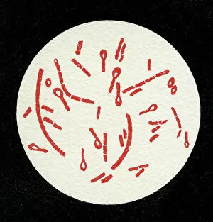
3501913 Colony of Clostridium tetani with spores, 1906 (litho) by German School, (20th century); Private Collection; (add.info.: Bacteria constitute a large domain of prokaryotic microorganisms)

Background imageCells Collection: Colony of Mycobacterium tuberculosis, 1906 (litho)

Colony of Mycobacterium tuberculosis, 1906 (litho)
3501910 Colony of Mycobacterium tuberculosis, 1906 (litho) by German School, (20th century); Private Collection; (add.info.: Bacteria constitute a large domain of prokaryotic microorganisms)

Background imageCells Collection: A colony of Staphylococcus pyogenes after cell division, 1906 (litho)

A colony of Staphylococcus pyogenes after cell division, 1906 (litho)
3501897 A colony of Staphylococcus pyogenes after cell division, 1906 (litho) by German School, (20th century); Private Collection; (add.info)

Background imageCells Collection: Colony of Streptococcus pyogenes, 1906 (litho)

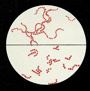
Colony of Streptococcus pyogenes, 1906 (litho)
3501894 Colony of Streptococcus pyogenes, 1906 (litho) by German School, (20th century); Private Collection; (add.info.: Bacteria constitute a large domain of prokaryotic microorganisms)

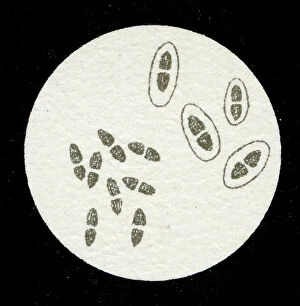
Background imageCells Collection: Colony of Yersinia pestis, 1906 (litho)

Colony of Yersinia pestis, 1906 (litho)
3501917 Colony of Yersinia pestis, 1906 (litho) by German School, (20th century); Private Collection; (add.info.: Bacteria constitute a large domain of prokaryotic microorganisms)

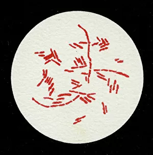
Background imageCells Collection: Colony of Salmonella Typhi showing Bacilli with flagella, 1906 (litho)

Colony of Salmonella Typhi showing Bacilli with flagella, 1906 (litho)
3501916 Colony of Salmonella Typhi showing Bacilli with flagella, 1906 (litho) by German School, (20th century); Private Collection; (add.info)

Background imageCells Collection: Colony of Salmonella typhi, 1906 (litho)

Colony of Salmonella typhi, 1906 (litho)
3501915 Colony of Salmonella typhi, 1906 (litho) by German School, (20th century); Private Collection; (add.info.: Bacteria constitute a large domain of prokaryotic microorganisms)


For sale as Licensed Images

Choose your image, Select your licence and Download the media

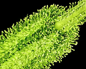
"Exploring the Intricacies of Cells: From Cerebellum Tissue to Geranium Anther" Delving into the complexity of cells, we examine cerebellum tissue through a captivating light micrograph. Behind the walls of Wandsworth Prison in southwest London, cells silently carry out their vital functions within inmates' bodies. Witnessing the intricate network of nerve and glial cells, a mesmerizing light micrograph reveals their interconnectedness. At the synapse nerve junction, captured by TEM imaging, we witness the fascinating communication between cells that allows our body to function seamlessly. Within Pentonville Prison in Islington, North London, hidden stories unfold as various cell types coexist amidst confinement. Shedding light on groundbreaking research, T lymphocytes and cancer they can magnified under SEM imaging - offering hope for future treatments (SEM C001 / 1679). Peering into hippocampus brain tissue unveils a world where countless cells work together to shape our memories and emotions. Glial cells take center stage in a confocal light micrograph - showcasing their crucial role in supporting and protecting neurons within our nervous system. HeLa cells come alive under scrutiny with an intriguing light micrograph (C017 / 8299), reminding us of Henrietta Lacks' immortal contribution to medical science. Journey back in time as Aylesbury Prison's historical significance intertwines with its cellular inhabitants from 1900 onwards. Exploring beyond human boundaries, dicotyledon plant stems reveal their cellular architecture through an enchanting light micrograph. Zooming closer than ever before, geranium anthers expose their delicate beauty under SEM imaging - unveiling nature's remarkable cellular structures.