Cells Collection (#6)

"Exploring the Intricacies of Cells: From Cerebellum Tissue to Geranium Anther" Delving into the complexity of cells

Background imageCells Collection: Plan of the Mazas Prison and cells for prisoners, Boulevard Mazas, Paris (w / c on paper)

Plan of the Mazas Prison and cells for prisoners, Boulevard Mazas, Paris (w / c on paper)
XIR437861 Plan of the Mazas Prison and cells for prisoners, Boulevard Mazas, Paris (w/c on paper) by French School, (19th century); Musee de la Ville de Paris, Musee Carnavalet, Paris

Background imageCells Collection: Electricity (engraving)

Electricity (engraving)
5207057 Electricity (engraving) by American School, (19th century); Private Collection; (add.info.: Electricity. Illustration for Farrows Military Encyclopedia by Edwards Farrow)

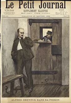
Background imageCells Collection: Cover of Le Petit Journal, 20 January 1895 (coloured engraving)

Cover of Le Petit Journal, 20 January 1895 (coloured engraving)
960254 Cover of Le Petit Journal, 20 January 1895 (coloured engraving) by French School, (19th century); Private Collection; (add.info.: Cover of Le Petit Journal, 20 January 1895)

Background imageCells Collection: Military Life on Board a Troopship (engraving)

Military Life on Board a Troopship (engraving)
1060252 Military Life on Board a Troopship (engraving) by Dollman, John Charles (1851-1934) (after); Private Collection; (add.info.: Military Life on Board a Troopship)

Background imageCells Collection: The Prisoner (colour litho)

The Prisoner (colour litho)
961230 The Prisoner (colour litho) by English School, (19th century); Private Collection; (add.info.: The Prisoner, with a man with his feet in shackles kissing a woman through the bars of his cell)

Background imageCells Collection: 1195038

1195038

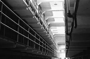
Background imageCells Collection: Interior of a cell block in Alcatraz prison, San Francisco Bay

Interior of a cell block in Alcatraz prison, San Francisco Bay. September 1979 The prison was originally built by the US Army in 1910

Background imageCells Collection: Bandleader Acker Bilk, Jazz money spinner prepared to go to prison for a 3rd time

Bandleader Acker Bilk, Jazz money spinner prepared to go to prison for a 3rd time, playing clarenet through bars May 1962

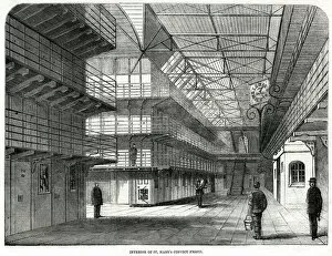
Background imageCells Collection: Interior of St Marys Convict Prision 1861

Interior of St Marys Convict Prision 1861
Interior of St Marys Island Convict Prision in Chatham. Date: 1861

Background imageCells Collection: PRIESTLEYS BATTERY

PRIESTLEYS BATTERY 64-cell electro-chemical battery, invented by Joseph Priestley (1733-1804), English clergyman and chemist. Date: 1810

Background imageCells Collection: FRENCH ELECTRIC BATTERY

FRENCH ELECTRIC BATTERY
Batterie electrique - an early French model Date: circa 1830

Background imageCells Collection: La Sante prison, Paris

La Sante prison, Paris
La Sante, Paris: guards checking the cells on the upper gallery. Date: 1870

Background imageCells Collection: White and red blood cells, illustration

White and red blood cells, illustration
3d illustration of white blood cells (leukocytes) in the human body

Background imageCells Collection: Fungus, illustration

Fungus, illustration

Background imageCells Collection: Tongue bacteria

Tongue bacteria. Coloured scanning electron micrograph (SEM) of bacteria on the surface of a human tongue. Large numbers of bacteria can form a visible layer on the surface of the tongue

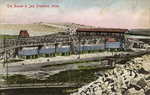
Background imageCells Collection: The Simmer and Jack Proprietary Gold Mines - South Africa

The Simmer and Jack Proprietary Gold Mines - South Africa
The Simmer and Jack Proprietary Gold Mines - east of Johannesburg, South Africa. The company which was founded in 1887 by the German August Simmer and Scotsman John Jack

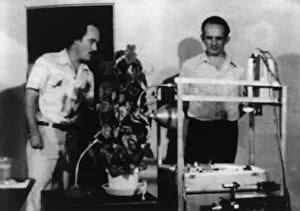
Background imageCells Collection: Cleve Backster experimenting on plant sensitivity

Cleve Backster experimenting on plant sensitivity
Grover Cleveland " Cleve" Backster (b.1924), American scientist, best known for his experiments with biocommunication in plant and animal cells

Background imageCells Collection: Human olfactory system, artwork C016 / 9378

Human olfactory system, artwork C016 / 9378
Human olfactory system. Computer artwork of a section through a human head, showing the olfactory receptor cells (brush-like), and their location in the olfactory bulb (sausage-shape)

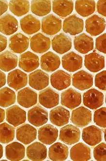
Background imageCells Collection: Uncapped honeycomb

Uncapped honeycomb. Close-up of honeycomb from a honey bee (Apis mellifera) colony. The individual cells contain honey

Background imageCells Collection: Anton van Leeuwenhoek, caricature

Anton van Leeuwenhoek, caricature
Anton van Leeuwenhoek. Caricature of the Dutch microbiologist Anton van Leeuwenhoek (1632-1723), looking through a magnifying glass to represent his discoveries in microbiology

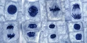
Background imageCells Collection: Plant cell mitosis, light micrograph

Plant cell mitosis, light micrograph
Plant cell mitosis. Light micrograph of root tip cells from an onion (Allium sp.) undergoing cell division (mitosis). From top left to bottom right

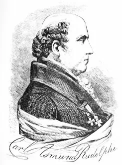
Background imageCells Collection: Karl Rudolphi, Swedish naturalist

Karl Rudolphi, Swedish naturalist. Historical artwork of Karl Asmund Rudolphi (1771-1832). Rudolphi is credited with being the father of helminthology, the study of worms

Background imageCells Collection: Inner ear hairs, SEM

Inner ear hairs, SEM
Inner ear hairs. Coloured scanning electron micrograph (SEM) of stereocilia, sensory hair-like projections, from hair cells of the cochlea of the inner ear

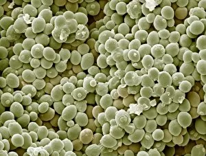
Background imageCells Collection: Yeast cells, SEM

Yeast cells, SEM
Yeast cells. Coloured scanning electron micrograph (SEM) of cells of bakers yeast (Saccharomyces cerevisiae) from part of a dried, commercial yeast pellet

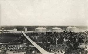
Background imageCells Collection: Presidio Modelo Prison, Isla de Pinos, Cuba

Presidio Modelo Prison, Isla de Pinos, Cuba
General view of the Presidio Modelo Prison on the Isla de Pinos (Isle of Pines), Cuba, now renamed Isla de la Juventud (Isle of Youth)

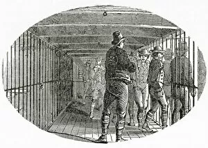
Background imageCells Collection: The Gallery on board The HMS York, a British prison hulk used to house the Tolpuddle Martyrs

The Gallery on board The HMS York, a British prison hulk used to house the Tolpuddle Martyrs. Prison hulks were decommissioned ships that authorities used as floating prisons in the 18th

Background imageCells Collection: The washing room on board The HMS York, a British prison hulk used to house the Tolpuddle Martyrs

The washing room on board The HMS York, a British prison hulk used to house the Tolpuddle Martyrs. Prison hulks were decommissioned ships that authorities used as floating prisons in the 18th

Background imageCells Collection: Orbicular Design

Orbicular Design

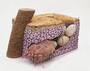
Background imageCells Collection: Cross-section model of Bedbug (Cimicidae) burrowed in skin, laying eggs

Cross-section model of Bedbug (Cimicidae) burrowed in skin, laying eggs

Background imageCells Collection: Finley Quaye singer Greenock Prison March 1998 played for prisoners inmates at

Finley Quaye singer Greenock Prison March 1998 played for prisoners inmates at Greenock prison

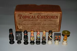
Background imageCells Collection: Suffragette Chess Set

Suffragette Chess Set
A painted figural chess set, entitled Topical Chessmen (Suffragettes v The Law). It comprises King and Queen, bishops as judges, rooks as burning buildings, prison cells

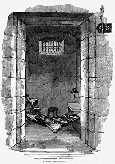
Background imageCells Collection: Pentonville prison cell interior

Pentonville prison cell interior
Pentonville prison in London: interior of cell with a hammock for sleeping. Date: 1843

Background imageCells Collection: Police Station at Cherat - NWFP

Police Station at Cherat - NWFP. Cherat is a hill station, immediately above the village Chapri and Saleh Khana in the Nowshera District of Khyber-Pakhtunkhwa in Pakistan

Background imageCells Collection: Picture No. 10876987

Picture No. 10876987
Scanning Electron Micrograph (SEM): Human skin section across vein showing red blood cells Date:

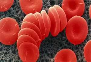
Background imageCells Collection: Coloured SEM of red blood cells, rouleau formation

Coloured SEM of red blood cells, rouleau formation
Red blood cells. Coloured scanning electron micrograph of red blood cells in rouleau formation. They often stack together in this way because of their shape

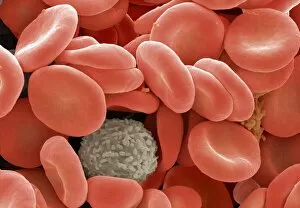
Background imageCells Collection: Red and white blood cells

Red and white blood cells. Coloured scanning electron micrograph (SEM) showing two types of human blood cells. Red blood cells (or erythrocytes, red) and a white blood cell (or leucocyte)

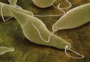
Background imageCells Collection: Euglena gracilis, SEM

Euglena gracilis, SEM
Euglena gracilis protists. Coloured scanning electron micrograph (SEM) of Euglena gracilis protists. These organisms possess a unique combination of plant and animal characters. Like the algae, E

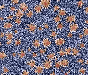
Background imageCells Collection: Nasal lining, SEM

Nasal lining, SEM
Nasal lining. Coloured scanning electron micrograph (SEM) of the olfactory epithelium that lines the nasal cavity, showing olfactory cells (red) surrounded by numerous cilia (hair-like projections)

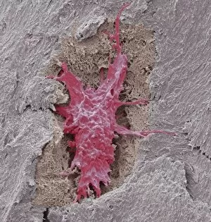
Background imageCells Collection: Osteocyte bone cell, SEM C016 / 9026

Osteocyte bone cell, SEM C016 / 9026
Osteocyte bone cell. Coloured scanning electron micrograph (SEM) of an osteocyte bone cell (red) surrounded by bone tissue (grey)

Background imageCells Collection: Helicobacter pylori bacteria, SEM C016 / 9136

Helicobacter pylori bacteria, SEM C016 / 9136
Helicobacter pylori bacteria. Coloured scanning electron micrograph (SEM) of Helicobacter pylori bacteria (green) in the stomach. H

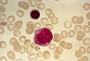
Background imageCells Collection: Myeloblast blood cell, light micrograph

Myeloblast blood cell, light micrograph
Myeloblast blood cell. Light micrograph of blood cells, including a myeloblast, a precursor for a type of white blood cell (leucocyte). Myeloblasts differentiate into granulocytes

Background imageCells Collection: Eosinophil white blood cell, TEM

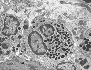
Eosinophil white blood cell, TEM
Eosinophil white blood cell. Transmission electron micrograph (TEM) of a section through an eosinophil white blood cell (leukocyte)

Background imageCells Collection: Pancreatic acinar cell

Pancreatic acinar cell. Transmission electron micrograph (TEM) of a section through an enzyme-secreting acinar cell in the human pancreas, showing part of the nucleus (round, far left)

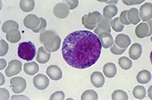
Background imageCells Collection: Promyelocyte blood cell, light micrograph

Promyelocyte blood cell, light micrograph. This blood cell (centre) is a precursor for a type of white blood cell called a granulocyte, formed by granulopoiesis in the bone marrow

Background imageCells Collection: Chronic lymphocytic leukaemia, micrograph

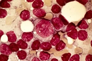
Chronic lymphocytic leukaemia, micrograph
Chronic lymphocytic leukaemia. Light micrograph of blood cells sampled from a lymphatic ganglion in a case of chronic lymphocytic leukaemia, also called chronic lymphoid leukaemia (CLL)


For sale as Licensed Images

Choose your image, Select your licence and Download the media

"Exploring the Intricacies of Cells: From Cerebellum Tissue to Geranium Anther" Delving into the complexity of cells, we examine cerebellum tissue through a captivating light micrograph. Behind the walls of Wandsworth Prison in southwest London, cells silently carry out their vital functions within inmates' bodies. Witnessing the intricate network of nerve and glial cells, a mesmerizing light micrograph reveals their interconnectedness. At the synapse nerve junction, captured by TEM imaging, we witness the fascinating communication between cells that allows our body to function seamlessly. Within Pentonville Prison in Islington, North London, hidden stories unfold as various cell types coexist amidst confinement. Shedding light on groundbreaking research, T lymphocytes and cancer they can magnified under SEM imaging - offering hope for future treatments (SEM C001 / 1679). Peering into hippocampus brain tissue unveils a world where countless cells work together to shape our memories and emotions. Glial cells take center stage in a confocal light micrograph - showcasing their crucial role in supporting and protecting neurons within our nervous system. HeLa cells come alive under scrutiny with an intriguing light micrograph (C017 / 8299), reminding us of Henrietta Lacks' immortal contribution to medical science. Journey back in time as Aylesbury Prison's historical significance intertwines with its cellular inhabitants from 1900 onwards. Exploring beyond human boundaries, dicotyledon plant stems reveal their cellular architecture through an enchanting light micrograph. Zooming closer than ever before, geranium anthers expose their delicate beauty under SEM imaging - unveiling nature's remarkable cellular structures.