Cells Collection (#9)

"Exploring the Intricacies of Cells: From Cerebellum Tissue to Geranium Anther" Delving into the complexity of cells

Background imageCells Collection: Carder-Bees Nest and Cells

Carder-Bees Nest and Cells

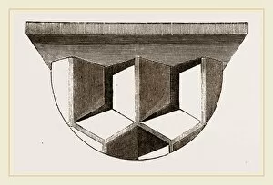
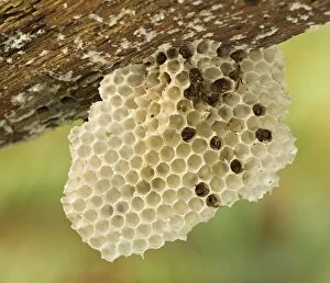
Background imageCells Collection: Cells of Honey-comb

Cells of Honey-comb

Background imageCells Collection: Solar panels and sky

Solar panels and sky

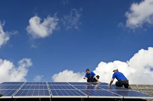
Background imageCells Collection: Installation of a solar collector, near Freiburg in Breisgau, Baden-Wuerttemberg, Germany, Europe

Installation of a solar collector, near Freiburg in Breisgau, Baden-Wuerttemberg, Germany, Europe

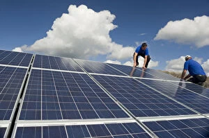
Background imageCells Collection: Installation of solar panels near Freiburg im Breisgau, Baden-Wuerttemberg, Germany, Europe

Installation of solar panels near Freiburg im Breisgau, Baden-Wuerttemberg, Germany, Europe

Background imageCells Collection: Solar panels a the roundabout island, Ost district, Woergl, Tyrol, Austria, Europe

Solar panels a the roundabout island, Ost district, Woergl, Tyrol, Austria, Europe

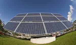
Background imageCells Collection: Solar plant near Freiburg im Breisgau, Baden-Wuerttemberg, Germany, Europe

Solar plant near Freiburg im Breisgau, Baden-Wuerttemberg, Germany, Europe

Background imageCells Collection: Picture No. 11092002

Picture No. 11092002
Old Wasps Nest underneath fallen branch Date:

Background imageCells Collection: Apple stem, light micrograph

Apple stem, light micrograph
Apple stem. Polarised light micrograph of a section through the stem of a young apple (Malus domestica) tree plant. Magnification: x100 when printed 10 centimetres wide

Background imageCells Collection: Picture No. 10873686

Picture No. 10873686
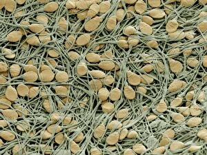
Human sperm cells Date:

Background imageCells Collection: Picture No. 10873675

Picture No. 10873675
Human White and Red Blood Cells Date:

Background imageCells Collection: Picture No. 10873703

Picture No. 10873703
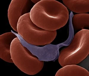
Sleeping Sickness Parasite in red blood cells (Trypanosoma sp.) Date:

Background imageCells Collection: Picture No. 10877012

Picture No. 10877012
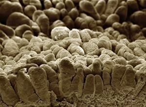
Scanning Electron Micrograph (SEM): human intestine showing villi Date:

Background imageCells Collection: Picture No. 10876996

Picture No. 10876996
Scanning Electron Micrograph (SEM): Human Retina showing photoreceptors rod and cone cells Date:

Background imageCells Collection: Picture No. 10876991

Picture No. 10876991
Scanning Electron Micrograph (SEM): Human testis tubes - Sperm cells forming in seminal tubes of testis Date:

Background imageCells Collection: Picture No. 10876993

Picture No. 10876993
Scanning Electron Micrograph (SEM): Human Retina showing photoreceptors rod and cone cells Date:

Background imageCells Collection: Picture No. 10876995

Picture No. 10876995
Scanning Electron Micrograph (SEM): Human Retina showing photoreceptors rod and cone cells Date:

Background imageCells Collection: Picture No. 10876994

Picture No. 10876994
Scanning Electron Micrograph (SEM): Human Retina showing photoreceptors rod and cone cells Date:

Background imageCells Collection: Picture No. 10876988

Picture No. 10876988
Scanning Electron Micrograph (SEM): Early human embryo, one cell removed for genetic analysis Date:

Background imageCells Collection: Picture No. 10876986

Picture No. 10876986
Scanning Electron Micrograph (SEM): Human Red Blood Cells Date:

Background imageCells Collection: Picture No. 10874514

Picture No. 10874514
Light Micrograph (LM): A light micrograph of the nuclei of plant cells Date:

Background imageCells Collection: Picture No. 10866606

Picture No. 10866606
Orchid - root on the bark of a tree. The whitish part is covered with cells capable of storing water like a sponge. Date:

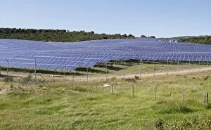
Background imageCells Collection: Solar panels on solar farm in open field, near Bonifacio, Corsica, France, April

Solar panels on solar farm in open field, near Bonifacio, Corsica, France, April

Background imageCells Collection: Oudemas or edemas erupting on a cabbage leaf. The cause is not know but is probably physiological

Oudemas or edemas erupting on a cabbage leaf. The cause is not know but is probably physiological

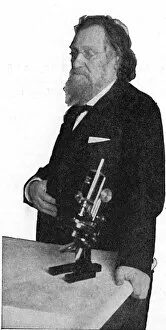
Background imageCells Collection: Ilya Ilich Mechnikov ( Elie Metchnikoff 1845-1916) Russian zoologist who continued

Ilya Ilich Mechnikov ( Elie Metchnikoff 1845-1916) Russian zoologist who continued Pasteurs work. Discovered phagocytes, cells which destroy infective organisms

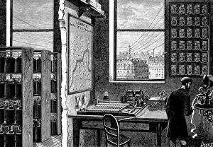
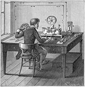
Background imageCells Collection: Main station of Exchange Telegraph Company, London, England, showing operator receiving

Main station of Exchange Telegraph Company, London, England, showing operator receiving a message on ticker-tape machine. Morse transmitting instrument connected directly to the Stock Exchange

Background imageCells Collection: Operator receiving a message in Morse code on printing telegraph. In box under table

Operator receiving a message in Morse code on printing telegraph. In box under table are the wet cells (batteries) supplying electricity. Wood engraving 1887

Background imageCells Collection: Operator sending a message on a Morse electric printing telegraph by tapping out code with key

Operator sending a message on a Morse electric printing telegraph by tapping out code with key. Under table are we cells (batteries) supplying electricity. Wood engraving 1887

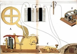
Background imageCells Collection: Morse electric printing telegraph. Front view of instrument showing roll of paper

Morse electric printing telegraph. Front view of instrument showing roll of paper for recording messages and the transmitting key at A. D are wet cells (batteries) providing electricity

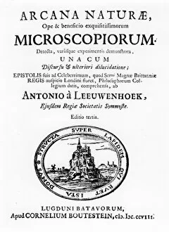
Background imageCells Collection: Anton van Leeuwenhoek (1632-1723), Dutch pioneer microscopist. Title page of Microscopium

Anton van Leeuwenhoek (1632-1723), Dutch pioneer microscopist. Title page of Microscopium by Anton van Leeuwenhoek, third edition, (Leyden, 1708)

Background imageCells Collection: I. The Faraday Entering Ballinskelligs Bay 2. Lower Lighthouse On The Great Skellig 3

I. The Faraday Entering Ballinskelligs Bay 2. Lower Lighthouse On The Great Skellig 3. The Bull Rock Off Dursey Head, Where The New Lighthouse Is Being Constructed 4

Background imageCells Collection: Zero emission hydrogen fuel cell vehicle parked below mountain peak, Cimon della Pala

Zero emission hydrogen fuel cell vehicle parked below mountain peak, Cimon della Pala, Pale di San Martino, Dolomites, Italian Alps, Italy, October

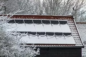
Background imageCells Collection: photo voltaic solar panels stop working when covered in winter snow

photo voltaic solar panels stop working when covered in winter snow

Background imageCells Collection: Simplified colourful model of DNA structure, front view

Simplified colourful model of DNA structure, front view

Background imageCells Collection: Slide showing cluster of a single celled organism called cyanobacteria

Slide showing cluster of a single celled organism called cyanobacteria

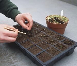
Background imageCells Collection: Apium graveolens, Celery seedlings being transferred into module

Apium graveolens, Celery seedlings being transferred into module, dibber being used to create holes in cells

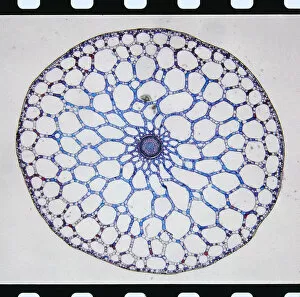
Background imageCells Collection: Micrograph through Stem of Mares Tail Plant

Micrograph through Stem of Mares Tail Plant

Background imageCells Collection: Stochastic gene expression, illustration C018 / 0906

Stochastic gene expression, illustration C018 / 0906
Stochastic gene expression, illustration. Every cell in an organism contains every single gene that makes up the organisms genome. However, they are not all active (expressed) in each cell

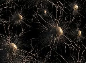
Background imageCells Collection: Astrocyte nerve cells, artwork C017 / 3425

Astrocyte nerve cells, artwork C017 / 3425
Astrocyte nerve cells, computer artwork. Astrocytes are a type of glial cell.They provide structural support and protection for neurons (nerve cells) and also supply them with nutrients and oxygen

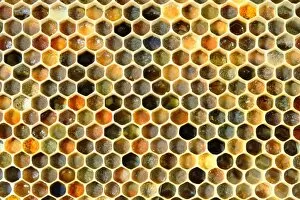
Background imageCells Collection: Honey bee comb C017 / 3713

Honey bee comb C017 / 3713
Honey bee comb. Close-up of a comb from the hive of a honey bee (Apis sp.). The cells of this comb are used to store pollen. The different colours are due to the different types of pollen being stored

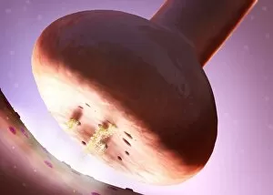
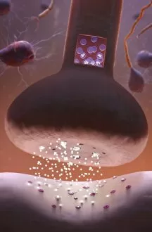
Background imageCells Collection: Nerve synapse, artwork C017 / 3427

Nerve synapse, artwork C017 / 3427
Nerve synapse. Computer artwork of of a junction, or synapse, between two nerve cells (neurons). As the electrical signal reaches the presynaptic end of a neuron it triggers the release of

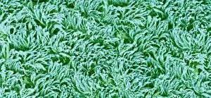
Background imageCells Collection: Trachea lining, SEM C016 / 9756

Trachea lining, SEM C016 / 9756
Trachea lining. Coloured scanning electron micrograph (SEM) of the lining of the trachea (wind pipe). The trachea links the larynx to the lungs

Background imageCells Collection: Nerve synapse, artwork C017 / 3428

Nerve synapse, artwork C017 / 3428
Nerve synapse. Computer artwork of of a junction, or synapse, between two nerve cells (neurons). As the electrical signal reaches the presynaptic end of a neuron it triggers the release of

Background imageCells Collection: Staphylococcus aureus bacteria, SEM C017 / 7137

Staphylococcus aureus bacteria, SEM C017 / 7137
Staphylococcus aureus bacteria, coloured scanning electron micrograph (SEM). These Gram-positive coccus (round) bacteria are found on the skin and mucous membranes of humans and many animals

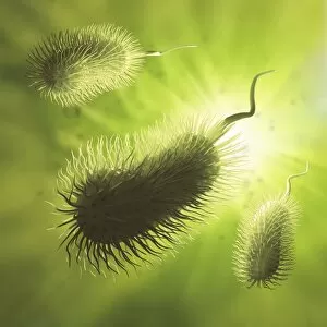
Background imageCells Collection: Bacteria, artwork F006 / 3717

Bacteria, artwork F006 / 3717
Bacteria, computer artwork

Background imageCells Collection: MRI brain scan and nerve cells F006 / 3595

MRI brain scan and nerve cells F006 / 3595
Computer artwork of a sagittal (side) view of a human head. The largest part of the brain is the cerebrum (upper part). This surrounds the limbic system (curved structures at upper centre)


For sale as Licensed Images

Choose your image, Select your licence and Download the media

"Exploring the Intricacies of Cells: From Cerebellum Tissue to Geranium Anther" Delving into the complexity of cells, we examine cerebellum tissue through a captivating light micrograph. Behind the walls of Wandsworth Prison in southwest London, cells silently carry out their vital functions within inmates' bodies. Witnessing the intricate network of nerve and glial cells, a mesmerizing light micrograph reveals their interconnectedness. At the synapse nerve junction, captured by TEM imaging, we witness the fascinating communication between cells that allows our body to function seamlessly. Within Pentonville Prison in Islington, North London, hidden stories unfold as various cell types coexist amidst confinement. Shedding light on groundbreaking research, T lymphocytes and cancer they can magnified under SEM imaging - offering hope for future treatments (SEM C001 / 1679). Peering into hippocampus brain tissue unveils a world where countless cells work together to shape our memories and emotions. Glial cells take center stage in a confocal light micrograph - showcasing their crucial role in supporting and protecting neurons within our nervous system. HeLa cells come alive under scrutiny with an intriguing light micrograph (C017 / 8299), reminding us of Henrietta Lacks' immortal contribution to medical science. Journey back in time as Aylesbury Prison's historical significance intertwines with its cellular inhabitants from 1900 onwards. Exploring beyond human boundaries, dicotyledon plant stems reveal their cellular architecture through an enchanting light micrograph. Zooming closer than ever before, geranium anthers expose their delicate beauty under SEM imaging - unveiling nature's remarkable cellular structures.