
Cells Collection (#2)
"Exploring the Intricacies of Cells: From Cerebellum Tissue to Geranium Anther" Delving into the complexity of cells


For sale as Licensed Images
Choose your image, Select your licence and Download the media
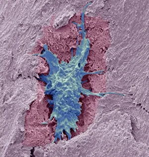
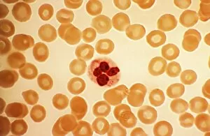
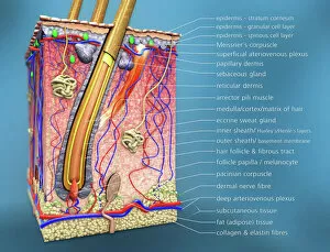
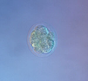
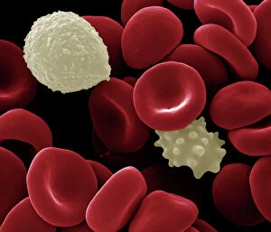
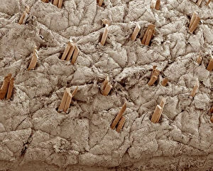
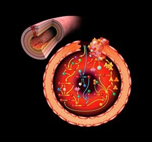
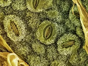
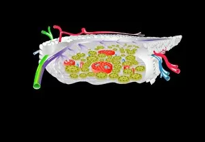
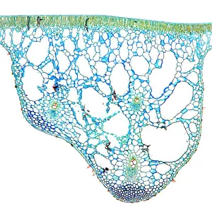
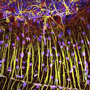
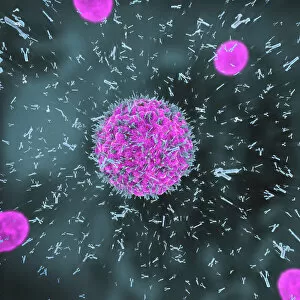
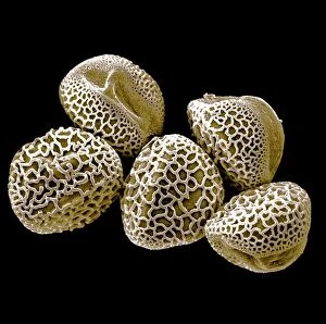
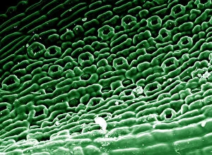
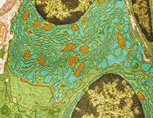
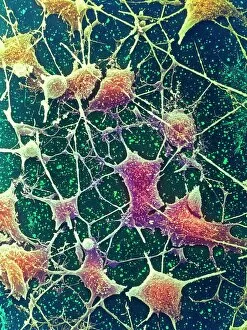
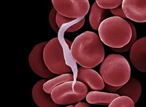
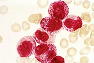
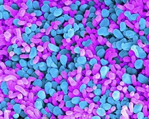
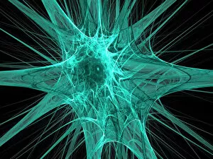
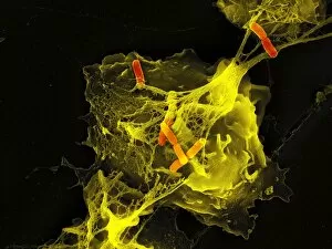
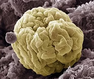
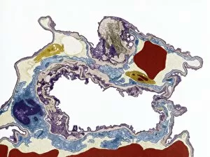
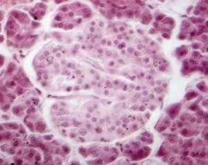
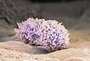
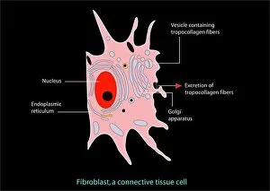
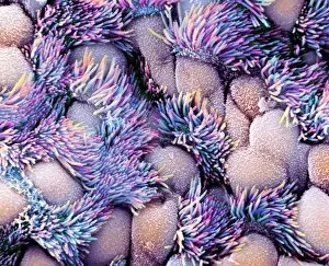
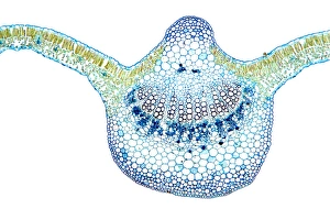
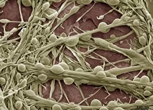
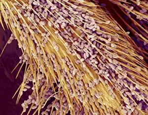
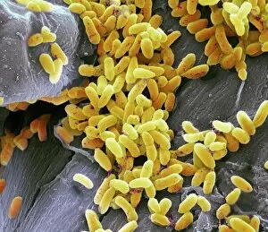
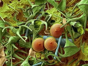
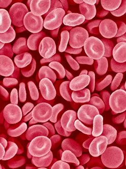
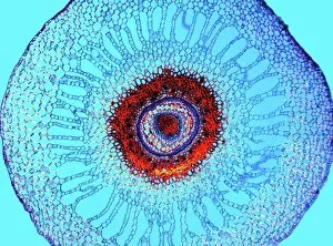
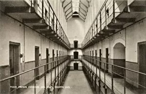
"Exploring the Intricacies of Cells: From Cerebellum Tissue to Geranium Anther" Delving into the complexity of cells, we examine cerebellum tissue through a captivating light micrograph. Behind the walls of Wandsworth Prison in southwest London, cells silently carry out their vital functions within inmates' bodies. Witnessing the intricate network of nerve and glial cells, a mesmerizing light micrograph reveals their interconnectedness. At the synapse nerve junction, captured by TEM imaging, we witness the fascinating communication between cells that allows our body to function seamlessly. Within Pentonville Prison in Islington, North London, hidden stories unfold as various cell types coexist amidst confinement. Shedding light on groundbreaking research, T lymphocytes and cancer they can magnified under SEM imaging - offering hope for future treatments (SEM C001 / 1679). Peering into hippocampus brain tissue unveils a world where countless cells work together to shape our memories and emotions. Glial cells take center stage in a confocal light micrograph - showcasing their crucial role in supporting and protecting neurons within our nervous system. HeLa cells come alive under scrutiny with an intriguing light micrograph (C017 / 8299), reminding us of Henrietta Lacks' immortal contribution to medical science. Journey back in time as Aylesbury Prison's historical significance intertwines with its cellular inhabitants from 1900 onwards. Exploring beyond human boundaries, dicotyledon plant stems reveal their cellular architecture through an enchanting light micrograph. Zooming closer than ever before, geranium anthers expose their delicate beauty under SEM imaging - unveiling nature's remarkable cellular structures.