Cross Section Collection (#83)

"Unveiling the Hidden World: Exploring Cross Sections in Nature and Architecture" Delving into the intricate world of honey bees

Background imageCross Section Collection: Conifer needle, SEM

Conifer needle, SEM
Conifer needle. Coloured scanning electron micrograph (SEM) of a section through a needle (leaf) from a conifer tree, showing the vascular bundle (large round cluster)

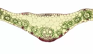
Background imageCross Section Collection: Leaf midrib, light micrograph

Leaf midrib, light micrograph
Leaf midrib. Light micrograph (LM) of a section through the midrib of a leaf from a monocotyledon plant. The midrib (midvein) is the continuation of a leafs stem along the centre of the leaf

Background imageCross Section Collection: Leaf bud, light micrograph

Leaf bud, light micrograph
Leaf bud. Light micrograph of a cross-section through a winter leaf bud of the sycamore tree (Acer pseudoplatanus). This trees leaves are arranged in an opposite and decussate manner

Background imageCross Section Collection: Poppy ovary with developing seeds, LM

Poppy ovary with developing seeds, LM
Poppy ovary with developing seeds. Light micrograph (LM) of a cross-section through the centre of a mature poppy ovary, showing placentas bearing developing seeds (red)

Background imageCross Section Collection: Sycamore bud, light micrograph

Sycamore bud, light micrograph
Sycamore bud. Light micrograph of a cross-section through a sycamore bud (Acer pseudo-plantanus). The unexpanded leaves (red) are surrounded by protective outer scale leaves covered in cork

Background imageCross Section Collection: Sycamore leaf bud, light micrograph

Sycamore leaf bud, light micrograph
Sycamore leaf bud (Acer pseudoplatanus). Light micrograph of a cross-section through a winter leaf bud from a sycamore tree. The leaf material (red) is surrounded by a protective layer of cork (pink)

Background imageCross Section Collection: Mature poppy ovary, light micrograph

Mature poppy ovary, light micrograph
Mature poppy ovary. Light micrograph (LM) of a cross-section through a mature poppy ovary, which is incompletely divided by septa (membranes) that do not meet in the centre

Background imageCross Section Collection: Section through a pine needle, LM

Section through a pine needle, LM
Section through a pine needle. Light micrograph (LM) of a section through the needle (leaf) of a pine tree, Pinus sp.. The centre of the needle is occupied by two vascular bundles

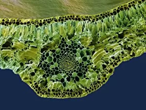
Background imageCross Section Collection: Leaf midrib, SEM

Leaf midrib, SEM
Leaf midrib. Coloured scanning electron micrograph (SEM) of a section through the midrib of a leaf from the Common Box (Buxus sempervirens)

Background imageCross Section Collection: Tulip anther, light micrograph

Tulip anther, light micrograph
Tulip anther. Light micrograph of a cross-section through the anther of a tulip bud (Tulipa sp.), showing a full anther sac with developing pollen grains (white)

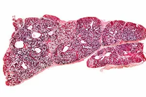
Background imageCross Section Collection: Breast in pregnancy, light micrograph

Breast in pregnancy, light micrograph
Breast in pregnancy. Light micrograph of a transverse section of the human mammary gland during pregnancy. The breast is normally made up of fibro-fatty connective tissue

Background imageCross Section Collection: Scalp tissue, light micrograph

Scalp tissue, light micrograph
Scalp tissue. Polarised light micrograph of a section through the human scalp. This section shows oblique and transverse sections through hair shafts (yellow-orange)

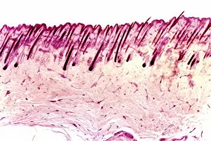
Background imageCross Section Collection: Skin tissue, light micrograph

Skin tissue, light micrograph
Skin tissue. Light micrograph of a transverse section through human skin. The skin is made up of an outer epidermis (pale purple, across top) with hair follicles (deep purple)

Background imageCross Section Collection: Compact bone, light micrograph

Compact bone, light micrograph
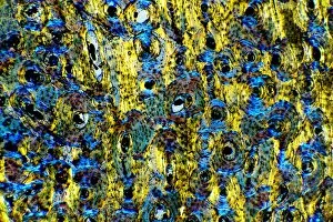
Compact bone. Polarised light micrograph of a transverse section through compact bone tissue, showing Haversian canals (circular regions)

Background imageCross Section Collection: Dogfish liver area, transverse section

Dogfish liver area, transverse section
Dogfish liver area. Light micrograph of a transverse section through the body of a lesser spotted dogfish (Scyliorhinus canicula), in the region of the liver (lower frame)

Background imageCross Section Collection: Dogfish head, transverse section

Dogfish head, transverse section
Dogfish head. Light micrograph of a transverse section through the head of a lesser spotted dogfish (Scyliorhinus canicula), in the region of the eyes (upper left and upper right)

Background imageCross Section Collection: Intervertebral disc, light micrograph

Intervertebral disc, light micrograph
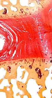
Intervertebral disc. Light micrograph of a transverse section through an intervertebral disc (red) and the surrounding tissue (brown)

Background imageCross Section Collection: Heel skin tissue, light micrograph

Heel skin tissue, light micrograph
Heel skin tissue. Polarised light micrograph of a transverse section through skin from the heel of a human foot. The sole of the foot has to withstand the weight of the body

Background imageCross Section Collection: Lamprey gill region, light micrograph

Lamprey gill region, light micrograph
Lamprey gill. Light micrograph of a transverse section through the gill region of a lamprey (Petromyzon marinus), a parasitic fish. The spinal area is at upper centre

Background imageCross Section Collection: Sheeps hoof, light micrograph

Sheeps hoof, light micrograph
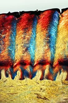
Sheeps hoof. Polarised light micrograph of a transverse section through the hoof of a sheep. In sheep and other hooved animals, the hoof is made of the same material as nails and claws

Background imageCross Section Collection: Sago cycad leaf, SEM

Sago cycad leaf, SEM
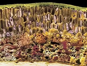
Sago cycad leaf. Coloured scanning electron micrograph (SEM) of a section through a sago cycad (Cycas revoluta) leaf. At lower centre and left are vascular bundles (orange clusters)

Background imageCross Section Collection: Spinal cord, transverse section

Spinal cord, transverse section
Spinal cord. Light micrograph of a transverse section through a human spinal cord. The spinal cord has a central canal around which are the dorsal and ventral horns (orange)

Background imageCross Section Collection: Dog tongue tissue, light micrograph

Dog tongue tissue, light micrograph
Dog tongue tissue. Light micrograph of a transverse section through the tongue of a dog, showing the tongues surface (across top)

Background imageCross Section Collection: Foot skin tissue, light micrograph

Foot skin tissue, light micrograph
Foot skin tissue. Light micrograph of a transverse section through skin from a human foot. The skin is made up of an outer epidermis (across top)

Background imageCross Section Collection: Skin sweat glands, light micrograph

Skin sweat glands, light micrograph
Skin sweat glands. Light micrograph of a transverse section through skin from a human foot. The skin is made up of an outer epidermis (across top) and a lower epidermis (deep purple)

Background imageCross Section Collection: Cat tongue tissue, light micrograph

Cat tongue tissue, light micrograph
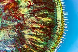
Cat tongue tissue. Polarised light micrograph of a transverse section through a cats tongue. The outer stratified epithelium (right) has spike-like (white) keratinised papillae

Background imageCross Section Collection: Tongue tissue, light micrograph

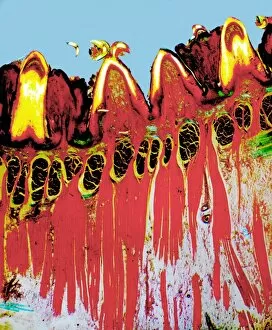
Tongue tissue, light micrograph
Tongue tissue. Polarised light micrograph of a transverse section through a tongue, showing the surface (across top). Here, there are four rounded fungiform papillae

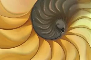
Background imageCross Section Collection: Chambered Nautilus (Nautilus pompilius) cross section of shell showing chambers

Chambered Nautilus (Nautilus pompilius) cross section of shell showing chambers

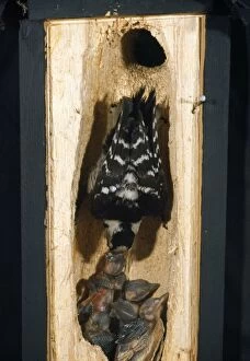
Background imageCross Section Collection: Lesser-spotted Woodpecker - cross section of tree into nest with adult feeding young

Lesser-spotted Woodpecker - cross section of tree into nest with adult feeding young
WOM-44 Lesser-spotted Woodpecker - cross section of tree into nest with adult feeding young Dendrocopos minor Wilhelm Moller Please note that prints are for personal display purposes only

Background imageCross Section Collection: Douglas Tree - Growth ring The Netherlands, Overijssel, forestry Staphorst

Douglas Tree - Growth ring The Netherlands, Overijssel, forestry Staphorst
VG-1601 Douglas Fir Tree - Growth ring The Netherlands, Overijssel, forestry Staphorst Pseudotsuga menziesii Paul Van Gaalen Please note that prints are for personal display purposes only

Background imageCross Section Collection: Fig - in studio

Fig - in studio
LA-4256 Fig - in studio Jean Michel Labat Please note that prints are for personal display purposes only and may not be reproduced in any way

Background imageCross Section Collection: Earthworm pulling leaf down into its burrow KAT01964

Earthworm pulling leaf down into its burrow KAT01964
AUS-1938 Earthworm - pulling leaf down into its burrow Kathie Atkinson / Auscape / ardea.com Auscape Please note that prints are for personal display purposes only

Background imageCross Section Collection: Nautilus - section

Nautilus - section
PJG-339-c Nautilus - section Pompilius philippians Peter Green Please note that prints are for personal display purposes only and may not be reproduced in any way

Background imageCross Section Collection: Light Micrograph (LM): plant cell chloroplasts - the site where photosynthesis takes place

Light Micrograph (LM): plant cell chloroplasts - the site where photosynthesis takes place
LRDS-192 Light Micrograph (LM): plant cell chloroplasts - the site where photosynthesis takes place David Spears (Last Refuge)

Background imageCross Section Collection: Light Micrograph (LM): Cells from a hair on the stamen of the common spiderwort (Tradescantia)

Light Micrograph (LM): Cells from a hair on the stamen of the common spiderwort (Tradescantia)
LRDS-171 Light Micrograph (LM): Cells from a hair on the stamen of the common spiderwort (Tradescantia) Tradescantia sp David Spears (Last Refuge)

Background imageCross Section Collection: Light Micrograph (LM): A transverse section of a Fig leaf); Magnification x15 (on 10)

Light Micrograph (LM): A transverse section of a Fig leaf); Magnification x15 (on 10. 5 cm width print)
LRDS-187 Light Micrograph (LM): A transverse section of a Fig leaf) Magnification x15 (on 10.5 cm width print) David Spears (Last Refuge)

Background imageCross Section Collection: Light Micrograph (LM): The cellular struture of a liverwort plant (Hepatica)

Light Micrograph (LM): The cellular struture of a liverwort plant (Hepatica); Magnification x1200 (on 10)
LRDS-190 Light Micrograph (LM): The cellular struture of a liverwort plant (Hepatica) Magnification x1200 (on 10.5 cm width print) Hepatica sp David Spears (Last Refuge)

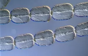
Background imageCross Section Collection: Light Micrograph (LM): A single cell from a hair on the stamen of the common spiderwort

Light Micrograph (LM): A single cell from a hair on the stamen of the common spiderwort (Tradescantia)
LRDS-170 Light Micrograph (LM): A single cell from a hair on the stamen of the common spiderwort (Tradescantia) Tradescantia sp David Spears (Last Refuge)

Background imageCross Section Collection: Fruit - Mandarin

Fruit - Mandarin
WAT-12573 Fruit - Mandarin M. Watson Please note that prints are for personal display purposes only and may not be reproduced in anyway. contact details: prints@ardea.com tel: + 44 (0) 20 8318 1401

Background imageCross Section Collection: Prickly Pear Cactus - showing fruit in section

Prickly Pear Cactus - showing fruit in section
SG-7708 Prickly Pear Cactus - showing fruit in section Ardea London Please note that prints are for personal display purposes only and may not be reproduced in anyway

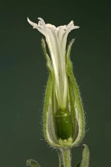
Background imageCross Section Collection: White Campion - cross-section of a female flower Europe

White Campion - cross-section of a female flower Europe
PPG-1490 White Campion - cross-section of a female flower Europe Silene alba Pascal Goetgheluck Please note that prints are for personal display purposes only and may not be reproduced in anyway

Background imageCross Section Collection: Walnuts - closeup of basket with single fruit opened to expose the edible walnut seed

Walnuts - closeup of basket with single fruit opened to expose the edible walnut seed
MAB-440 Walnuts - closeup of basket with single fruit opened to expose the edible walnut seed. Juglans regia Mark Boulton Please note that prints are for personal display purposes only

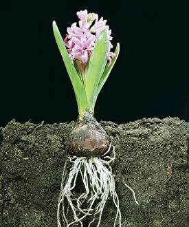
Background imageCross Section Collection: Hyacinth LA 102 Cross Section in earth showing roots © J. M. Labat ARDEA LONDON

Hyacinth LA 102 Cross Section in earth showing roots © J. M. Labat ARDEA LONDON
LA-102 HYACINTH - DEVELOPMENT STAGE 4 Cross Section in earth showing roots Jean Michel Labat Please note that prints are for personal display purposes only and may not be reproduced in anyway

Background imageCross Section Collection: Microscopic Fish Gill - tranverse section through gill region of fish. Fish anatomy

Microscopic Fish Gill - tranverse section through gill region of fish. Fish anatomy
JC-429 Microscopic Fish Gill - tranverse section through gill region of fish. Fish anatomy. John Clegg Please note that prints are for personal display purposes only

Background imageCross Section Collection: Light Micrograph (LM): A Tranverse section of a stem of a Common or European Ash tree

Light Micrograph (LM): A Tranverse section of a stem of a Common or European Ash tree (Fraxinus excelsior)
LRDS-258 Light Micrograph (LM): A Tranverse section of a stem of a Common or European Ash tree Magnification x12 (on 10.5 cm width print) Fraxinus excelsior David Spears (Last Refuge)

Background imageCross Section Collection: Light Micrograph (LM): Onion skin cells; Magnification x600 (on 10. 5 cm width print)

Light Micrograph (LM): Onion skin cells; Magnification x600 (on 10. 5 cm width print)
LRDS-252 Light Micrograph (LM): Onion skin cells; Magnification x600 (on 10.5 cm width print) David Spears (Last Refuge) / ardea.com Last Refuge Please note that prints are for personal display

Background imageCross Section Collection: Light Micrograph (LM): Longitudinal section of stem of Crown of Thorns (Euphorbia splendens)

Light Micrograph (LM): Longitudinal section of stem of Crown of Thorns (Euphorbia splendens)
LRDS-248 Light Micrograph (LM): Longitudinal section of stem of Crown of Thorns Magnification x1200 (on 10.5 cm width print) Euphorbia splendens David Spears (Last Refuge)


For sale as Licensed Images

Choose your image, Select your licence and Download the media

"Unveiling the Hidden World: Exploring Cross Sections in Nature and Architecture" Delving into the intricate world of honey bees, witness the mesmerizing cross section of a honeycomb revealing their remarkable life cycle. 🐝🍯 Journey deep within a Honey Bee (Apis mellifera) to discover its internal anatomy through a fascinating cross-section exploration. Marvel at the complexity of human hair follicles and skin as we unveil their hidden secrets through an enlightening cross-sectional view. Embark on a captivating expedition through the human brain, where a cross section exposes the enigmatic limbic system and primitive forebrain. As twilight descends upon Kathmandu's Boudhanath Stupa, immerse yourself in its beauty with an awe-inspiring cross-sectional perspective. Witness nature's underground marvels as we reveal a mole devouring worms beneath our feet—a captivating glimpse into their subterranean world. Step back in time to admire the architectural genius behind Florence Cathedral's dome, revealed through an intricate plan and cutaway drawing from the 16th century. Prayer flags flutter alongside Nepal's iconic Boudhanath Stupa—experience this spiritual haven from an extraordinary cross-sectional viewpoint. Encounter Australia's vibrant wildlife like never before as you explore the inner workings of a Green Tree Python through its stunning cross section. Take flight aboard Pan-American Clipper Boeing 314 with an exclusive cutaway view that unveils every detail of this magnificent aircraft from c1940. Explore Tab X’s mysterious contents by peering into its intriguing cross-section—an invitation to unravel its enigmatic nature.