Fibres Collection (#11)

"Fibres: Unraveling the Intricacies of the Human Brain and Beyond" Exploring the intricate network of brain fibres through DTI MRI scans (C017 / 7099

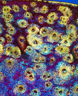
Background imageFibres Collection: Beech tree stem, light micrograph

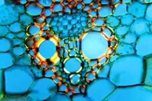
Beech tree stem, light micrograph
Beech tree stem. Polarised light micrograph of a section through the woody stem of a common beech tree (Fagus sylvatica), showing diffuse-porous xylem vessels (dark blue)

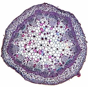
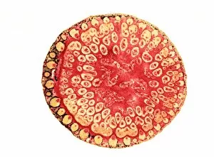
Background imageFibres Collection: Geranium stem, light micrograph

Geranium stem, light micrograph
Geranium stem. Light micrograph of a section through a young stem of a geranium (Pelargonium sp.) plant. The outer stem is covered with a thin epidermis (red) which has stomata

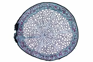
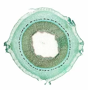
Background imageFibres Collection: Oak root, light micrograph

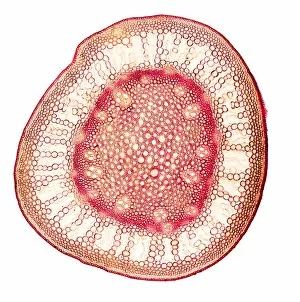
Oak root, light micrograph
Oak root. Light micrograph of a section through a secondary root from an oak (Quercus sp.) tree. The primary cortex (outer layer) has been shed by the formation of a circular meristem, the periderm

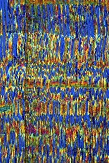
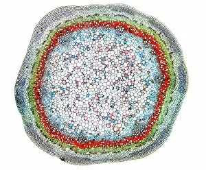
Background imageFibres Collection: African mahogany stem, light micrograph

African mahogany stem, light micrograph
African mahogany stem. Polarised light micrograph of a section through the stem of an African mahogany (Khaya sp.) tree. The multicoloured cells are fibres, and fibre tracheids, which carry water

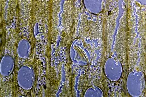
Background imageFibres Collection: African teak stem, light micrograph

African teak stem, light micrograph
African teak stem. Light micrograph of a section through the woody stem of an African teak (Pericopsis elata) plant, showing its diffuse-porous xylem tissue

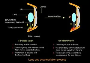
Background imageFibres Collection: Eye lens and accommodation, diagram

Eye lens and accommodation, diagram
Eye lens and accommodation. Diagram showing the process by which the eye changes the shape of the lens when changing between close vision and distant vision. This process is known as accommodation

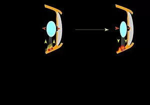
Background imageFibres Collection: Eye lens and accommodation, artwork

Eye lens and accommodation, artwork
Eye lens and accommodation. Artwork showing the process by which the eye changes the shape of the lens when changing between close vision and distant vision. This process is known as accommodation

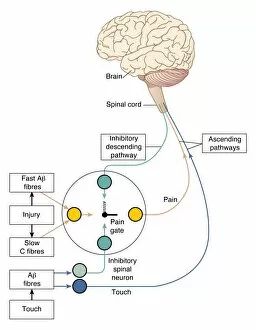
Background imageFibres Collection: Gate control theory of pain, artwork

Gate control theory of pain, artwork
Gate control theory of pain. Computer artwork of the neural circuit diagram used to represent the gate control theory of pain

Background imageFibres Collection: Antimicrobial wound dressing, SEM

Antimicrobial wound dressing, SEM
Antimicrobial wound dressing. Coloured scanning electron micrograph (SEM) of fibres (diagonal) from an Aquacel Ag wound dressing

Background imageFibres Collection: Plant cell wall, diagram

Plant cell wall, diagram

Background imageFibres Collection: Nerve fibre, artwork

Nerve fibre, artwork
Nerve fibre, computer artwork

Background imageFibres Collection: Muscle structure, artwork

Muscle structure, artwork
Muscle structure. Computer artwork showing the protein structure of myofibrils and how they are bound together to form muscle fibres

Background imageFibres Collection: Muscle anatomy, artwork

Muscle anatomy, artwork
Muscle anatomy. Computer artwork of a cross-section through a skeletal muscle. Single muscle fibres (top right) are made up of many myofibrils surrounded by endomysium connective tissue (orange)

Background imageFibres Collection: Keratinocyte skin cells, light micrograph

Keratinocyte skin cells, light micrograph
Keratinocyte skin cells. Fluorescent light micrograph of the cytoskeleton of human keratinocyte skin cells. Cell nuclei are oval. The rest of the cells contents have been biochemically extracted

Background imageFibres Collection: Polycotton fabric, SEM

Polycotton fabric, SEM
Polycotton fabric, coloured scanning electron micrograph (SEM). This material is made from polyester and cotton fibres woven together

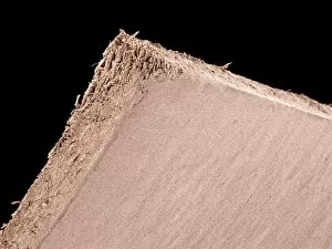
Background imageFibres Collection: Paper, SEM

Paper, SEM
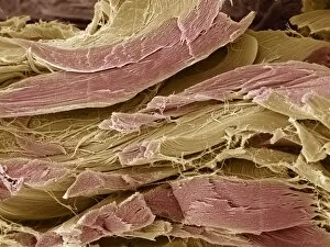
Paper, coloured scanning electron micrograph (SEM). Paper mostly consists of a mesh of softened and pulped wood fibres

Background imageFibres Collection: Muscle anatomy

Muscle anatomy. Cutaway artwork showing the anatomy and internal structure of a muscle, from the macroscopic (left) to the microscopic (right) level

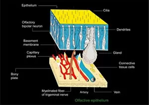
Background imageFibres Collection: Olfactory epithelium, artwork

Olfactory epithelium, artwork
Olfactory epithelium. Computer artwork showing the structure of the specialised layer of tissue that lines the inside of the nasal cavity and is involved in smell

Background imageFibres Collection: Damaged cardiac tissue, artwork

Damaged cardiac tissue, artwork
Damaged cardiac tissue. Artwork of damaged heart fibres and cells in heart muscle tissue (myocardium). This can occur due to reduced blood supply (ischaemia)

Background imageFibres Collection: Cardiac muscle, artwork

Cardiac muscle, artwork
Cardiac muscle. Computer artwork of cardiac muscle fibres. Cardiac muscle is under subconscious control and continuously contracts to pump blood around the body without tiring

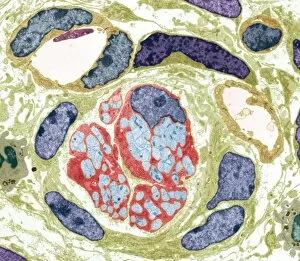
Background imageFibres Collection: Unmyelinated nerve bundle, TEM

Unmyelinated nerve bundle, TEM
Unmyelinated nerve bundle. Coloured transmission electron micrograph (TEM) of a section through a group of unmylinated nerve fibres

Background imageFibres Collection: Collagen, artwork

Collagen, artwork
Collagen. Computer artwork showing the structure of collagen fibres (grey). Each fibre consists of many molecules of collagen

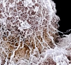
Background imageFibres Collection: Nucleolus, SEM

Nucleolus, SEM
Nucleolus, coloured scanning electron micrograph (SEM). The nucleolus is responsible for producing components of ribosomes, the cells protein-manufacturing organelles

Background imageFibres Collection: Nerve damage, artwork

Nerve damage, artwork
Nerve damage, computer artwork. The protective covering of the nerve has been lost and the structure of the actual nerve itself (centre) has degraded

Background imageFibres Collection: Severed nerve, artwork

Severed nerve, artwork
Severed nerve, computer artwork. The protective covering of the nerve has been lost and the structure of the actual nerve itself (centre) has degraded

Background imageFibres Collection: Collagen, SEM

Collagen, SEM
Collagen fibres, coloured scanning electron micrograph (SEM). Collagen, a type of connective tissue, is the main structural protein in the body

Background imageFibres Collection: Myelinated nerves, SEM

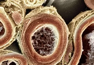
Myelinated nerves, SEM
Myelinated nerves. Coloured scanning electron micrograph (SEM) of a section through myelinated nerve fibres and Schwann cells

Background imageFibres Collection: Umbrella grass stem, light micrograph

Umbrella grass stem, light micrograph
Umbrella grass stem. Polarised light micrograph of a section through a umbrella grass stem (Cyperus alternifolius). Vascular bundles (brown) can be seen containing xylem (larger openings)

Background imageFibres Collection: Purple moor grass stem, light micrograph

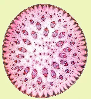
Purple moor grass stem, light micrograph
Purple moor grass stem. Polarised light micrograph of a section through a purple moor grass stem (Molinia caerula). Vascular bundles (round orange structures)

Background imageFibres Collection: Bamboo stem, light micrograph

Bamboo stem, light micrograph
Bamboo stem. Polarised light micrograph of a section through a bamboo stem (Bambusa vulgaris). Vascular bundles can be seen (yellow/green) containing xylem (larger openings)

Background imageFibres Collection: Maize stem, light micrograph

Maize stem, light micrograph
Maize stem. Polarised light micrograph of a section through a maize plant stem (Zea mays). A vascular bundles (brown) can be seen containing xylem (larger openings)

Background imageFibres Collection: Spiny rush stem, light micrograph

Spiny rush stem, light micrograph
Spiny rush stem. Polarised light micrograph of a section through a spiny rush stem (Juncus actus). Vascular bundles can be seen (purple/red) containing xylem (larger openings)

Background imageFibres Collection: Sugar cane stem, light micrograph

Sugar cane stem, light micrograph
Sugar cane stem. Polarised light micrograph of a section through a sugar cane stem (Saccharum officinareum). Vascular bundles (round blue/purple structures)

Background imageFibres Collection: Reed canary grass stem, light micrograph

Reed canary grass stem, light micrograph
Reed canary grass stem. Light micrograph of a section through a reed canary grass stem (Phalaris arundinacea). Vascular bundles (oval yellow and red structures)

Background imageFibres Collection: Dragon tree stem, light micrograph

Dragon tree stem, light micrograph
Dragon tree stem. Light micrograph of a section through a dragon tree stem (Dracaena draco). Vascular bundles (round dark structures) can be seen containing xylem (larger openings)

Background imageFibres Collection: Lilac stem, light micrograph

Lilac stem, light micrograph
Lilac stem. Light micrograph of a transverse section through the young woody stem of a lilac (Fraxinus excelsior) tree. The thick epidermis (solid green) is being sloughed (pushed/broken)

Background imageFibres Collection: Common broom stem, light micrograph

Common broom stem, light micrograph
Common broom stem. Light micrograph of a transverse section through the stem of a common broom (Salicornia europaea) plant

Background imageFibres Collection: Ash stem, light micrograph

Ash stem, light micrograph
Ash stem. Light micrograph of a transverse section through the woody stem of an ash (Fraxinus excelsior) tree showing four growth rings (concentric circles)

Background imageFibres Collection: Elder tree stem, light micrograph

Elder tree stem, light micrograph
Elder tree stem. Light micrograph of a transverse section through the very young woody stem of an elder (Sambuca nigra) tree

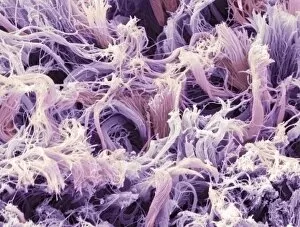
Background imageFibres Collection: Tendon fibres, SEM

Tendon fibres, SEM
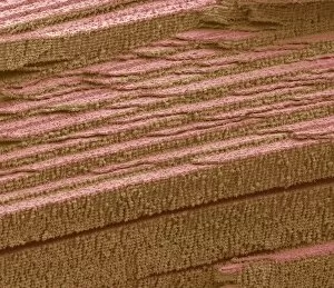
Tendon fibres. Coloured freeze-fracture scanning electron micrograph (SEM) of tendon fibres. These fibres are made from collagen

Background imageFibres Collection: Japanese sago palm leaf, light micrograph

Japanese sago palm leaf, light micrograph
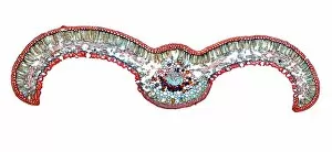
Japanese sago palm leaf. Light micrograph of a transverse section through the leaf (pinna) of a Japanese sago palm, cycad, (Cycas revoluta)
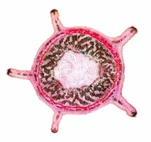
Background imageFibres Collection: Recycled materials

Recycled materials. Close-up of cardboard sheets and the label on a bag showing that it is made from 100 percent recycled material
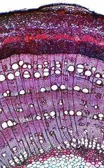
Background imageFibres Collection: Eye lens fibres, SEM

Eye lens fibres, SEM
Eye lens fibres. Coloured scanning electron micrograph (SEM) of a freeze-fracture of a lens of an eye, showing the regular arrangement of fibres

Background imageFibres Collection: Stem of a dracaena tree, micrograph

Stem of a dracaena tree, micrograph
Stem of a Dracaena. Light micrograph of the stem of a Dracaena (Darcaena terminalis) of the Agavaceae (Monocotyledons), showing two vascular bundles containing fibres, found in the ground tissue

Background imageFibres Collection: Bamboo stem, SEM

Bamboo stem, SEM
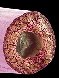
Bamboo stem. Coloured scanning electron micrograph (SEM) of a section through a bamboo stem (Phyllostachys sp.). Vascular bundles can be seen (bright yellow) containing xylem (larger openings)


For sale as Licensed Images

Choose your image, Select your licence and Download the media

"Fibres: Unraveling the Intricacies of the Human Brain and Beyond" Exploring the intricate network of brain fibres through DTI MRI scans (C017 / 7099). The mesmerizing artwork depicting white matter fibres and their connection to the brain (C015 / 1930). Delving deeper into understanding brain fibres with DTI MRI scans (C017 / 7035). Illuminating neurons and astrocytes through immunofluorescent LM, revealing the complexity of brain fibres. Peering into the white matter fibres of the human brain, unlocking its mysteries (C014 / 5666). Witnessing nerve cells regenerate under a TEM microscope, showcasing nature's resilience. Marveling at the intricate structure of a lime tree stem captured in a light micrograph. Discovering beauty in unexpected places - castor oil stem captured in a mesmerizing light micrograph. Unveiling myelination's role in enhancing nerve fibre function through TEM imaging. Witnessing cell division unfold under fluorescent micrographs, highlighting life's continuous renewal process. Celebrating nature's ability to heal as we observe regenerating nerve cells under TEM microscopy once again. Exploring further into white matter fibres within our complex human brains (C014/5668). In this captivating journey, we unravel the wonders – from delicate neural connections within our brains to resilient plant stems that provide sustenance for life itself.