Internal Anatomy Collection (#10)

"Exploring the Intricacies: Unveiling Internal Anatomy through Art and Science" Delving into the hidden world of internal anatomy

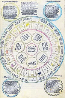
Background imageInternal Anatomy Collection: Fasciculus medicinae

Fasciculus medicinae
2626474 Fasciculus medicinae; (add.info.: Illustration from Fasciculus medicinae Venice; 1494 the first printed book to contain anatomical illustrations)

Background imageInternal Anatomy Collection: Les oeuures d'Ambroise Pare?

Les oeuures d'Ambroise Pare?
2626406 Les oeuures d'Ambroise Pare?; (add.info.: Illustration from Les oeuures d'Ambroise Pare? 1585 by Ambroise Pare?, 1510-1590 French surgeon and anatomist.); Universal History Archive/UIG

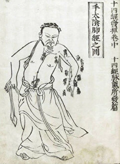
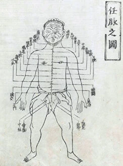
Background imageInternal Anatomy Collection: Shi si jing fa hui

Shi si jing fa hui
2626558 Shi si jing fa hui; (add.info.: Illustration from the Japanese & Chinese edition (1716) of Shi si jing fa hui, (Routes of the Fourteen Meridians and their Functions)

Background imageInternal Anatomy Collection: Equine anatomical Illustration

Equine anatomical Illustration
2626311 Equine anatomical Illustration; (add.info.: Equine anatomical Illustration from Anatomia del cavallo, infermita, et suoi rimedii')

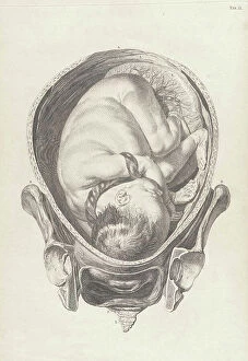
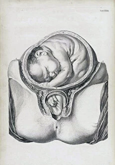
Background imageInternal Anatomy Collection: A set of anatomical tables, with explanations, and an abridgment, of the practice of midwifery

A set of anatomical tables, with explanations, and an abridgment, of the practice of midwifery, 18th (engraving)
2626256 A set of anatomical tables, with explanations, and an abridgment, of the practice of midwifery, 18th (engraving); (add.info)

Background imageInternal Anatomy Collection: Shi si jing fa hui

Shi si jing fa hui
2626540 Shi si jing fa hui; (add.info.: Illustration from the Japanese & Chinese edition (1716) of Shi si jing fa hui, (Routes of the Fourteen Meridians and their Functions)

Background imageInternal Anatomy Collection: A set of anatomical tables, with explanations, and an abridgment, of the practice of midwifery

A set of anatomical tables, with explanations, and an abridgment, of the practice of midwifery, 18th (engraving)
2626219 A set of anatomical tables, with explanations, and an abridgment, of the practice of midwifery, 18th (engraving); (add.info)

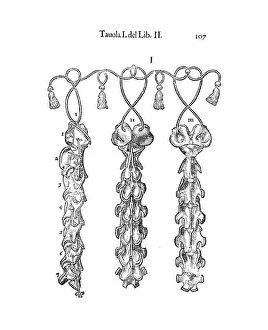
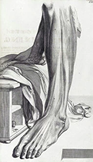
Background imageInternal Anatomy Collection: The anatomy textbook Ontleding des Menschelyken Lichaams, 1690 (engraving)

The anatomy textbook Ontleding des Menschelyken Lichaams, 1690 (engraving)
2625813 The anatomy textbook Ontleding des Menschelyken Lichaams, 1690 (engraving); (add.info.: Illustrations by Govard Bidloo, from the anatomy textbook Ontleding des menschelyken Lichaams')

Background imageInternal Anatomy Collection: Les oeuures d'Ambroise Pare?

Les oeuures d'Ambroise Pare?
2626404 Les oeuures d'Ambroise Pare?; (add.info.: Illustration from Les oeuures d'Ambroise Pare? 1585 by Ambroise Pare?, 1510-1590 French surgeon and anatomist.); Universal History Archive/UIG

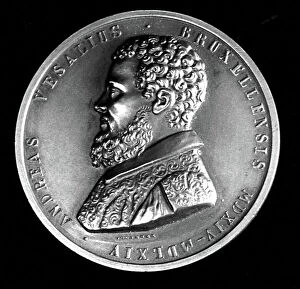
Background imageInternal Anatomy Collection: Medal depicting Andreas Vesalius

Medal depicting Andreas Vesalius
3586027 Medal depicting Andreas Vesalius; (add.info.: Medal depicting Andreas Vesalius (1514-1564) a Flemish/Netherlandish anatomist, physician, and author of books on human anatomy

Background imageInternal Anatomy Collection: William Henry Flower (1831-1899)

William Henry Flower (1831-1899)
865073 William Henry Flower (1831-1899); (add.info.: William Henry Flower (1831-1899) English surgeon and zoologist, born at Stratford-on-Avon, Warwickshire, England

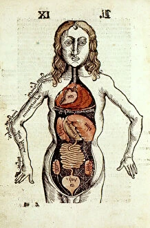
Background imageInternal Anatomy Collection: Internal organs of the human body. From Gregor Reisch Margarita Philosophica, Basle, 1508
Internal organs of the human body. From Gregor Reisch Margarita Philosophica, Basle, 1508
527495 Internal organs of the human body. From Gregor Reisch Margarita Philosophica, Basle, 1508. ; Universal History Archive/UIG

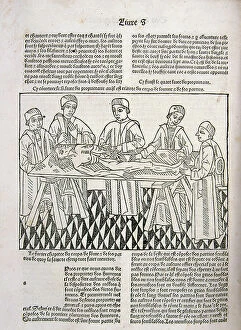
Background imageInternal Anatomy Collection: A dissection in progress. From Le livre de la propriete des choses (On the Properites of Things)

A dissection in progress. From Le livre de la propriete des choses (On the Properites of Things)
527490 A dissection in progress. From Le livre de la propriete des choses (On the Properites of Things) written by the English Franciscan monk Bartholomew Glanville, called Bartolomaeus Anglicus

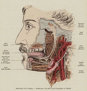
Background imageInternal Anatomy Collection: Nerves involved in the sense of taste (coloured engraving)

Nerves involved in the sense of taste (coloured engraving)
8655717 Nerves involved in the sense of taste (coloured engraving) by French School, (19th century) (after); Private Collection; (add.info.: Nerves involved in the sense of taste)

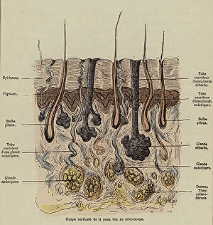
Background imageInternal Anatomy Collection: Cross-section of the skin (coloured engraving)

Cross-section of the skin (coloured engraving)
8655716 Cross-section of the skin (coloured engraving) by French School, (19th century) (after); Private Collection; (add.info.: Cross-section of the skin)

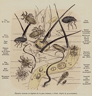
Background imageInternal Anatomy Collection: Parasites of the human skin (coloured engraving)

Parasites of the human skin (coloured engraving)
8655715 Parasites of the human skin (coloured engraving) by French School, (19th century) (after); Private Collection; (add.info.: Parasites of the human skin)

Background imageInternal Anatomy Collection: Muscular system of the human body (coloured engraving)

Muscular system of the human body (coloured engraving)
8655713 Muscular system of the human body (coloured engraving) by French School, (19th century) (after); Private Collection; (add.info.: Muscular system of the human body (front view)

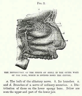
Background imageInternal Anatomy Collection: The distribution of the nerve of smell in the outer wall of the nose

The distribution of the nerve of smell in the outer wall of the nose, which is divided down the centre (engraving)
8646012 The distribution of the nerve of smell in the outer wall of the nose, which is divided down the centre (engraving) by English School

Background imageInternal Anatomy Collection: Glottis and vocal cords (engraving)

Glottis and vocal cords (engraving)
8655777 Glottis and vocal cords (engraving) by French School, (19th century) (after); Private Collection; (add.info.: Glottis and vocal cords)

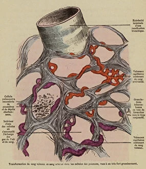
Background imageInternal Anatomy Collection: Oxygenation of blood in the lungs (coloured engraving)

Oxygenation of blood in the lungs (coloured engraving)
8655756 Oxygenation of blood in the lungs (coloured engraving) by French School, (19th century) (after); Private Collection; (add.info.: Oxygenation of blood in the lungs)

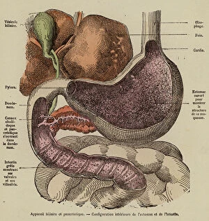
Background imageInternal Anatomy Collection: Organs of the digestive system (coloured engraving)

Organs of the digestive system (coloured engraving)
8655752 Organs of the digestive system (coloured engraving) by French School, (19th century) (after); Private Collection; (add.info.: Organs of the digestive system)

Background imageInternal Anatomy Collection: Muscles of the jaw and lips and their role in eating food (coloured engraving)

Muscles of the jaw and lips and their role in eating food (coloured engraving)
8655748 Muscles of the jaw and lips and their role in eating food (coloured engraving) by French School, (19th century) (after); Private Collection; (add.info)

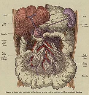
Background imageInternal Anatomy Collection: Organs of absorption in the digestive system (coloured engraving)

Organs of absorption in the digestive system (coloured engraving)
8655745 Organs of absorption in the digestive system (coloured engraving) by French School, (19th century) (after); Private Collection; (add.info.: Organs of absorption in the digestive system)

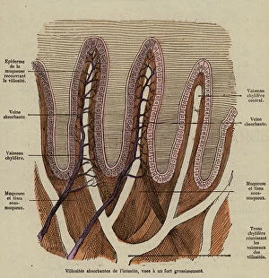
Background imageInternal Anatomy Collection: Intestinal villi (coloured engraving)

Intestinal villi (coloured engraving)
8655743 Intestinal villi (coloured engraving) by French School, (19th century) (after); Private Collection; (add.info.: Intestinal villi)

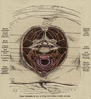
Background imageInternal Anatomy Collection: Cross-section of the neck at the leval of the sixth cervical vertebra (coloured engraving)

Cross-section of the neck at the leval of the sixth cervical vertebra (coloured engraving)
8655734 Cross-section of the neck at the leval of the sixth cervical vertebra (coloured engraving) by French School, (19th century) (after); Private Collection; (add.info)

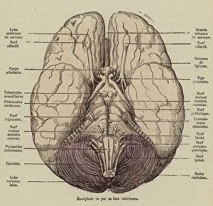
Background imageInternal Anatomy Collection: Brain seen from below (coloured engraving)

Brain seen from below (coloured engraving)
8655729 Brain seen from below (coloured engraving) by French School, (19th century) (after); Private Collection; (add.info.: Brain seen from below)

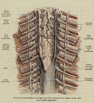
Background imageInternal Anatomy Collection: Section of the spinal cord showing the roots of the spinal nerves (coloured engraving)

Section of the spinal cord showing the roots of the spinal nerves (coloured engraving)
8655727 Section of the spinal cord showing the roots of the spinal nerves (coloured engraving) by French School, (19th century) (after); Private Collection; (add.info)

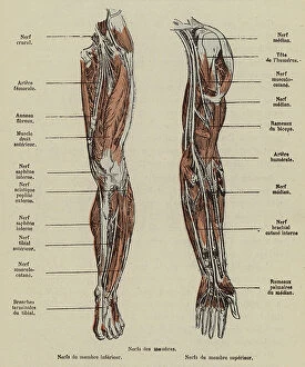
Background imageInternal Anatomy Collection: Nerves of the arm and leg (coloured engraving)

Nerves of the arm and leg (coloured engraving)
8655725 Nerves of the arm and leg (coloured engraving) by French School, (19th century) (after); Private Collection; (add.info.: Nerves of the arm and leg)

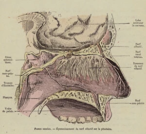
Background imageInternal Anatomy Collection: Nasal cavity and olfactory tract (coloured engraving)

Nasal cavity and olfactory tract (coloured engraving)
8655719 Nasal cavity and olfactory tract (coloured engraving) by French School, (19th century) (after); Private Collection; (add.info.: Nasal cavity and olfactory tract)

Background imageInternal Anatomy Collection: Dr Frank Truth. Nerve specialist (colour litho)

Dr Frank Truth. Nerve specialist (colour litho)
8656808 Dr Frank Truth. Nerve specialist (colour litho) by English School, (20th century); Private Collection; (add.info.: "Dr Frank Truth. Nerve specialist)

Background imageInternal Anatomy Collection: Elbow Joint, Inner Aspect (colour litho)

Elbow Joint, Inner Aspect (colour litho)
8667654 Elbow Joint, Inner Aspect (colour litho) by English School, (20th century); Private Collection; (add.info.: Elbow Joint, Inner Aspect)

Background imageInternal Anatomy Collection: Deep Dissection of the Front of the Forearm and Hand (colour litho)

Deep Dissection of the Front of the Forearm and Hand (colour litho)
8667628 Deep Dissection of the Front of the Forearm and Hand (colour litho) by English School, (20th century); Private Collection; (add.info.: Deep Dissection of the Front of the Forearm and Hand)

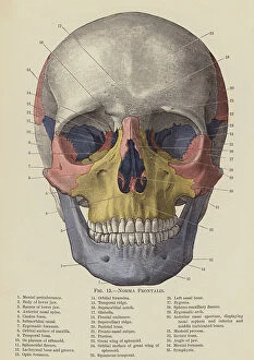
Background imageInternal Anatomy Collection: Norma Frontalis (colour litho)

Norma Frontalis (colour litho)
8667623 Norma Frontalis (colour litho) by English School, (20th century); Private Collection; (add.info.: Norma Frontalis)

Background imageInternal Anatomy Collection: Andre Vesale discovers the origin of blood vessels, in Paris (engraving)

Andre Vesale discovers the origin of blood vessels, in Paris (engraving)
5944552 Andre Vesale discovers the origin of blood vessels, in Paris (engraving); Private Collection; (add.info.: Engraving It was during his stay in Paris that Andr Vsale (1514 - 1564)

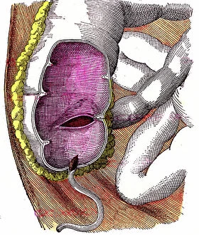
Background imageInternal Anatomy Collection: Digestive tract. The caecum or cecum and the appendix ileo cecal

Digestive tract. The caecum or cecum and the appendix ileo cecal. Book by Dr Jules Rengade "Life and Health"
7295213 Digestive tract. The caecum or cecum and the appendix ileo cecal. Book by Dr Jules Rengade "Life and Health", Paris 1881; Private Collection; Photo ©Jaime Abecasis

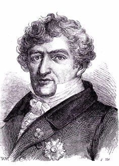
Background imageInternal Anatomy Collection: Georges CUVIER, french anatomist and paleontologist. 1869 (engraving)

Georges CUVIER, french anatomist and paleontologist. 1869 (engraving)
6426200 Georges CUVIER, french anatomist and paleontologist. 1869 (engraving); Private Collection; (add.info.: Georges CUVIER (1769-1832), french anatomist and paleontologist

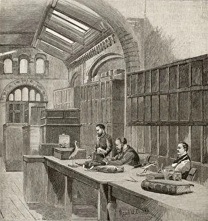
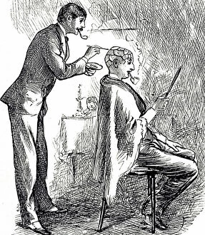
Background imageInternal Anatomy Collection: A cartoon commenting on the fashionable revival of phrenology, 19th century

A cartoon commenting on the fashionable revival of phrenology, 19th century
5311366 A cartoon commenting on the fashionable revival of phrenology, 19th century; (add.info.: Date: 1878); Universal History Archive/UIG

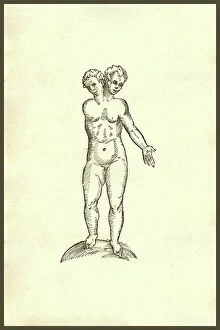
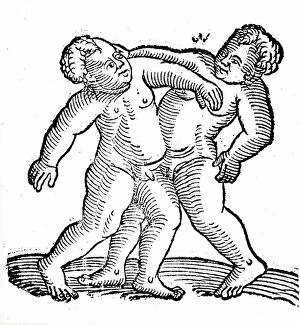
Background imageInternal Anatomy Collection: Siamese twins, one boy and one girl, 16th century

Siamese twins, one boy and one girl, 16th century
5311328 Siamese twins, one boy and one girl, 16th century; Universal History Archive/UIG

Background imageInternal Anatomy Collection: Infans Biceps unico Brachio : Infans biceps unico brachio, two heads on one body, siamese twins

Infans Biceps unico Brachio : Infans biceps unico brachio, two heads on one body, siamese twins
UIG5327230 Infans Biceps unico Brachio : Infans biceps unico brachio, two heads on one body, siamese twins. From the 1642 book Monstrorum Historia by Ulisse Aldrovandi (Bologna, 1522-1605)

Background imageInternal Anatomy Collection: Monstrosi Piscis Volantis : Monster fish Creature. From the 1642 book Monstrorum Historia by

Monstrosi Piscis Volantis : Monster fish Creature. From the 1642 book Monstrorum Historia by Ulisse Aldrovandi
UIG5327195 Monstrosi Piscis Volantis : Monster fish Creature. From the 1642 book Monstrorum Historia by Ulisse Aldrovandi (Bologna, 1522-1605). He is considered the founder of modern Natural History

Background imageInternal Anatomy Collection: Ancient Monsters : Monsters! From the 1642 book Monstrorum Historia by Ulisse Aldrovandi (Bologna)

Ancient Monsters : Monsters! From the 1642 book Monstrorum Historia by Ulisse Aldrovandi (Bologna, 1522-1605)
UIG5327190 Ancient Monsters : Monsters! From the 1642 book Monstrorum Historia by Ulisse Aldrovandi (Bologna, 1522-1605). He is considered the founder of modern Natural History

Background imageInternal Anatomy Collection: Rana piscatrix & Echeneis Five Remora : Rana piscatrix Georgij Fabricij

Rana piscatrix & Echeneis Five Remora : Rana piscatrix Georgij Fabricij, frog- or toad-like fish with a large
UIG5327178 Rana piscatrix & Echeneis Five Remora : Rana piscatrix Georgij Fabricij, frog- or toad-like fish with a large, toothed maw along with an echeneis five remora

Background imageInternal Anatomy Collection: Infans partibus inferioribus caninis : Infans partibus inferioribus caninis

Infans partibus inferioribus caninis : Infans partibus inferioribus caninis, child with a dog's lower body
UIG5327175 Infans partibus inferioribus caninis : Infans partibus inferioribus caninis, child with a dog's lower body

Background imageInternal Anatomy Collection: Infans Auribus Leporinis : Infans auribus leporinis: a child with rabbit ears

Infans Auribus Leporinis : Infans auribus leporinis: a child with rabbit ears
UIG5327166 Infans Auribus Leporinis : Infans auribus leporinis: a child with rabbit ears. From the 1642 book Monstrorum Historia by Ulisse Aldrovandi (Bologna, 1522-1605)

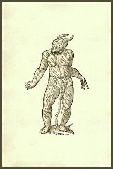
Background imageInternal Anatomy Collection: Puer Cornutus Ore Patulo : Infans cornutus ore patulo, child with flaky and cornificated skin

Puer Cornutus Ore Patulo : Infans cornutus ore patulo, child with flaky and cornificated skin
UIG5327163 Puer Cornutus Ore Patulo : Infans cornutus ore patulo, child with flaky and cornificated skin. From the 1642 book Monstrorum Historia by Ulisse Aldrovandi (Bologna, 1522-1605)

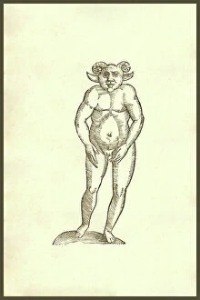
Background imageInternal Anatomy Collection: Monstrum Humanum Coribus Arietinis : Monstrum Humanum Coribus Arietinis

Monstrum Humanum Coribus Arietinis : Monstrum Humanum Coribus Arietinis. From the 1642 book Monstrorum Historia by
UIG5327156 Monstrum Humanum Coribus Arietinis : Monstrum Humanum Coribus Arietinis. From the 1642 book Monstrorum Historia by Ulisse Aldrovandi (Bologna, 1522-1605)

Background imageInternal Anatomy Collection: Vitulus capite fere humano : Vitulus capite fere humano, calf with human head and hands

Vitulus capite fere humano : Vitulus capite fere humano, calf with human head and hands
UIG5327150 Vitulus capite fere humano : Vitulus capite fere humano, calf with human head and hands. From the 1642 book Monstrorum Historia by Ulisse Aldrovandi (Bologna, 1522-1605)

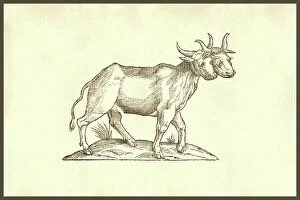
Background imageInternal Anatomy Collection: Bos Biceps : Bos biceps, cow with one body and two heads. From the 1642 book Monstrorum Historia

Bos Biceps : Bos biceps, cow with one body and two heads. From the 1642 book Monstrorum Historia by Ulisse Aldrovandi
UIG5327128 Bos Biceps : Bos biceps, cow with one body and two heads. From the 1642 book Monstrorum Historia by Ulisse Aldrovandi (Bologna, 1522-1605)


For sale as Licensed Images

Choose your image, Select your licence and Download the media

"Exploring the Intricacies: Unveiling Internal Anatomy through Art and Science" Delving into the hidden world of internal anatomy, we are captivated by the wonders that lie beneath our skin. From the delicate cross-section of a Honey Bee (Apis mellifera) to the meticulous illustrations of the Channel Tunnel Project, each piece unravels a unique perspective on this captivating subject. Intriguingly, even centuries ago, artists like Giovanni Battista Bianchi skillfully captured human form in terracotta sculptures, reminding us of our intricate inner workings. Meanwhile, Andreas Vesalius revolutionized anatomical understanding with his groundbreaking studies during the Renaissance era. As we journey through time and mediums, Waldeck's original artwork takes us back to Mesoamerica in 1831. The prints from "La nature et l'homme" by Rengade depict twin fetuses in their ideal position during gemellar pregnancy – a testament to both artistry and scientific observation. The diversity within these artworks is astounding; from portraits such as Lucrezia Panciatichi's enigmatic gaze immortalized in oil on wood panel to Giulia di Alessandro de Medici holding a book – perhaps symbolizing knowledge about one's own body. Even photography joins this exploration as two male nudes perch upon a rock in an evocative black-and-white image from 1890. This photograph captures not only physicality but also invites contemplation on vulnerability and strength intertwined within our internal structures. From ancient times to modern endeavors like the Channel Tunnel Project illustration from 1906, humanity has sought to unravel its own mysteries. Through art and science alike, we continue to marvel at our internal anatomy – an ever-evolving tapestry waiting for discovery.