Joints Collection (#2)

"Exploring the Intricacies of Joints: A Journey through History and Anatomy" In this captivating journey, we delve into the world of joints

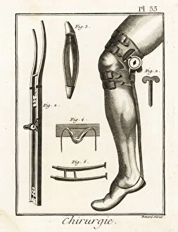
Background imageJoints Collection: 18th century surgical knee pad and dislocation machine

18th century surgical knee pad and dislocation machine
18th century surgical knee pad in place on a leg 1, Jean Louis Petits machine for dislocated joints 2-5. Copperplate engraving by Robert Benard from Denis Diderots Encyclopedia, Pellet, Geneva, 1779

Background imageJoints Collection: The Slaughtered Ox, 1635-55 (oil on canvas)

The Slaughtered Ox, 1635-55 (oil on canvas)
3704070 The Slaughtered Ox, 1635-55 (oil on canvas) by Hecken, Abraham van den (fl.1645); 114x98 cm; Rijksmuseum, Amsterdam, The Netherlands; (add.info.: ); Flemish, out of copyright

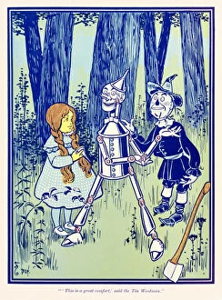
Background imageJoints Collection: 'This is a great comfort, said the Tin Woodman

"This is a great comfort, said the Tin Woodman
3614736 A" A This is a great comfort, A said the Tin Woodman.A" from A The Wonderful Wizard of OzA by L. Frank Baum by Denslow

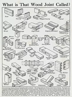
Background imageJoints Collection: Wood joints (litho)

Wood joints (litho)
3642511 Wood joints (litho) by English School, (20th century); Private Collection; (add.info.: Wood joints. Illusration for Everybodys Enquire Within (Amalgamated Press)

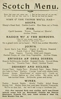
Background imageJoints Collection: Scotch Menu (litho)

Scotch Menu (litho)
3643458 Scotch Menu (litho) by English School, (20th century); Private Collection; (add.info.: Scotch Menu. Postcard, early 20th century.); © Look and Learn

Background imageJoints Collection: Anatomical Study of a Hand (pencil on paper)

Anatomical Study of a Hand (pencil on paper)
XOT361269 Anatomical Study of a Hand (pencil on paper) by Bandinelli, Clemente (1534-1555); Gabinetto dei Disegni e Stampe, Galleria Degli Uffizi, Florence, Tuscany

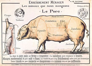
Background imageJoints Collection: Cuts of Pork, illustration from a French Domestic Science Manual by H

Cuts of Pork, illustration from a French Domestic Science Manual by H
CHT289133 Cuts of Pork, illustration from a French Domestic Science Manual by H. de Puytorac, published by Editions Fernand Nathan, late 19th century (colour litho) by French School

Background imageJoints Collection: Chiropractic adjustment, illustration from The Works of Hippocrates

Chiropractic adjustment, illustration from The Works of Hippocrates
STC445967 Chiropractic adjustment, illustration from The Works of Hippocrates, 1934 (colour litho) by Kuhn-Regnier, Joseph (1873-1940); Private Collection; (add.info)

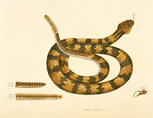
Background imageJoints Collection: Viper Caudison Snake, Rattlesnake, plate 41, vol. 1, from the

Viper Caudison Snake, Rattlesnake, plate 41, vol. 1, from the
NBY335514 Viper Caudison Snake, Rattlesnake, plate 41, vol. 1, from the Natural History of Carolina, Florida and the Bahamas (colour litho) by Catesby, Mark (1679-1749); Newberry Library, Chicago

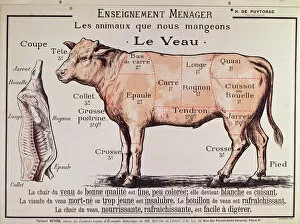
Background imageJoints Collection: Veal: diagram depicting the different cuts of meat (colour litho)

Veal: diagram depicting the different cuts of meat (colour litho)
CHT210241 Veal: diagram depicting the different cuts of meat (colour litho) by French School, (20th century); Private Collection; (add.info)

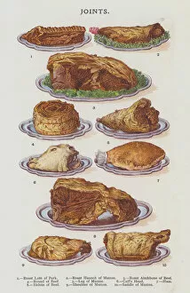
Background imageJoints Collection: Joints (chromolitho)

Joints (chromolitho)
3649855 Joints (chromolitho) by English School, (19th century); Private Collection; (add.info.: Joints. Illustration for Mrs Beetons Book of Household Management (Ward Lock)

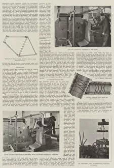
Background imageJoints Collection: Mr Hooleys Hydraulic Joint (b / w photo)

Mr Hooleys Hydraulic Joint (b / w photo)
1615195 Mr Hooleys Hydraulic Joint (b/w photo) by English Photographer, (19th century); Private Collection; (add.info.: Mr Hooleys Hydraulic Joint)

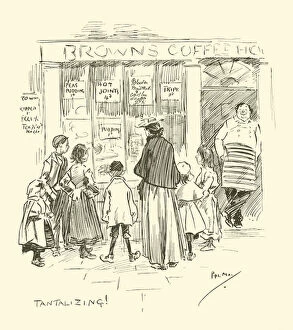
Background imageJoints Collection: Illustration for Phil Mays Gutter-Snipes (lithograph)

Illustration for Phil Mays Gutter-Snipes (lithograph)
3098241 Illustration for Phil Mays Gutter-Snipes (lithograph) by May, Phil (1864-1903); Private Collection; (add.info.: Illustration for Phil Mays Gutter-Snipes (Leadenhall Press, 1896)

Background imageJoints Collection: Making dolls: manufacturing ball joints and stuffing (chromolitho)

Making dolls: manufacturing ball joints and stuffing (chromolitho)
1093604 Making dolls: manufacturing ball joints and stuffing (chromolitho) by European School, (19th century); Private Collection; (add.info.: Making dolls: manufacturing ball joints and stuffing)

Background imageJoints Collection: Joints (chromolitho)

Joints (chromolitho)
3649857 Joints (chromolitho) by English School, (19th century); Private Collection; (add.info.: Joints. Illustration for Mrs Beetons Book of Household Management (Ward Lock)

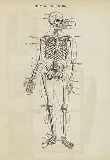
Background imageJoints Collection: Human Skeleton (engraving)

Human Skeleton (engraving)
2802021 Human Skeleton (engraving) by English School, (19th century); Private Collection; (add.info.: Human Skeleton. Illustration for An Illustrated Vocabulary For The Use Of The Deaf And Dumb)

Background imageJoints Collection: Advertisement for Doctor Parmenters Magnetic Oil, 1840s (colour litho)

Advertisement for Doctor Parmenters Magnetic Oil, 1840s (colour litho)
PNP383893 Advertisement for Doctor Parmenters Magnetic Oil, 1840s (colour litho) by American School, (19th century); Private Collection; (add.info.: Stop, friend! Read, reason and reflect)

Background imageJoints Collection: Separate Pieces of a Knights Armour, plate 18 from

Separate Pieces of a Knights Armour, plate 18 from
STC98440 Separate Pieces of a Knights Armour, plate 18 from The History of the Nations, engraved by G. Lago (engraving) by Italian School

Background imageJoints Collection: Joinery, Joints and mouldings (litho)

Joinery, Joints and mouldings (litho)
6013475 Joinery, Joints and mouldings (litho) by English School, (20th century); Private Collection; (add.info.: Joinery, Joints and mouldings)

Background imageJoints Collection: Still Life with Pigs Head, Pigs Knuckles and Sausage, c. 1650 (oil on panel)

Still Life with Pigs Head, Pigs Knuckles and Sausage, c. 1650 (oil on panel)
3704071 Still Life with Pigs Head, Pigs Knuckles and Sausage, c.1650 (oil on panel) by Monogrammist JVR (fl.1650); 55x72 cm; Rijksmuseum, Amsterdam, The Netherlands; (add.info.: ); Dutch

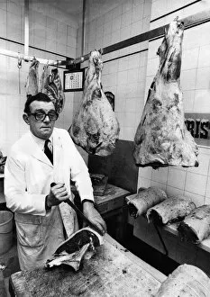
Background imageJoints Collection: Butcher Bert Sibbet cutting joints in his shop. November 1974

Butcher Bert Sibbet cutting joints in his shop. November 1974

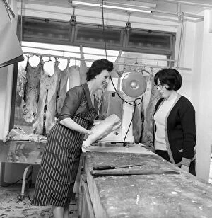
Background imageJoints Collection: A female butcher seen here at work in her shop with her colleague. 1963

A female butcher seen here at work in her shop with her colleague. 1963

Background imageJoints Collection: A female butcher seen here at work in her shop, serving a customer at the counter. 1963

A female butcher seen here at work in her shop, serving a customer at the counter. 1963

Background imageJoints Collection: A female butcher seen here at work in her shop, cutting up a joint of meat. 1963

A female butcher seen here at work in her shop, cutting up a joint of meat. 1963

Background imageJoints Collection: A female butcher seen here at work in her shop. 1963

A female butcher seen here at work in her shop. 1963

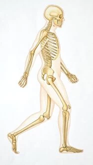
Background imageJoints Collection: Human skeleton showing all the joints in walking position

Human skeleton showing all the joints in walking position

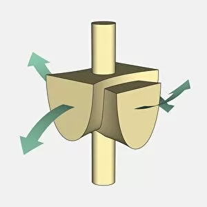
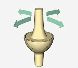
Background imageJoints Collection: Illustration, ball and socket joint, arrows indicating possible directions of movement

Illustration, ball and socket joint, arrows indicating possible directions of movement

Background imageJoints Collection: Illustration, hinge joint, arrows indicating possible directions of movement

Illustration, hinge joint, arrows indicating possible directions of movement

Background imageJoints Collection: Illustration, saddle joint, arrows indicating possible directions of movement

Illustration, saddle joint, arrows indicating possible directions of movement

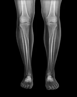
Background imageJoints Collection: Normal legs, X-rays

Normal legs, X-rays
Normal legs. Coloured frontal X-rays of the healthy legs, of a 35-year-old

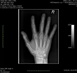
Background imageJoints Collection: Normal hand, digital X-ray

Normal hand, digital X-ray
Normal hand. Digital X-ray of the normal right hand of a patient in their late twenties. Various sets of data relating to the image can be seen around the edges of the X-ray

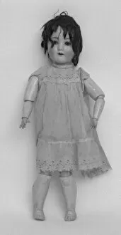
Background imageJoints Collection: German doll from Titanic

German doll from Titanic
Philomena, a German bisque doll rescued from the sea after the sinking of the Titanic. It was at first mistaken for a childs body

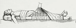
Background imageJoints Collection: Mode of support for a fractured thigh in old persons. From The Household Physician, published c

Mode of support for a fractured thigh in old persons. From The Household Physician, published c. 1898
Mode of support for a fractured thigh in old persons. From The Household Physician, published c.1898

Background imageJoints Collection: The application of Listons Long Splint. Long leg splinting is used to stabilize injuries by

The application of Listons Long Splint. Long leg splinting is used to stabilize injuries by decreasing movement and providing support, thus preventing further damage. Robert Liston, 1794 - 1847

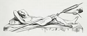
Background imageJoints Collection: Reduction of dislocated thigh using pulleys. From The Household Physician, published c. 1898

Reduction of dislocated thigh using pulleys. From The Household Physician, published c. 1898
Reduction of dislocated thigh using pulleys. From The Household Physician, published c.1898

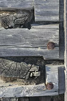
Background imageJoints Collection: Detail of the dovetailed corner a log cabin, Nulato, Interior Alaska, Alaska, USA

Detail of the dovetailed corner a log cabin, Nulato, Interior Alaska, Alaska, USA
Detail of the dovetailed corner construction from a weathered log cabin in winter; Nulato, Interior Alaska, Alaska, United States of America

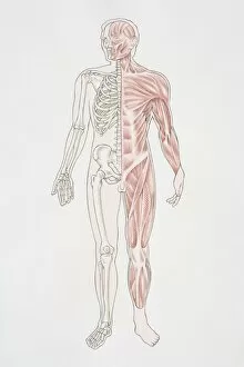
Background imageJoints Collection: Diagram illustrating the human musculo-skeletal system

Diagram illustrating the human musculo-skeletal system

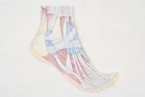
Background imageJoints Collection: Cross-section diagram of human foot, side view

Cross-section diagram of human foot, side view

Background imageJoints Collection: Illustration, ellipsoidal joint, arrows indicating possible directions of movement

Illustration, ellipsoidal joint, arrows indicating possible directions of movement

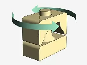
Background imageJoints Collection: Illustration, pivot joint, arrows indicating possible directions of movement

Illustration, pivot joint, arrows indicating possible directions of movement

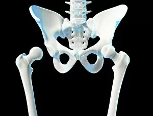
Background imageJoints Collection: Hip joint bones and anatomy, artwork C014 / 2032

Hip joint bones and anatomy, artwork C014 / 2032
Hip joint bones and anatomy. Artwork of the bones that form the two hip joints (right and left). The hip is a ball-and-socket joint that provides the mobility needed for walking and running

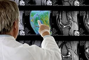
Background imageJoints Collection: Radiologist examining knee MRI scans C014 / 1300

Radiologist examining knee MRI scans C014 / 1300
MODEL RELEASED. Radiologist examining knee MRI (magnetic resonance imaging) scans

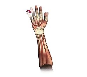
Background imageJoints Collection: Fist clenching movement, artwork C016 / 6795

Fist clenching movement, artwork C016 / 6795
Fist clenching movement. Artwork of the muscles of the forearm and hand from the front, with red arrows showing the direction of movement of the fingers during flexion to form a fist

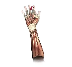
Background imageJoints Collection: Little and ring finger flexion, artwork C016 / 6793

Little and ring finger flexion, artwork C016 / 6793
Little and ring finger flexion. Artwork of the muscles of the forearm and hand from the front, with red arrows showing the direction of movement of the little and ring (fourth and third)

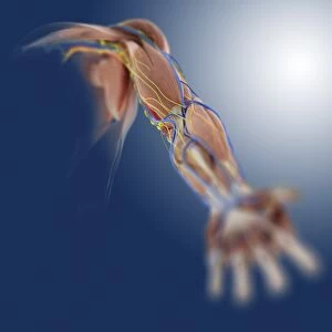
Background imageJoints Collection: Arm anatomy, artwork C013 / 4583

Arm anatomy, artwork C013 / 4583
Arm anatomy. Computer artwork of an oblique anterior view looking up a human arm, showing muscles and tendons (brown and white), bones and ligaments (white), arteries and veins (red and blue)

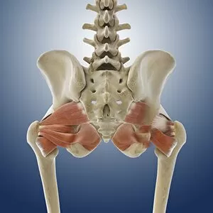
Background imageJoints Collection: Buttock muscles, artwork C013 / 4414

Buttock muscles, artwork C013 / 4414
Buttock muscles. Computer artwork of a posterior view of the hips showing some of the muscles (red) of the buttocks. These are shown attached to the femurs (thigh bones) and the pelvis bones

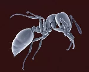
Background imageJoints Collection: Ant, SEM

Ant, SEM
Ant (family Formicidae), coloured scanning electron micrograph (SEM). One of its compound eyes (black) is seen, as well as two long antennae that are mounted on the head between the eyes

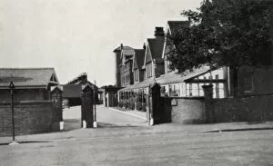
Background imageJoints Collection: Princess Marys Hospital, Margate, Kent

Princess Marys Hospital, Margate, Kent
Entrance to the Princess Marys Hospital, Wilderness Road, Margate, Kent. The site, originally known as Eastcliff House, was acquired in 1898 by the Metropolitan Asylums Board as a seaside hospital


For sale as Licensed Images

Choose your image, Select your licence and Download the media

"Exploring the Intricacies of Joints: A Journey through History and Anatomy" In this captivating journey, we delve into the world of joints, those remarkable connectors that enable movement and grace. Just like a palmistry map of the hand reveals secrets etched in our skin, so do joints hold tales of strength and resilience. Traveling back to 1855, we encounter a beef cuts diagram that showcases the intricate network within these joints. Stepping into a bustling butchers shop on High Street in Shoreham-by-Sea, Sussex during the 1920s, we witness a trailblazing woman butcher skillfully dissecting meat with precision. Zooming even closer under an electron microscope's lens, an ant's joint appears as a marvelously engineered masterpiece. Meanwhile, X-ray images expose bunions lurking beneath layers of flesh – silent reminders that even our they are bear burdens. Conceptual artworks emerge next; knee pain depicted as swirling colors and shapes evokes empathy for those grappling with discomfort. Joint pain takes form in abstract strokes on canvas - capturing both physical anguish and emotional strain. A black-and-white photograph from 1802 brings forth engravings showcasing beef, veal, pork, and mutton cuts – each slice revealing unique joint structures designed by nature herself. Historical artwork delves deeper into biomechanics - unraveling how forces interact within these pivotal connections. Conceptual art resurfaces once more to shed light on running injuries; brushstrokes depict shattered dreams amidst vibrant hues. Yet hope remains as another artwork illustrates leg muscles engaged in running - reminding us that healing is possible when nurtured with care. As we conclude this enlightening expedition through time and anatomy alike, let us appreciate the beauty hidden within every joint. For they are not mere hinges but rather gateways to mobility and vitality – connecting us all in our shared human experience.