Joints Collection (#7)

"Exploring the Intricacies of Joints: A Journey through History and Anatomy" In this captivating journey, we delve into the world of joints

Background imageJoints Collection: Ant feet, SEM

Ant feet, SEM
Ant feet. Coloured scanning electron micrograph (SEM) of the tip of two legs from an ant (family Formicidae). The end of an insect leg consists of the final segment, which is called the tarsus

Background imageJoints Collection: Hip bones, X-ray

Hip bones, X-ray
Hip joints. Coloured X-ray of a womans groin and upper legs, revealing the bones that form her hip joints. The left hip (identical to the right one) is at upper left in this rear view

Background imageJoints Collection: Female pelvis, X-ray

Female pelvis, X-ray
Female pelvis. Coloured X-ray of the pelvis of a woman. The lowest lumbar vertebra of the spinal column (top centre) forms a joint with the sacrum

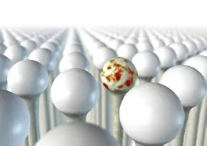
Background imageJoints Collection: Immune system components

Immune system components
Immune system. Conceptual computer artwork of a man standing with his arms outstretched surrounded by components of the immune system. The skeletal system is seen, with the joints highlighted in red

Background imageJoints Collection: Butchers shop

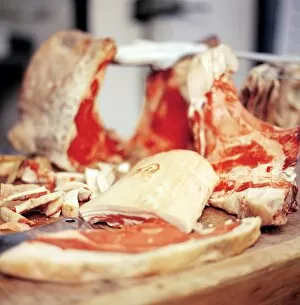
Butchers shop. Butcher preparing meat to be sold

Background imageJoints Collection: Spondylitis, X-ray

Spondylitis, X-ray
Spondylitis. Coloured X-ray of the lower spine and pelvis of a patient with spondylitis, an inflammation of the synovial joints of the backbone

Background imageJoints Collection: Thalassaemia, X-ray

Thalassaemia, X-ray
Thalassaemia. Coloured hand X-ray of a patient with thalassaemia, also known as Cooleys anaemia. The bones of the hand show demineralization (lighter patches)

Background imageJoints Collection: Potts disease

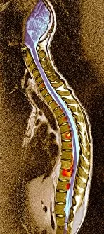
Potts disease. Coloured X-ray of the spine of a patient with Potts disease, or tuberculosis (TB) of the spine. The vertebrae (brown blocks) have become compacted and inflamed (red, yellow)

Background imageJoints Collection: Healthy hip bones, X-ray

Healthy hip bones, X-ray
Healthy hip bones. Coloured X-ray of the pelvis of a 49 year old woman, showing bones of the lower spine, hips and thighs

Background imageJoints Collection: Healthy ankle, X-ray

Healthy ankle, X-ray
Healthy ankle. Coloured X-ray of the ankle joint of an adult woman, in side view. The bones which form the ankle joint are the shin bone (tibia, at top) with the smaller fibula (behind it)

Background imageJoints Collection: Pelvis

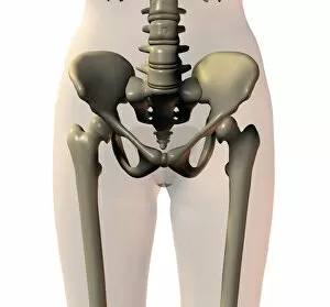
Pelvis. Computer artwork showing the bones of a healthy pelvis. At top is the spine, which consists of blocks of bone (vertebrae) separated by spongy intervertebral disks

Background imageJoints Collection: Knee bones and ligaments

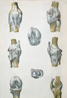
Knee bones and ligaments. Historical anatomical artwork of knee bones (yellow) and ligaments (pale blue). Ligaments are bands of fibrous tissue that hold bones together at joints

Background imageJoints Collection: Hand bones and ligaments

Hand bones and ligaments. Historical anatomical artwork of the bones (yellow) & ligaments (white) of the hand. Ligaments are bands of fibrous tissue that hold bones together at their joints

Background imageJoints Collection: Lower arm bones and ligaments

Lower arm bones and ligaments
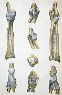
Lower arm bones & ligaments. Historical anatomical artwork of lower arm bones (yellow) and ligaments (pale blue). Ligaments are bands of fibrous tissue that hold bones together at joints

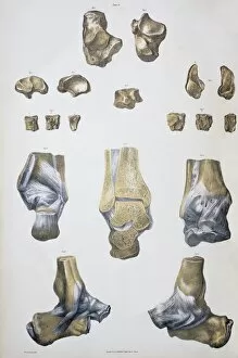
Background imageJoints Collection: Ankle bones and ligaments

Ankle bones and ligaments. Historical anatomical artwork of ankle bones (yellow) and ligaments (pale blue). Ligaments are bands of fibrous tissue that hold bones together at their joints

Background imageJoints Collection: Foot bones and ligaments

Foot bones and ligaments. Historical anatomical artwork of foot bones (yellow) and ligaments (pale blue). Ligaments are bands of fibrous tissue that hold bones together at their joints

Background imageJoints Collection: Lower leg bones and ligaments

Lower leg bones and ligaments. Historical anatomical artwork of lower leg bones (yellow) and ligaments (pale blue). Ligaments are bands of fibrous tissue that hold bones together at their joints

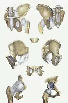
Background imageJoints Collection: Pelvis bones and ligaments

Pelvis bones and ligaments. Historical anatomical artwork of pelvis bones (yellow) and ligaments (white). Ligaments are bands of fibrous tissue that hold bones together at their joints

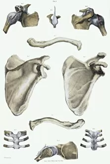
Background imageJoints Collection: Shoulder bones and ligaments

Shoulder bones and ligaments. Historical anatomical artwork of shoulder bones (yellow) and ligaments (white). Ligaments are bands of fibrous tissue that hold bones together at their joints

Background imageJoints Collection: Spinal bones and ligaments

Spinal bones and ligaments. Historical anatomical artwork of thoracic (upper) spinal bones (yellow, vertebrae) and ligaments (white)

Background imageJoints Collection: Osteoarthritis of hip joints, X-ray

Osteoarthritis of hip joints, X-ray
Hip joint osteoarthritis. Coloured X-ray of the pelvis of a 72-year-old woman showing areas of osteoarthritis of the hip joints (orange)

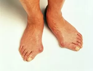
Background imageJoints Collection: Bunions

Bunions. Each of these fluid-filled pads covers the joint of the big toe. The majority of bunions are caused by the condition hallux valgus

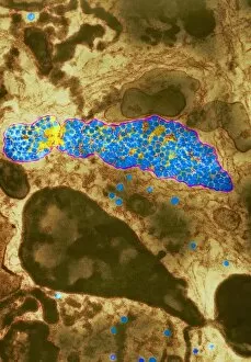
Background imageJoints Collection: Dengue fever virus particles, TEM

Dengue fever virus particles, TEM

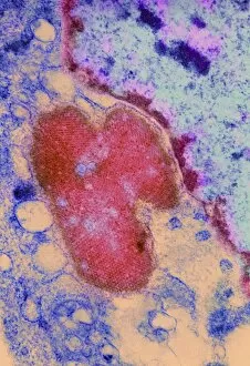
Background imageJoints Collection: Dengue fever virus replication, TEM

Dengue fever virus replication, TEM

Background imageJoints Collection: X-ray diagnosis

X-ray diagnosis
MODEL RELEASED. X-ray diagnosis. Doctor examining an X-ray of a hand

Background imageJoints Collection: Osteoarthritic pelvis, X-ray

Osteoarthritic pelvis, X-ray
Osteoarthritis. Coloured X-ray of the pelvis of a 52-year-old man showing areas of osteoarthritis (orange). The two femurs (upper leg bones)

Background imageJoints Collection: Arthritic joint

Arthritic joint. Conceptual image of an arthritic joint (red and white) surrounded by healthy joints (white). The healthy joints have smooth, well- defined edges

Background imageJoints Collection: Arthritis joints

Arthritis joints
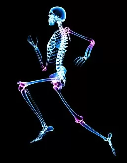
Arthritic joints. Computer artwork of a skeleton, showing arthritic joints in pink. Arthritis is a degenerative disease that causes the loss of the protective cartilage between joints

Background imageJoints Collection: Broken arm and dislocated wrist

Broken arm and dislocated wrist. Coloured X-ray showing a fracture (centre) of the radius bone in the forearm and a dislocation of the wrist joint at the end of the ulna bone (centre left)

Background imageJoints Collection: Dislocated finger

Dislocated finger

Background imageJoints Collection: Osteoporosis of the hip, X-ray

Osteoporosis of the hip, X-ray
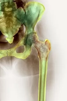
Osteoporosis. Coloured X-ray of the hip of a 43 year old woman showing reduced bone density (brown patchy area) in the head of the thigh bone (femur, centre right)

Background imageJoints Collection: Marfans syndrome

Marfans syndrome

Background imageJoints Collection: Arthritic hand

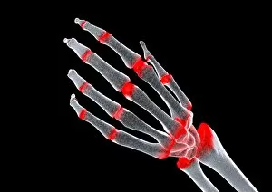
Arthritic hand, computer artwork. The finger and wrist joints are inflamed and swollen. Rheumatoid arthritis is caused by the immune system attacking the bodys own tissues

Background imageJoints Collection: Arthritic hand, conceptual artwork

Arthritic hand, conceptual artwork. Arthritis is a degenerative disease that results in the loss of cartilage between joints (red), causing pain and inflammation

Background imageJoints Collection: Arthritic hands, X-ray

Arthritic hands, X-ray
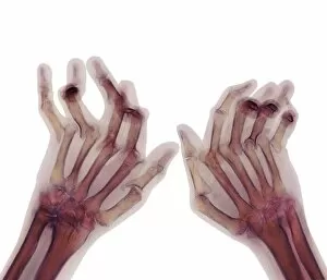
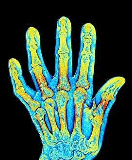
Arthritic hands. Coloured X-ray of the deformed hands of a patient suffering from rheumatoid arthritis. The patients fingers are abnormally bent because of damage to the joints

Background imageJoints Collection: Arthritic hand bones

Arthritic hand bones
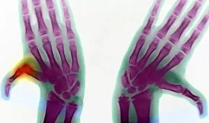
Arthritis. Coloured X-ray of arthritis affecting the joints of the hand. The thumb joint is seen to be both rougher and misaligned compared to a normal joint

Background imageJoints Collection: Ankylosing spondylitis X-ray

Ankylosing spondylitis X-ray
Ankylosing spondylitis. Coloured X-ray of the spine of a patient with ankylosing spondylitis, a form of arthritis. This is the lumbar region in the lower back

Background imageJoints Collection: Bony spurs in neck, X-ray

Bony spurs in neck, X-ray
Bony spurs in neck, coloured X-ray. Bony spurs (osteophytes, left) grow from the vertebrae as a normal part of the aging process

Background imageJoints Collection: Raw meat

Raw meat in a butchers shop

Background imageJoints Collection: Disarticulated skull, computer artwork

Disarticulated skull, computer artwork
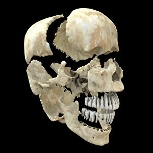
Skull. Computer artwork of a view of a disarticulated human skull. An adult skull is normally made up of 22 bones and can be subdivided into two parts: the cranium and the mandible (lower jaw bone)

Background imageJoints Collection: Skull, computer artwork

Skull, computer artwork
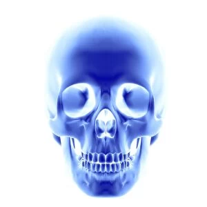
Skull. Computer artwork of an opaque view of a human skull. An adult skull is normally made up of 22 bones and can be subdivided into two parts: the cranium and the mandible (lower jaw bone)


For sale as Licensed Images

Choose your image, Select your licence and Download the media

"Exploring the Intricacies of Joints: A Journey through History and Anatomy" In this captivating journey, we delve into the world of joints, those remarkable connectors that enable movement and grace. Just like a palmistry map of the hand reveals secrets etched in our skin, so do joints hold tales of strength and resilience. Traveling back to 1855, we encounter a beef cuts diagram that showcases the intricate network within these joints. Stepping into a bustling butchers shop on High Street in Shoreham-by-Sea, Sussex during the 1920s, we witness a trailblazing woman butcher skillfully dissecting meat with precision. Zooming even closer under an electron microscope's lens, an ant's joint appears as a marvelously engineered masterpiece. Meanwhile, X-ray images expose bunions lurking beneath layers of flesh – silent reminders that even our they are bear burdens. Conceptual artworks emerge next; knee pain depicted as swirling colors and shapes evokes empathy for those grappling with discomfort. Joint pain takes form in abstract strokes on canvas - capturing both physical anguish and emotional strain. A black-and-white photograph from 1802 brings forth engravings showcasing beef, veal, pork, and mutton cuts – each slice revealing unique joint structures designed by nature herself. Historical artwork delves deeper into biomechanics - unraveling how forces interact within these pivotal connections. Conceptual art resurfaces once more to shed light on running injuries; brushstrokes depict shattered dreams amidst vibrant hues. Yet hope remains as another artwork illustrates leg muscles engaged in running - reminding us that healing is possible when nurtured with care. As we conclude this enlightening expedition through time and anatomy alike, let us appreciate the beauty hidden within every joint. For they are not mere hinges but rather gateways to mobility and vitality – connecting us all in our shared human experience.