
Joints Collection (#4)
"Exploring the Intricacies of Joints: A Journey through History and Anatomy" In this captivating journey, we delve into the world of joints


For sale as Licensed Images
Choose your image, Select your licence and Download the media
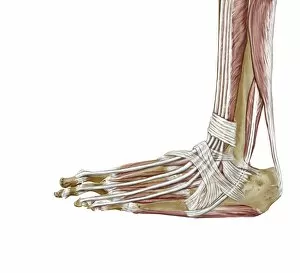
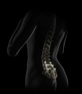
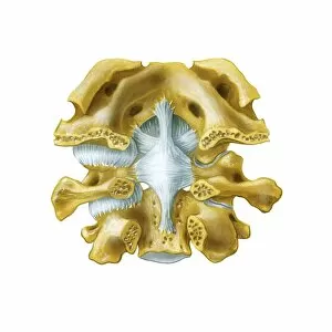
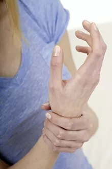
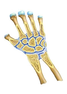
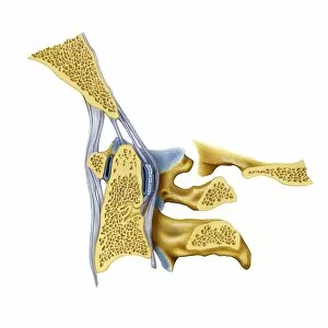
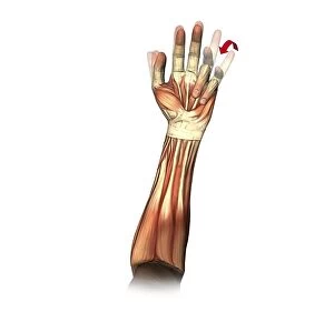
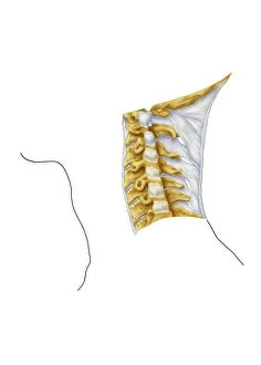
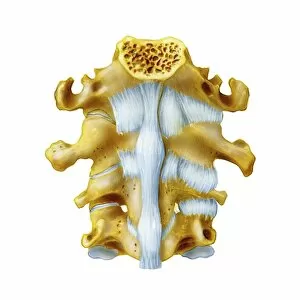
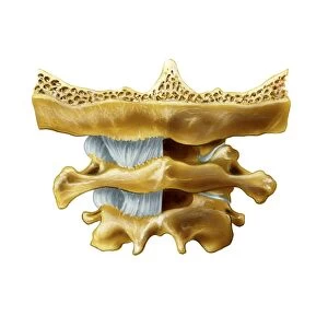
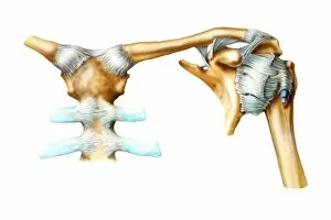
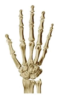
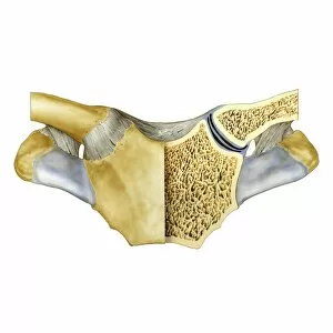
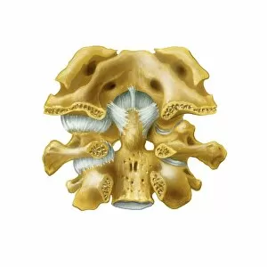
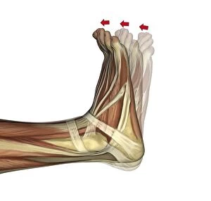
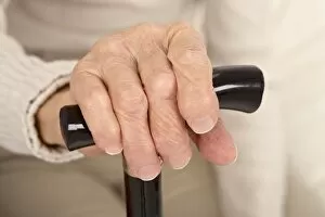
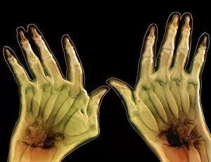
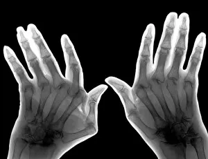
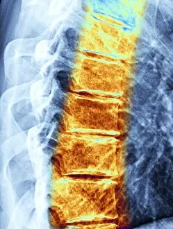
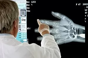
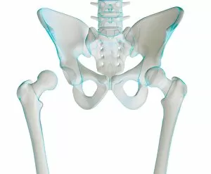
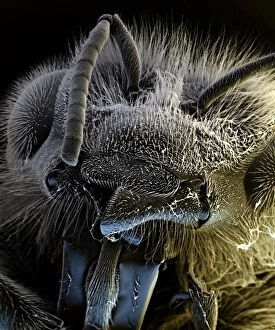
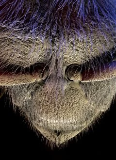
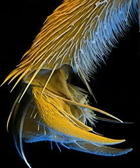
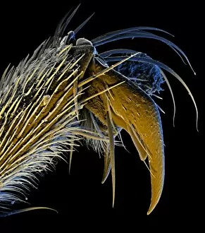
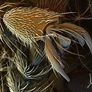
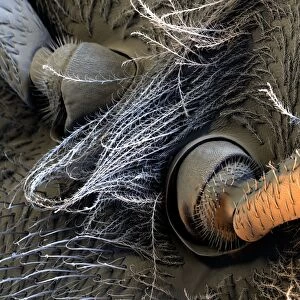
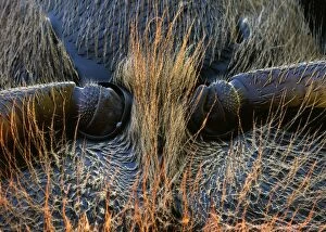
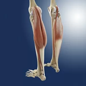
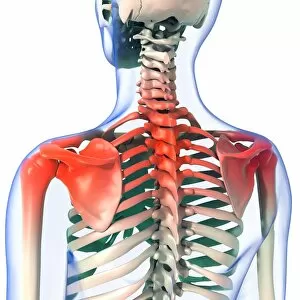
"Exploring the Intricacies of Joints: A Journey through History and Anatomy" In this captivating journey, we delve into the world of joints, those remarkable connectors that enable movement and grace. Just like a palmistry map of the hand reveals secrets etched in our skin, so do joints hold tales of strength and resilience. Traveling back to 1855, we encounter a beef cuts diagram that showcases the intricate network within these joints. Stepping into a bustling butchers shop on High Street in Shoreham-by-Sea, Sussex during the 1920s, we witness a trailblazing woman butcher skillfully dissecting meat with precision. Zooming even closer under an electron microscope's lens, an ant's joint appears as a marvelously engineered masterpiece. Meanwhile, X-ray images expose bunions lurking beneath layers of flesh – silent reminders that even our they are bear burdens. Conceptual artworks emerge next; knee pain depicted as swirling colors and shapes evokes empathy for those grappling with discomfort. Joint pain takes form in abstract strokes on canvas - capturing both physical anguish and emotional strain. A black-and-white photograph from 1802 brings forth engravings showcasing beef, veal, pork, and mutton cuts – each slice revealing unique joint structures designed by nature herself. Historical artwork delves deeper into biomechanics - unraveling how forces interact within these pivotal connections. Conceptual art resurfaces once more to shed light on running injuries; brushstrokes depict shattered dreams amidst vibrant hues. Yet hope remains as another artwork illustrates leg muscles engaged in running - reminding us that healing is possible when nurtured with care. As we conclude this enlightening expedition through time and anatomy alike, let us appreciate the beauty hidden within every joint. For they are not mere hinges but rather gateways to mobility and vitality – connecting us all in our shared human experience.