Joints Collection (#6)

"Exploring the Intricacies of Joints: A Journey through History and Anatomy" In this captivating journey, we delve into the world of joints

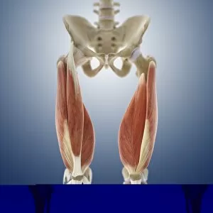
Background imageJoints Collection: Thigh muscles, artwork C013 / 4568

Thigh muscles, artwork C013 / 4568
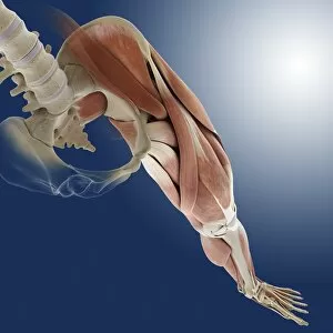
Thigh muscles. Computer artwork of an anterior view of some of the muscles and tendons (red and white) of the thighs and their attachment to the bones of the leg

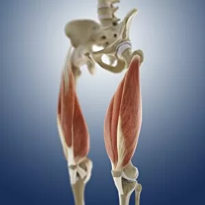
Background imageJoints Collection: Thigh muscles, artwork C013 / 4569

Thigh muscles, artwork C013 / 4569
Thigh muscles. Computer artwork of an oblique anterior view of some of the muscles and tendons (red and white) of the thighs and their attachment to the bones of the leg

Background imageJoints Collection: Shoulder muscles, artwork C013 / 4513

Shoulder muscles, artwork C013 / 4513
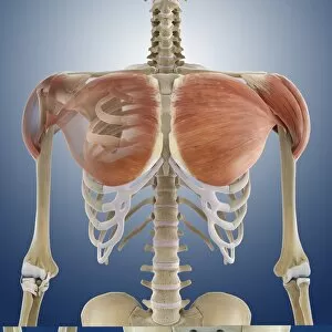
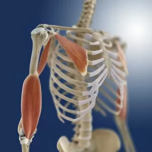
Shoulder muscles. Computer artwork of an anterior view of two of the muscles (red) of the shoulders and their attachment to the bones of the back and upper arms

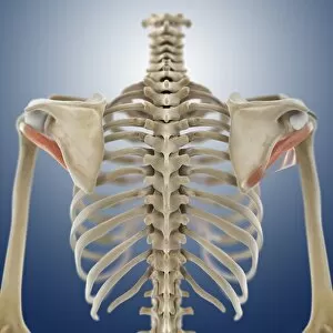
Background imageJoints Collection: Shoulder muscles, artwork C013 / 4515

Shoulder muscles, artwork C013 / 4515
Shoulder muscles. Computer artwork of a posterior view of two of the muscles (red) of the shoulders and their attachment to the bones of the back and upper arms

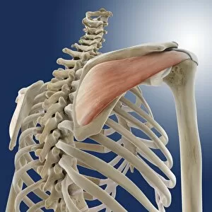
Background imageJoints Collection: Shoulder muscle, artwork C013 / 4508

Shoulder muscle, artwork C013 / 4508
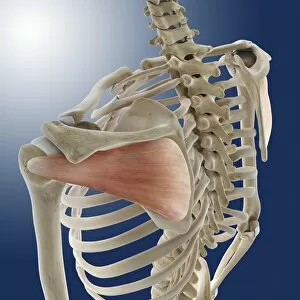
Shoulder muscle. Computer artwork of an oblique posterior view from below of one of the muscles (red) of the shoulders and its attachment to the bones of the back and upper arms

Background imageJoints Collection: Shoulder muscles, artwork C013 / 4504

Shoulder muscles, artwork C013 / 4504
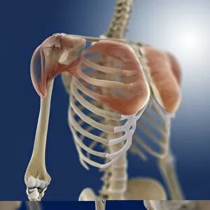
Shoulder muscles. Computer artwork of an oblique anterior view of two of the muscles (red) of the shoulders and their attachments to the bones of the chest, back and upper arms

Background imageJoints Collection: Shoulder muscle, artwork C013 / 4507

Shoulder muscle, artwork C013 / 4507
Shoulder muscle. Computer artwork of an oblique posterior view from above of one of the muscles (red) of the shoulders and its attachment to the bones of the back and upper arms

Background imageJoints Collection: Shoulder muscles, artwork C013 / 4505

Shoulder muscles, artwork C013 / 4505
Shoulder muscles. Computer artwork of an anterior view of two of the muscles (red) of the shoulders and their attachments to the bones of the chest, back and upper arms

Background imageJoints Collection: Shoulder muscles, artwork C013 / 4506

Shoulder muscles, artwork C013 / 4506
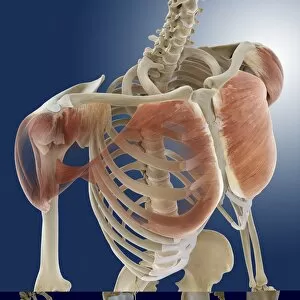
Shoulder muscles. Computer artwork of an oblique anterior view of two of the muscles (red) of the shoulders and their attachments to the bones of the chest, back and upper arms

Background imageJoints Collection: Shoulder muscles, artwork C013 / 4500

Shoulder muscles, artwork C013 / 4500
Shoulder muscles. Computer artwork of an anterior view of some of the muscles (red) of the shoulders and their attachments to the bones of the chest, back and upper arms

Background imageJoints Collection: Shoulder muscles, artwork C013 / 4501

Shoulder muscles, artwork C013 / 4501
Shoulder muscles. Computer artwork of an oblique anterior view of some of the muscles (red) of the shoulders and their attachments to the bones of the chest, back and upper arms

Background imageJoints Collection: Leg anatomy, artwork C013 / 4497

Leg anatomy, artwork C013 / 4497
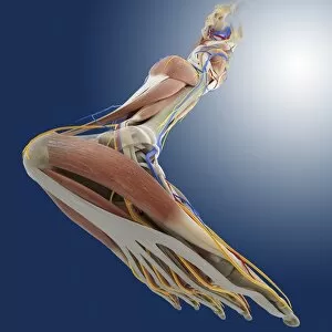
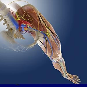
Leg anatomy. Computer artwork of an oblique posterior view looking up a human leg, showing muscles and tendons (brown and white), bones and ligaments (white), arteries and veins (red and blue)

Background imageJoints Collection: Leg anatomy, artwork C013 / 4495

Leg anatomy, artwork C013 / 4495
Leg anatomy. Computer artwork of an oblique side view looking down a human leg, showing muscles and tendons (brown and white), bones and ligaments (white), arteries and veins (red and blue)

Background imageJoints Collection: Leg anatomy, artwork C013 / 4496

Leg anatomy, artwork C013 / 4496
Leg anatomy. Computer artwork of an oblique anterior view looking up a human leg, showing muscles and tendons (brown and white), bones and ligaments (white), arteries and veins (red and blue)

Background imageJoints Collection: Leg anatomy, artwork C013 / 4493

Leg anatomy, artwork C013 / 4493
Leg anatomy. Computer artwork of an oblique anterior view looking down a human leg, showing muscles and tendons (brown and white), bones and ligaments (white), arteries and veins (red and blue)

Background imageJoints Collection: Leg anatomy, artwork C013 / 4494

Leg anatomy, artwork C013 / 4494
Leg anatomy. Computer artwork of an oblique anterior view looking down a human leg, showing muscles and tendons (brown and white), bones and ligaments (white), arteries and veins (red and blue)

Background imageJoints Collection: Leg ligaments, artwork

Leg ligaments, artwork
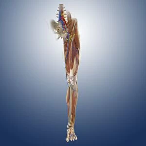
Leg anatomy. Computer artwork of an anterior view of a human leg, showing the muscles and tendons (brown and white), bones and ligaments (white), arteries and veins (red and blue), nerves (yellow)

Background imageJoints Collection: Foot muscles, artwork C013 / 4487

Foot muscles, artwork C013 / 4487
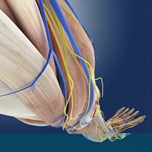
Foot muscles. Computer artwork of a view looking down on some of the muscles and tendons (red and white) of the upper surface of the foot

Background imageJoints Collection: Foot muscles, artwork C013 / 4486

Foot muscles, artwork C013 / 4486
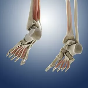
Foot muscles. Computer artwork of an oblique anterior view of some of the muscles and tendons (red and white) of the upper surface of the foot

Background imageJoints Collection: Foot muscles, artwork C013 / 4485

Foot muscles, artwork C013 / 4485
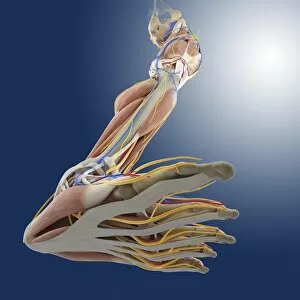
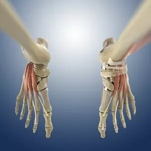
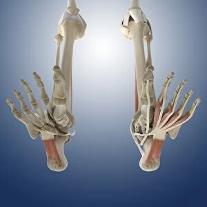
Foot muscles. Computer artwork of a view from below of some of the muscles and tendons (red and white) of the sole of the foot

Background imageJoints Collection: Foot muscles, artwork C013 / 4483

Foot muscles, artwork C013 / 4483
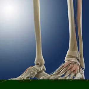
Foot muscles. Computer artwork of an oblique view from below and behind of some of the muscles and tendons (red and white) of the sole of the foot

Background imageJoints Collection: Outer ankle ligaments, artwork C013 / 4456

Outer ankle ligaments, artwork C013 / 4456
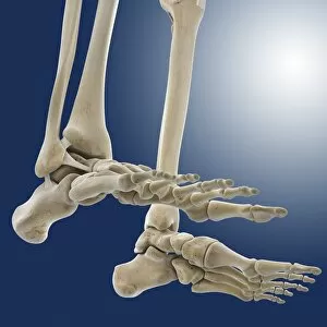
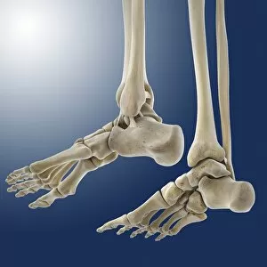
Outer ankle ligaments. Computer artwork of the bones and ligaments (white) of the feet and ankles from an oblique side view, with the outer side of the right foot at left

Background imageJoints Collection: Outer ankle ligaments, artwork C013 / 4455

Outer ankle ligaments, artwork C013 / 4455
Outer ankle ligaments. Computer artwork of the bones and ligaments (white) of the feet and ankles from an oblique side view, with the outer side of the left foot at upper centre

Background imageJoints Collection: Hip ligaments, artwork C013 / 4434

Hip ligaments, artwork C013 / 4434
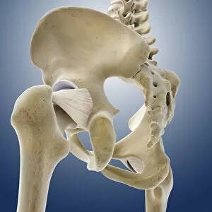
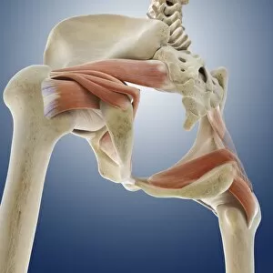
Hip ligaments. Computer artwork of a posterior oblique view of the hip bones and ligaments (white). At upper right the lumbar spine ends in the sacrum and coccyx (tailbone)

Background imageJoints Collection: Hip ligaments, artwork C013 / 4433

Hip ligaments, artwork C013 / 4433
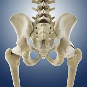
Hip ligaments. Computer artwork of a posterior view of the hip bones and ligaments (white). At centre the lumbar spine ends in the sacrum and coccyx (tailbone)

Background imageJoints Collection: Hip ligaments, artwork C013 / 4432

Hip ligaments, artwork C013 / 4432
Hip ligaments. Computer artwork of an anterior view of the hip bones and ligaments (white). At centre the lumbar spine ends in the sacrum and coccyx (tailbone)

Background imageJoints Collection: Hip anatomy, artwork C013 / 4430

Hip anatomy, artwork C013 / 4430
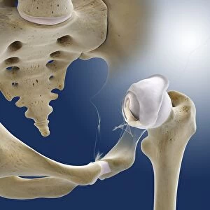
Hip anatomy. Computer artwork of the head of the left femur (centre right) articulating with the left-hand side of the pelvis to form the hip joint

Background imageJoints Collection: Leg muscles, artwork C013 / 4429

Leg muscles, artwork C013 / 4429
Leg muscles. Computer artwork of the bones and outer muscles of the left leg, seen from the front (anterior view) at an oblique angle looking downwards

Background imageJoints Collection: Thigh hamstring muscles, artwork C013 / 4428

Thigh hamstring muscles, artwork C013 / 4428
Thigh hamstring muscles. Computer artwork of a posterior view of two of the three hamstring muscles (red) of the thighs. The muscles are the semimembranosus (inner) and semitendinosus (outer)

Background imageJoints Collection: Iliopsoas muscles, artwork C013 / 4425

Iliopsoas muscles, artwork C013 / 4425
Iliopsoas muscles. Computer artwork of an anterior (frontal) view of the hips, showing the inner hip muscles (red), the ilioposas

Background imageJoints Collection: Buttock muscles, artwork C013 / 4423

Buttock muscles, artwork C013 / 4423
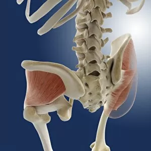
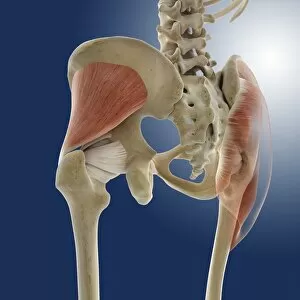
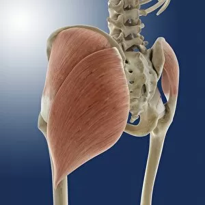
Buttock muscles. Computer artwork of a posterior oblique view of the hips, showing some of the muscles (red) of the buttocks

Background imageJoints Collection: Buttock muscles, artwork C013 / 4424

Buttock muscles, artwork C013 / 4424
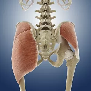
Buttock muscles. Computer artwork of a posterior view of the hips showing some of the muscles (red) of the buttocks. These are shown attached to the femurs (thigh bones) and the pelvis bones

Background imageJoints Collection: Buttock muscles, artwork C013 / 4422

Buttock muscles, artwork C013 / 4422
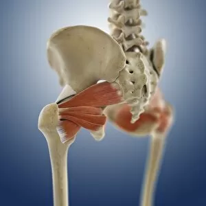
Buttock muscles. Computer artwork of a posterior oblique view of the hips showing some of the muscles (red) of the buttocks. These are shown attached to the femurs (thigh bones) and the pelvis bones

Background imageJoints Collection: Buttock muscles, artwork C013 / 4420

Buttock muscles, artwork C013 / 4420
Buttock muscles. Computer artwork of a posterior view of the hips showing some of the muscles (red) of the buttocks. These are shown attached to the femurs (thigh bones) and the pelvis bones

Background imageJoints Collection: Buttock muscles, artwork C013 / 4421

Buttock muscles, artwork C013 / 4421
Buttock muscles. Computer artwork of a side view (front of body at left) of the hips showing some of the muscles (red) of the buttocks

Background imageJoints Collection: Buttock muscles, artwork C013 / 4419

Buttock muscles, artwork C013 / 4419
Buttock muscles. Computer artwork of a posterior oblique view of the hips showing some of the muscles (red) of the buttocks. These are shown attached to the femurs (thigh bones) and the pelvis bones

Background imageJoints Collection: Buttock muscles, artwork C013 / 4415

Buttock muscles, artwork C013 / 4415
Buttock muscles. Computer artwork of a posterior oblique view of the hips showing some of the muscles (red) of the buttocks. These are shown attached to the femurs (thigh bones) and the pelvis bones

Background imageJoints Collection: Buttock muscles, artwork C013 / 4416

Buttock muscles, artwork C013 / 4416
Buttock muscles. Computer artwork of a posterior oblique view of the hips showing some of the muscles (red) of the buttocks. These are shown attached to the femurs (thigh bones) and the pelvis bones

Background imageJoints Collection: Section of track showing dipped joints

Section of track showing dipped joints, 1977 Date: 1977

Background imageJoints Collection: Butcher prepares meat

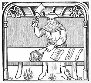
Butcher prepares meat
A 15th century butcher prepares cuts of meat, including ribs and hooves. Date: 15th century

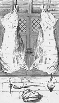
Background imageJoints Collection: Beef cuts, 1857

Beef cuts, 1857
Town and country methods of cutting up an ox. The head and hooves ar shown, as well as the carcass. Date: 1857

Background imageJoints Collection: Ant feet, SEM

Ant feet, SEM
Ant feet. Coloured scanning electron micrograph (SEM) of the tip of two legs from an ant (family Formicidae). The end of an insect leg consists of the final segment, which is called the tarsus

Background imageJoints Collection: Hip bones, X-ray

Hip bones, X-ray
Hip joints. Coloured X-ray of a womans groin and upper legs, revealing the bones that form her hip joints. The left hip (identical to the right one) is at upper left in this rear view

Background imageJoints Collection: Female pelvis, X-ray

Female pelvis, X-ray
Female pelvis. Coloured X-ray of the pelvis of a woman. The lowest lumbar vertebra of the spinal column (top centre) forms a joint with the sacrum

Background imageJoints Collection: Immune system components

Immune system components
Immune system. Conceptual computer artwork of a man standing with his arms outstretched surrounded by components of the immune system. The skeletal system is seen, with the joints highlighted in red

Background imageJoints Collection: Butchers shop

Butchers shop. Butcher preparing meat to be sold

Background imageJoints Collection: Spondylitis, X-ray

Spondylitis, X-ray
Spondylitis. Coloured X-ray of the lower spine and pelvis of a patient with spondylitis, an inflammation of the synovial joints of the backbone

Background imageJoints Collection: Thalassaemia, X-ray

Thalassaemia, X-ray
Thalassaemia. Coloured hand X-ray of a patient with thalassaemia, also known as Cooleys anaemia. The bones of the hand show demineralization (lighter patches)


For sale as Licensed Images

Choose your image, Select your licence and Download the media

"Exploring the Intricacies of Joints: A Journey through History and Anatomy" In this captivating journey, we delve into the world of joints, those remarkable connectors that enable movement and grace. Just like a palmistry map of the hand reveals secrets etched in our skin, so do joints hold tales of strength and resilience. Traveling back to 1855, we encounter a beef cuts diagram that showcases the intricate network within these joints. Stepping into a bustling butchers shop on High Street in Shoreham-by-Sea, Sussex during the 1920s, we witness a trailblazing woman butcher skillfully dissecting meat with precision. Zooming even closer under an electron microscope's lens, an ant's joint appears as a marvelously engineered masterpiece. Meanwhile, X-ray images expose bunions lurking beneath layers of flesh – silent reminders that even our they are bear burdens. Conceptual artworks emerge next; knee pain depicted as swirling colors and shapes evokes empathy for those grappling with discomfort. Joint pain takes form in abstract strokes on canvas - capturing both physical anguish and emotional strain. A black-and-white photograph from 1802 brings forth engravings showcasing beef, veal, pork, and mutton cuts – each slice revealing unique joint structures designed by nature herself. Historical artwork delves deeper into biomechanics - unraveling how forces interact within these pivotal connections. Conceptual art resurfaces once more to shed light on running injuries; brushstrokes depict shattered dreams amidst vibrant hues. Yet hope remains as another artwork illustrates leg muscles engaged in running - reminding us that healing is possible when nurtured with care. As we conclude this enlightening expedition through time and anatomy alike, let us appreciate the beauty hidden within every joint. For they are not mere hinges but rather gateways to mobility and vitality – connecting us all in our shared human experience.