Joints Collection (#3)

"Exploring the Intricacies of Joints: A Journey through History and Anatomy" In this captivating journey, we delve into the world of joints

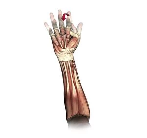
Background imageJoints Collection: Fist clenching movement, artwork C016 / 6795

Fist clenching movement, artwork C016 / 6795
Fist clenching movement. Artwork of the muscles of the forearm and hand from the front, with red arrows showing the direction of movement of the fingers during flexion to form a fist

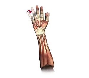
Background imageJoints Collection: Little and ring finger flexion, artwork C016 / 6793

Little and ring finger flexion, artwork C016 / 6793
Little and ring finger flexion. Artwork of the muscles of the forearm and hand from the front, with red arrows showing the direction of movement of the little and ring (fourth and third)

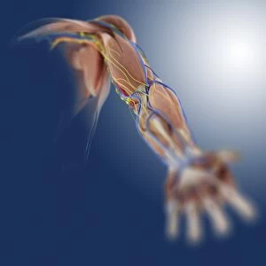
Background imageJoints Collection: Arm anatomy, artwork C013 / 4583

Arm anatomy, artwork C013 / 4583
Arm anatomy. Computer artwork of an oblique anterior view looking up a human arm, showing muscles and tendons (brown and white), bones and ligaments (white), arteries and veins (red and blue)

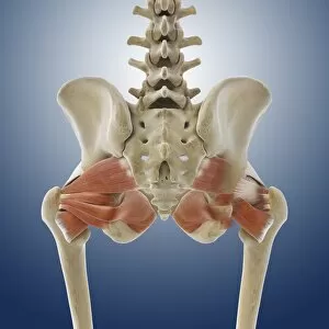
Background imageJoints Collection: Buttock muscles, artwork C013 / 4414

Buttock muscles, artwork C013 / 4414
Buttock muscles. Computer artwork of a posterior view of the hips showing some of the muscles (red) of the buttocks. These are shown attached to the femurs (thigh bones) and the pelvis bones

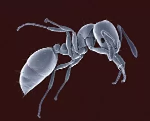
Background imageJoints Collection: Ant, SEM

Ant, SEM
Ant (family Formicidae), coloured scanning electron micrograph (SEM). One of its compound eyes (black) is seen, as well as two long antennae that are mounted on the head between the eyes

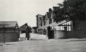
Background imageJoints Collection: Princess Marys Hospital, Margate, Kent

Princess Marys Hospital, Margate, Kent
Entrance to the Princess Marys Hospital, Wilderness Road, Margate, Kent. The site, originally known as Eastcliff House, was acquired in 1898 by the Metropolitan Asylums Board as a seaside hospital

Background imageJoints Collection: British soldiers putting joints into the oven to roast

British soldiers putting joints into the oven to roast
Photograph: British soldiers putting joints into the oven to roast.One of 193 British and Allied Official photographs taken on the Western Front during World War One.Mounted on card

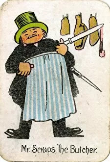
Background imageJoints Collection: The Round Game of Happy Families, Mr Scraps

The Round Game of Happy Families, Mr Scraps
Chad Valley, The Round Game of Happy Families, Mr Scraps the Butcher. circa 1910

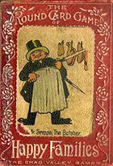
Background imageJoints Collection: The Round Game of Happy Families, box design

The Round Game of Happy Families, box design
Chad Valley, The Round Game of Happy Families, box design, showing Mr Scraps the Butcher. circa 1910

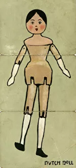
Background imageJoints Collection: Misfitz Dolls - Dutch Doll

Misfitz Dolls - Dutch Doll. Date: early 20th century

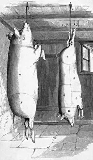
Background imageJoints Collection: Bacon Joints 1857

Bacon Joints 1857
A diagram showing bacon joints. Date: 1857

Background imageJoints Collection: Cartoon, Cost of food and drink, WW1

Cartoon, Cost of food and drink, WW1
Cartoon illustrating the cost of food and drink during the First World War. Various tradesmen comment on the cost of their produce. Date: 1916

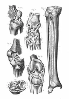
Background imageJoints Collection: Knee foot joint anatomy engraving 1866

Knee foot joint anatomy engraving 1866
Atlas d anatomie descriptive du corps humain C. Bonamy - Paul Broca Victor Masson et Fils Paris 1866

Background imageJoints Collection: Knee anatomy engraving 1866

Knee anatomy engraving 1866
Atlas d anatomie descriptive du corps humain C. Bonamy - Paul Broca Victor Masson et Fils Paris 1866

Background imageJoints Collection: Foot joint anatomy engraving 1866

Foot joint anatomy engraving 1866
Atlas d anatomie descriptive du corps humain C. Bonamy - Paul Broca Victor Masson et Fils Paris 1866

Background imageJoints Collection: Diagram showing removal of affected head of a femur bone

Diagram showing removal of affected head of a femur bone

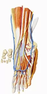
Background imageJoints Collection: Human foot, internal anatomy

Human foot, internal anatomy

Background imageJoints Collection: Interior of a dining hall, 1866

Interior of a dining hall, 1866
BAL84600 Interior of a dining hall, 1866 by Yeames, William Frederick (1835-1918); Roy Miles Fine Paintings; English, out of copyright

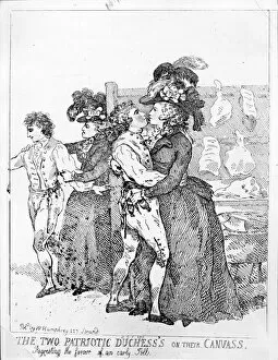
Background imageJoints Collection: The Two Patriotic Duchesss on their Canvass, 1784 (engraving)

The Two Patriotic Duchesss on their Canvass, 1784 (engraving)
XJF473767 The Two Patriotic Duchesss on their Canvass, 1784 (engraving) by Rowlandson, Thomas (1756-1827); Private Collection; (add.info)

Background imageJoints Collection: Christmas Joints, Uk

Christmas Joints, Uk
CHRISTMAS JOINTS, UK

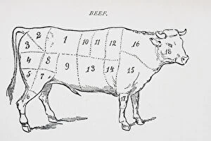
Background imageJoints Collection: Drawing of a bullock marked to show eighteen different cuts of meat (litho)

Drawing of a bullock marked to show eighteen different cuts of meat (litho)
KW271361 Drawing of a bullock marked to show eighteen different cuts of meat (litho) by English School, (19th century); Private Collection; Ken Welsh; English, out of copyright

Background imageJoints Collection: Picture No. 10875822

Picture No. 10875822
Human skeleton - body structure showing bone joints Date:

Background imageJoints Collection: Sandblasting the joints of a bicycle frame: operator wears helmet with breathing tube

Sandblasting the joints of a bicycle frame: operator wears helmet with breathing tube and a protective tunic: France. Wood engraving Paris 1896

Background imageJoints Collection: Christmas Joints

Christmas Joints, UK

Background imageJoints Collection: Sirloin joints of beef in abattoir, Yorkshire, England, February

Sirloin joints of beef in abattoir, Yorkshire, England, February

Background imageJoints Collection: Worker with sirloin joints of beef in abattoir, Yorkshire, England, February

Worker with sirloin joints of beef in abattoir, Yorkshire, England, February

Background imageJoints Collection: Ginger Rock formation on eroded coastal rocks with tourists, Yehliu Geopark, Yehliu Promontory

Ginger Rock formation on eroded coastal rocks with tourists, Yehliu Geopark, Yehliu Promontory, Taiwan, April

Background imageJoints Collection: Pouring water into gaps between patio slabs, close-up

Pouring water into gaps between patio slabs, close-up

Background imageJoints Collection: Arthritic hand, X-ray C017 / 7171

Arthritic hand, X-ray C017 / 7171
Arthritic hand. X-ray of the hand and wrist of a 56 year old patient with rheumatoid arthritis. Rheumatoid arthritis is caused by the immune system attacking the bodys own tissues

Background imageJoints Collection: Osteoarthritis of the hip, X-ray F006 / 3744

Osteoarthritis of the hip, X-ray F006 / 3744
Osteoarthritis of the hip. Coloured frontal X-ray (front view) of the pelvis in a 59 year old patient showing osteoarthritis of both hip joints

Background imageJoints Collection: Osteoarthritis of the hip, X-ray F006 / 3745

Osteoarthritis of the hip, X-ray F006 / 3745
Osteoarthritis of the hip. Coloured frontal X-ray (front view) of the pelvis in a 59 year old patient showing osteoarthritis of both hip joints

Background imageJoints Collection: Osteoarthritis of the thoracic spine C017 / 0697

Osteoarthritis of the thoracic spine C017 / 0697
Osteoarthritis. X-ray of the thorarcic spine of a 64 year old man with slight osteoarthritis. The arthritic vertebrae are closer together and have rough and ragged edges

Background imageJoints Collection: Hip pain, artwork C013 / 5917

Hip pain, artwork C013 / 5917
Hip pain, computer artwork

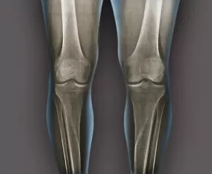
Background imageJoints Collection: Normal knees, X-rays

Normal knees, X-rays
Normal knees. Coloured frontal X-rays of the healthy knees of a 35-year-old

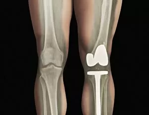
Background imageJoints Collection: Total knee replacement, X-ray

Total knee replacement, X-ray
Total knee replacement. X-ray of the legs of a 49-year-old patient who has had a total knee replacement (white) of the left knee (right)

Background imageJoints Collection: Hand pain C016 / 9657

Hand pain C016 / 9657
MODEL RELEASED. Hand pain

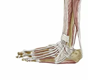
Background imageJoints Collection: Foot ligaments and tendons, artwork C016 / 7010

Foot ligaments and tendons, artwork C016 / 7010
Foot ligaments and tendons. Artwork of the bones, ligaments (white, between bones), and tendons (white, muscle-bone) of the foot and the ankle joint from the side

Background imageJoints Collection: Elbow pain C016 / 9661

Elbow pain C016 / 9661
MODEL RELEASED. Elbow pain

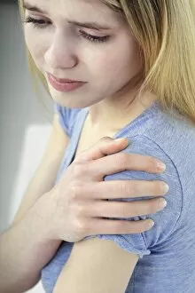
Background imageJoints Collection: Shoulder pain C016 / 9662

Shoulder pain C016 / 9662
MODEL RELEASED. Shoulder pain

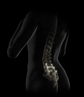
Background imageJoints Collection: Lumbar spine fusion, artwork C016 / 6993

Lumbar spine fusion, artwork C016 / 6993
Lumbar spine fusion. Artwork of pins and screws inserted into a patients lumbar spine in a case of spinal fusion. The pins hold together the patients vertebrae

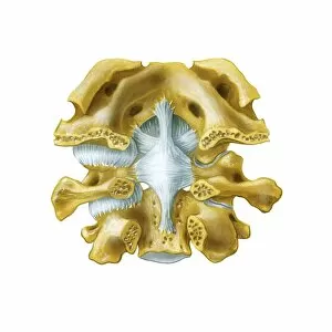
Background imageJoints Collection: Atlanto-occipital joint, artwork C016 / 6554

Atlanto-occipital joint, artwork C016 / 6554
Atlanto-occipital joint. Artwork of the bones (yellow) and ligaments (blue) of the atlanto-occipital joint, as seen from behind (posterior view)

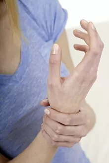
Background imageJoints Collection: Wrist pain C016 / 9660

Wrist pain C016 / 9660
MODEL RELEASED. Wrist pain

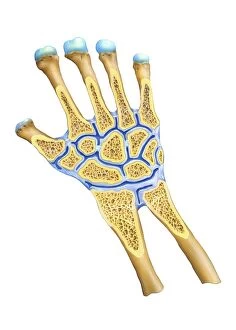
Background imageJoints Collection: Wrist joint anatomy, artwork C016 / 6549

Wrist joint anatomy, artwork C016 / 6549
Wrist joint anatomy. Artwork of the bones (yellow) and ligaments (blue) of the wrist joint, in longitudinal section. The left hand has been sectioned in a palmar aspect to expose the synovial

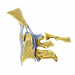
Background imageJoints Collection: Atlanto-occipital joint, artwork C016 / 6557

Atlanto-occipital joint, artwork C016 / 6557
Atlanto-occipital joint. Artwork of the bones (yellow) and ligaments (blue) of the atlanto-occipital joint, as seen from the side in a medial sagittal section. The rear of the spine is at right

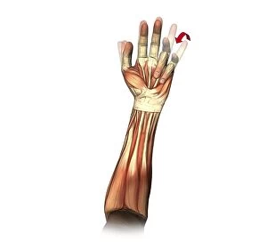
Background imageJoints Collection: Little and ring finger flexion, artwork C016 / 6794

Little and ring finger flexion, artwork C016 / 6794
Little and ring finger flexion. Artwork of the muscles of the forearm and hand from the front, with red arrows showing the direction of movement of the little and ring (fourth and third)


For sale as Licensed Images

Choose your image, Select your licence and Download the media

"Exploring the Intricacies of Joints: A Journey through History and Anatomy" In this captivating journey, we delve into the world of joints, those remarkable connectors that enable movement and grace. Just like a palmistry map of the hand reveals secrets etched in our skin, so do joints hold tales of strength and resilience. Traveling back to 1855, we encounter a beef cuts diagram that showcases the intricate network within these joints. Stepping into a bustling butchers shop on High Street in Shoreham-by-Sea, Sussex during the 1920s, we witness a trailblazing woman butcher skillfully dissecting meat with precision. Zooming even closer under an electron microscope's lens, an ant's joint appears as a marvelously engineered masterpiece. Meanwhile, X-ray images expose bunions lurking beneath layers of flesh – silent reminders that even our they are bear burdens. Conceptual artworks emerge next; knee pain depicted as swirling colors and shapes evokes empathy for those grappling with discomfort. Joint pain takes form in abstract strokes on canvas - capturing both physical anguish and emotional strain. A black-and-white photograph from 1802 brings forth engravings showcasing beef, veal, pork, and mutton cuts – each slice revealing unique joint structures designed by nature herself. Historical artwork delves deeper into biomechanics - unraveling how forces interact within these pivotal connections. Conceptual art resurfaces once more to shed light on running injuries; brushstrokes depict shattered dreams amidst vibrant hues. Yet hope remains as another artwork illustrates leg muscles engaged in running - reminding us that healing is possible when nurtured with care. As we conclude this enlightening expedition through time and anatomy alike, let us appreciate the beauty hidden within every joint. For they are not mere hinges but rather gateways to mobility and vitality – connecting us all in our shared human experience.