
Malignant Collection
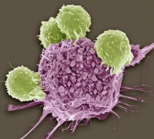
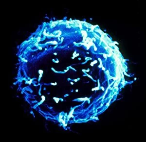
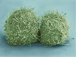
"Malignant: Unleashing the Battle Within" In a microscopic world, T lymphocytes stand as valiant warriors against cancer cells


For sale as Licensed Images
Choose your image, Select your licence and Download the media
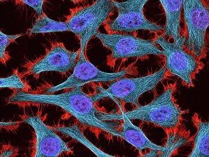
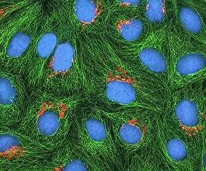
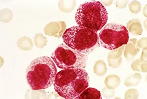
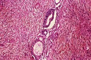
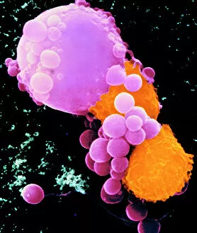
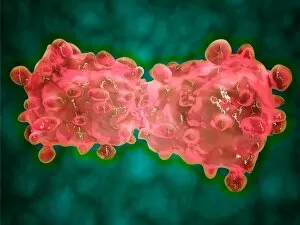
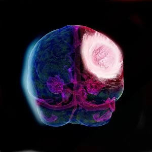
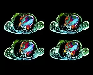
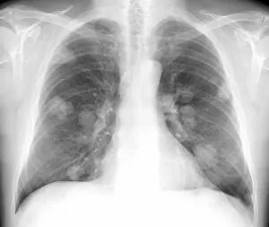
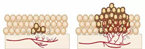
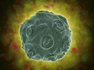
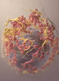
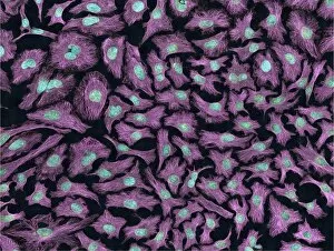
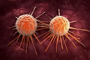
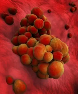
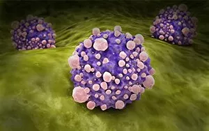
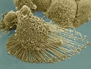
"Malignant: Unleashing the Battle Within" In a microscopic world, T lymphocytes stand as valiant warriors against cancer cells, their relentless pursuit captured in SEM C001 / 1679. Meanwhile, HeLa cells reveal their sinister nature under the watchful eye of a light micrograph in C017 / 8299 and C017 / 8298. A mesmerizing scene unfolds as coloured SEM showcases lymphocytes fearlessly attacking cancer cells, painting a vivid picture of hope amidst adversity. Acute promyelocytic leukemia reveals its haunting presence through an ominous micrograph while ovarian cancer exposes its treacherous grip in light micrograph C015 / 7103. Witnessing the battle at a cellular level is awe-inspiring; a coloured SEM image captures the moment when leukaemic white blood cells are confronted head-on by nanorobots determined to eradicate this malignant threat (M132 / 0488). The fight against cancer takes on otherworldly dimensions reminiscent of Amazing Stories sci-fi magazine covers featuring giant plants and fantastical creatures. Yet not all battles are fought within our bodies; sometimes they transcend into realms beyond. Like an exorcism performed by a cleric casting spells against evil spirits, we confront malignancy with unwavering determination. Just as history has witnessed struggles such as "The Condition of Ireland under the 'No Rent' Policy, " or "Johnnys Defeat at the Dock, " we too face adversity head-on. Amidst these hints lie tales of resilience and courage that remind us to never surrender to darkness. Malignant forces may try to overpower us, but united we stand ready to conquer them - be it through scientific advancements or sheer willpower. Together, let us rewrite the narrative and triumph over malignancy's grasp for brighter tomorrows await those who dare to fight back.