Physiological Collection (#4)

"Exploring the Intricacies of Physiology: From Airpumps to Blood Cells" Delving into the world of physiology

Background imagePhysiological Collection: Suspended animation, artwork

Suspended animation, artwork
Suspended animation. Computer artwork of futuristic humans in suspended animation. This is a state where fundamental processes, such as breathing and heart rate, are slowed, which extends lifespan

Background imagePhysiological Collection: Genders of Nobel laureates, artwork

Genders of Nobel laureates, artwork
Genders of Nobel laureates. Artwork showing the gender divide between women (left) and men (right) who have won Nobel Prizes

Background imagePhysiological Collection: Anterior pituitary gland, TEM

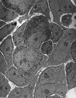
Anterior pituitary gland, TEM
Anterior pituitary gland. Transmission electron micrograph (TEM) of a section through cells of the anterior pituitary. The anterior pituitary, or adenohypophysis

Background imagePhysiological Collection: Olfactory bulb anatomy, artwork

Olfactory bulb anatomy, artwork
Olfactory bulb anatomy. Artwork of a human nose, showing the anatomy of the olfactory bulb, the organ of smell. This lies above the anterior aspect of the nasal cavity on the ethmoid bone
Background imagePhysiological Collection: Calcium ATPase ion pump C015 / 7143

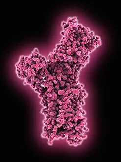
Calcium ATPase ion pump C015 / 7143
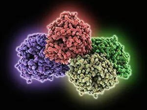
Calcium ATPase ion pump, molecular model. This enzyme is found in muscle cell membranes, where it pumps calcium in and out of muscle cells and controls muscle contractions

Background imagePhysiological Collection: Liver, SEM C015 / 9924

Liver, SEM C015 / 9924
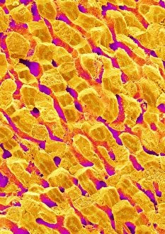
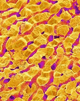
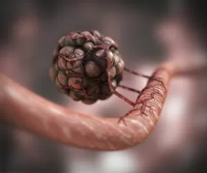
Liver vein. Coloured scanning electron micrograph (SEM) of liver tissue. The functions of the liver include destroying old red blood cells

Background imagePhysiological Collection: Liver, SEM C015 / 9922

Liver, SEM C015 / 9922
Liver vein. Coloured scanning electron micrograph (SEM) of liver tissue showing a large vein (pink, up centre). The functions of the liver include destroying old red blood cells

Background imagePhysiological Collection: Thrombin complexed with fibrinogen C015 / 7148

Thrombin complexed with fibrinogen C015 / 7148
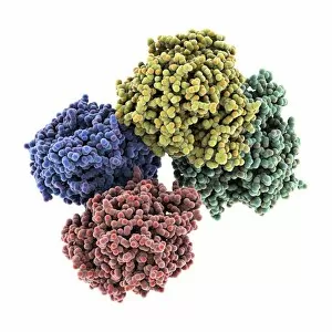
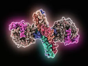
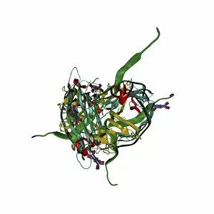
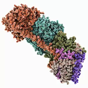
Thrombin complexed with fibrinogen, molecular model. The thrombin molecules (left and right, purple and green) are bound to the central part of the fibrinogen molecule (centre, multiple colours)

Background imagePhysiological Collection: Retinal-producing oxygenase enzyme C015 / 7150

Retinal-producing oxygenase enzyme C015 / 7150
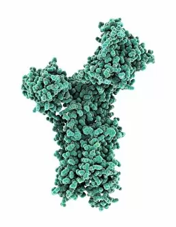
Retinal-producing oxygenase enzyme, molecular model. This enzyme, an oxygenase, plays a role in the production and metabolism of retinal and other apocarotenoids

Background imagePhysiological Collection: Liver, SEM C015 / 9923

Liver, SEM C015 / 9923
Liver vein. Coloured scanning electron micrograph (SEM) of liver tissue. The functions of the liver include destroying old red blood cells

Background imagePhysiological Collection: Thrombin complexed with fibrinogen C015 / 7149

Thrombin complexed with fibrinogen C015 / 7149
Thrombin complexed with fibrinogen, molecular model. The thrombin molecules (left and right, brown and pink) are bound to the central part of the fibrinogen molecule (centre, multiple colours)

Background imagePhysiological Collection: Calcium ATPase ion pump C015 / 7142

Calcium ATPase ion pump C015 / 7142
Calcium ATPase ion pump, molecular model. This enzyme is found in muscle cell membranes, where it pumps calcium in and out of muscle cells and controls muscle contractions

Background imagePhysiological Collection: Liver, SEM C015 / 9925

Liver, SEM C015 / 9925
Liver vein. Coloured scanning electron micrograph (SEM) of liver tissue. The functions of the liver include destroying old red blood cells

Background imagePhysiological Collection: Retinal-producing oxygenase enzyme C015 / 7151

Retinal-producing oxygenase enzyme C015 / 7151
Retinal-producing oxygenase enzyme, molecular model. This enzyme, an oxygenase, plays a role in the production and metabolism of retinal and other apocarotenoids

Background imagePhysiological Collection: Torn muscle, illustration C018 / 0806

Torn muscle, illustration C018 / 0806
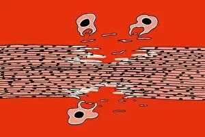
Torn muscle. Illustration of a tear in a skeletal muscle. Muscle tears can either result in muscle growth or injury depending on the circumstances

Background imagePhysiological Collection: Retina layers, SEM C018 / 0521

Retina layers, SEM C018 / 0521
Retina layers, coloured scanning electron micrograph (SEM). The retina of the human eye is found on the inside of the eyeball and is formed from a number of layers

Background imagePhysiological Collection: Physical fitness test C014 / 1104

Physical fitness test C014 / 1104
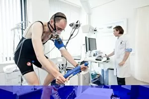
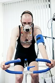
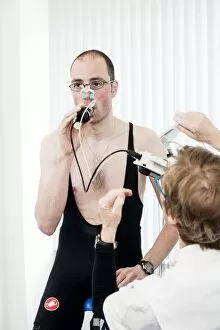
Physical fitness test. Athlete riding an exercise bike while his oxygen consumption is measured with a face mask. Electrodes on his chest monitor his heart rate

Background imagePhysiological Collection: Eye muscle, TEM C014 / 1467

Eye muscle, TEM C014 / 1467
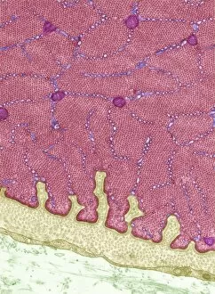
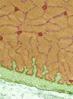
Eye muscle. Transmission electron micrograph (TEM) of a section through a striated muscle cell from the ciliary muscle of a human eye

Background imagePhysiological Collection: Heart and brain vascular system, artwork

Heart and brain vascular system, artwork
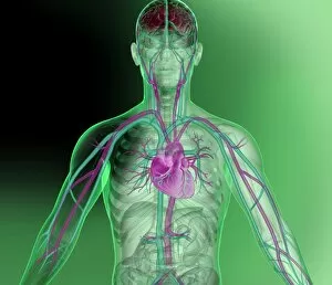
Heart and brain vascular system. Artwork showing the arteries (pink) and veins (blue) of the torso, arms, neck, head and brain. The brain is at top (red) and the heart at centre

Background imagePhysiological Collection: Eye muscle, TEM C014 / 1466

Eye muscle, TEM C014 / 1466
Eye muscle. Transmission electron micrograph (TEM) of a section through a striated muscle cell from the ciliary muscle of a human eye

Background imagePhysiological Collection: Psychic brain, conceptual image C014 / 1480

Psychic brain, conceptual image C014 / 1480
Psychic brain, conceptual image. Artwork showing the human brain from the side, surrounded by flames, with the pineal gland shown

Background imagePhysiological Collection: Physical fitness test C014 / 1101

Physical fitness test C014 / 1101
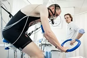
Physical fitness test. Athlete riding an exercise bike while his oxygen consumption is measured with a face mask. Electrodes on his chest monitor his heart rate

Background imagePhysiological Collection: Physical fitness test C014 / 1107

Physical fitness test C014 / 1107
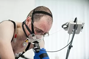
Physical fitness test. Athlete riding an exercise bike while his oxygen consumption is measured with a face mask. Electrodes on his chest monitor his heart rate

Background imagePhysiological Collection: Physical fitness test C014 / 1102

Physical fitness test C014 / 1102
Physical fitness test. Athlete riding an exercise bike while his oxygen consumption is measured with a face mask. Electrodes on his chest monitor his heart rate

Background imagePhysiological Collection: Physical fitness test C014 / 1100

Physical fitness test C014 / 1100
Physical fitness test. Athlete undergoing a physical examination. Here his lung capacity is being tested using a spirometer, which measures the volume and speed of air that he can exhale and inhale

Background imagePhysiological Collection: Nerve fibre node, TEM C014 / 1458

Nerve fibre node, TEM C014 / 1458
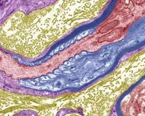
Nerve fibre node. Transmission electron micrograph (TEM) of a longitudinal section through a nerve fibre at a node of Ranvier, showing the nerve tissue (red) and the myelin sheath (dark blue)

Background imagePhysiological Collection: Psychic brain, conceptual image C014 / 1478

Psychic brain, conceptual image C014 / 1478
Psychic brain, conceptual image. Artwork of an anterior (frontal) view of the human brain. The cerebrum (the main visible area) is divided into two hemispheres

Background imagePhysiological Collection: Psychic brain, conceptual image C014 / 1479

Psychic brain, conceptual image C014 / 1479
Psychic brain, conceptual image. Artwork of an anterior (frontal) view of the human brain. The cerebrum (the main visible area) is divided into two hemispheres

Background imagePhysiological Collection: Saliva chemicals, molecular model

Saliva chemicals, molecular model
Saliva chemicals. Molecular structure of a group of saliva molecules. These include the antibody immunoglobulin A (blue, double-y shape, see C014/5652)

Background imagePhysiological Collection: Human muscle fibres, diagram

Human muscle fibres, diagram
Human muscle fibres. Diagram showing the structure of human muscles from the cellular to the macroscopic level. At lower right, muscle cells contain many myofibrils

Background imagePhysiological Collection: Human lymphatic system, artwork

Human lymphatic system, artwork
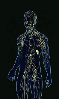
Human lymphatic system. Artwork of a front view of a human, showing the lymphatic system (yellow). Also seen are associated organs including the spleen (centre right)

Background imagePhysiological Collection: Jean Fernel, French physician

Jean Fernel, French physician
Jean Francois Fernel (1497-1558), French physician. Fernel, the son of an innkeeper, lived in Paris and became personal physician to King Henry II of France

Background imagePhysiological Collection: Lining of the duodenum, endoscopic view C016 / 8321

Lining of the duodenum, endoscopic view C016 / 8321
Lining of the duodenum. Endoscopic view of the lining of the duodenum - the first section of the small intestine - showing lipid (fat, white) filled villi

Background imagePhysiological Collection: Blood vessel formation, artwork

Blood vessel formation, artwork
Blood vessel formation. Artwork showing malignant (cancerous) tumour cells promoting the formation of new blood vessels, a process known as angiogenesis

Background imagePhysiological Collection: Lungs anatomy, artwork

Lungs anatomy, artwork
Lungs anatomy, computer artwork. At top left is the trachea (windpipe, white), which splits into two bronchi, one for each lung (blue)

Background imagePhysiological Collection: Insulin production, artwork

Insulin production, artwork
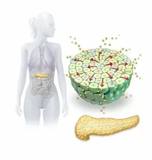
Insulin production. Computer artwork of the human pancreas (yellow) and an islet of Langerhans (green) producing the hormone insulin (small spheres)

Background imagePhysiological Collection: Ovulation and fertilisation, artwork

Ovulation and fertilisation, artwork
Ovulation and fertilisation. Computer artwork of the female reproductive system, showing the development of an ovum (egg, small circle) inside an ovary (oval)

Background imagePhysiological Collection: Lactating breast tissue, light micrograph C015 / 7102

Lactating breast tissue, light micrograph C015 / 7102
Lactating breast tissue. Light micrograph of a section through healthy breast (mammary gland) tissue during lactation. Glandular tissue in the breast has proliferated to produce spaces (alveoli)

Background imagePhysiological Collection: Breathing and body temperature, artwork C013 / 5938

Breathing and body temperature, artwork C013 / 5938
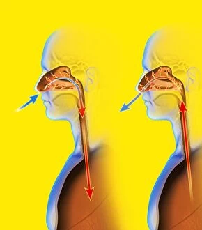
Breathing and body temperature. Computer artwork showing how breathing through the nose helps to regulate body temperature

Background imagePhysiological Collection: Oxidoreductase enzyme complex C015 / 6554

Oxidoreductase enzyme complex C015 / 6554
Oxidoreductase enzyme complex, molecular model. This is the membrane-bound domain formed from of a complex of NADH-quinone oxidoreductase subunits

Background imagePhysiological Collection: Oxidoreductase enzyme complex C015 / 6553

Oxidoreductase enzyme complex C015 / 6553
Oxidoreductase enzyme complex, molecular model. This is the membrane-bound domain formed from of a complex of NADH-quinone oxidoreductase subunits

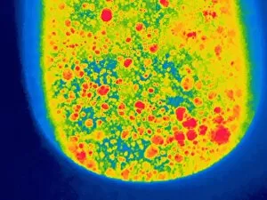
Background imagePhysiological Collection: Urine in a toilet, thermogram C016 / 7563

Urine in a toilet, thermogram C016 / 7563
Urine in a toilet. Thermogram of recently excreted urine mixing with the water in a toilet bowl. The colours show variation in temperature

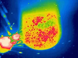
Background imagePhysiological Collection: Urine in a toilet, thermogram C016 / 7561

Urine in a toilet, thermogram C016 / 7561
Urine in a toilet. Thermogram of recently excreted urine mixing with the water in a toilet bowl. The colours show variation in temperature

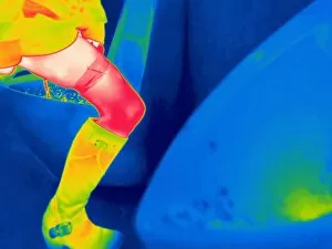
Background imagePhysiological Collection: Woman using a urinal, thermogram C016 / 7560

Woman using a urinal, thermogram C016 / 7560
Woman using a urinal. Thermogram of a woman urinating in a urinal. The colours show variation in temperature. The scale runs from black (coldest) through purple, pink, red, orange and yellow

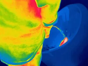
Background imagePhysiological Collection: Man urinating, thermogram C016 / 7554

Man urinating, thermogram C016 / 7554
Man urinating, thermogram. The colours show variation in temperature. The scale runs from black (coldest) through purple, pink, red, orange and yellow, to white (warmest)

Background imagePhysiological Collection: Man urinating, thermogram C016 / 7553

Man urinating, thermogram C016 / 7553
Man urinating. Thermogram of a man urinating into a urinal. The colours show variation in temperature. The scale runs from black (coldest) through purple, pink, red, orange and yellow

Background imagePhysiological Collection: Man urinating, thermogram C016 / 7551

Man urinating, thermogram C016 / 7551
Man urinating. Thermogram of a man urinating into a toilet. The colours show variation in temperature. The scale runs from black (coldest) through purple, pink, red, orange and yellow

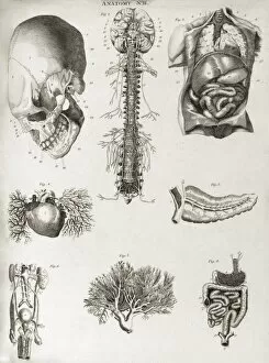
Background imagePhysiological Collection: Human anatomy, 1823 C017 / 8057

Human anatomy, 1823 C017 / 8057
Human anatomy. 19th-century illustrations showing the anatomy of a human skull (upper left), brain and spinal column (upper centre), chest and abdominal organs (lungs, heart, liver, stomach)


For sale as Licensed Images

Choose your image, Select your licence and Download the media

"Exploring the Intricacies of Physiology: From Airpumps to Blood Cells" Delving into the world of physiology, we uncover a fascinating array of scientific discoveries and visual wonders. Joseph Wright's masterpiece, "The Airpump, " captures the essence of early physiological experiments, showcasing humanity's thirst for knowledge. Intricate synapse nerve junctions depicted through Transmission Electron Microscopy (TEM) reveal the intricate communication network within our bodies. These microscopic connections enable seamless transmission of signals between neurons, shaping our thoughts and actions. Moving upwards to the head, we encounter an astonishing view of the muscular system. The complexity and precision required for even simple movements become apparent as ligaments intertwine with bones in perfect harmony. Venturing deeper into female physiology, a Scanning Electron Microscope (SEM) reveals the uterus lining during menstruation. This glimpse into nature's cyclical process sheds light on one aspect of womanhood that has both fascinated and perplexed scientists throughout history. Stepping away from biology but still within the realm of physiology, lie detector tests emerge as tools to decipher truth from deception. By monitoring physiological responses such as heart rate and perspiration levels, these tests attempt to unravel hidden truths buried within our bodies. Returning to cellular structures through TEM imaging once again unveils rough endoplasmic reticulum - a vital component responsible for protein synthesis in cells. Its intricate web-like structure showcases nature's elegance at work. Tracing back centuries ago brings us to Descartes' optics theory from the 17th century - an exploration into how vision is perceived by our eyes and interpreted by our brains. This groundbreaking theory laid foundations for understanding human perception today. Transitioning towards physicality itself leads us to skeletons intertwined with ligaments - providing structural support while allowing flexibility in movement. These silent heroes ensure smooth locomotion throughout life's journey.