Physiological Collection (#2)

"Exploring the Intricacies of Physiology: From Airpumps to Blood Cells" Delving into the world of physiology

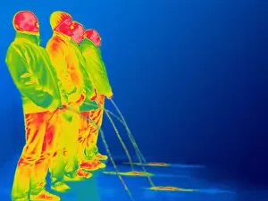
Background imagePhysiological Collection: Men urinating, thermogram C016 / 7556

Men urinating, thermogram C016 / 7556
Men urinating, thermogram. The colours show variation in temperature. The scale runs from black (coldest) through purple, pink, red, orange and yellow, to white (warmest)

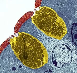
Background imagePhysiological Collection: Goblet cells

Goblet cells. Coloured transmission electron micrograph (TEM) of a section through goblet cells in the lining of the small intestine, part of the digestive tract. They are full of mucus (yellow)

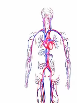
Background imagePhysiological Collection: Human circulatory system

Human circulatory system, computer artwork. Veins (blue) carry deoxygenated blood towards the heart. Arteries (red) carry oxygenated blood from the heart around the body

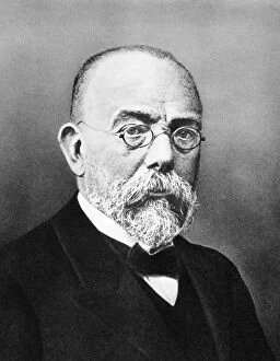
Background imagePhysiological Collection: Robert Koch, German bacteriologist

Robert Koch, German bacteriologist
Robert Koch (1843-1910), German bacteriologist. Along with Louis Pasteur, Koch is considered the founder of modern medical bacteriology

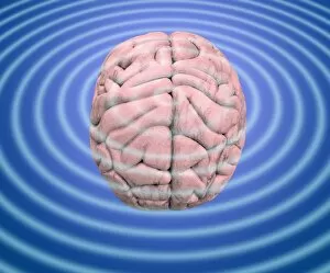
Background imagePhysiological Collection: Brain waves, conceptual image

Brain waves, conceptual image
Brain waves. Conceptual image of waves emanating from a human brain. This can represent the electrical activity of the brain, as recorded on an electroencephalogram (EEG)

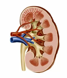
Background imagePhysiological Collection: Kidney anatomy, artwork

Kidney anatomy, artwork
Kidney anatomy. Cutaway artwork showing the internal structure of a kidney. The kidney, one of a pair of such organs, is part of the human excretory system
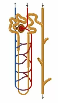
Background imagePhysiological Collection: Nephron structure, artwork

Nephron structure, artwork
Nephron structure. Artwork showing the structure of a nephron, the functional filtering unit of a kidney. The function of the kidney is to filter waste products such as urea from the blood
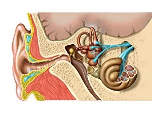
Background imagePhysiological Collection: Human ear anatomy

Human ear anatomy. Computer artwork of the structure of the human ear, showing the outer ear (left), middle ear and inner ear (right)
Background imagePhysiological Collection: Italian physician and physicist. Line engraving from Galvanis De Viribus Electricitatis, 1792
Italian physician and physicist. Line engraving from Galvanis De Viribus Electricitatis, 1792
LUIGI GALVANI (1737-1798). Italian physician and physicist. Line engraving from Galvanis De Viribus Electricitatis, 1792

Background imagePhysiological Collection: Luigi Galvanis production of an electric current between two metals and a frogs legs

Luigi Galvanis production of an electric current between two metals and a frogs legs
GALVANI: GALVANISM, 1791. Luigi Galvanis production of an electric current between two metals and a frogs legs, the graphic demonstration of galvanism

Background imagePhysiological Collection: Oudemas or edemas erupting on a cabbage leaf. The cause is not know but is probably physiological

Oudemas or edemas erupting on a cabbage leaf. The cause is not know but is probably physiological

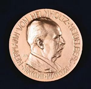
Background imagePhysiological Collection: Hermann Ludwig Ferdinand von Helmholtz, 1907. Helmholtz (1821-1894), German physicist

Hermann Ludwig Ferdinand von Helmholtz, 1907. Helmholtz (1821-1894), German physicist and physiologist, inventor of the ophthalmascope (1850)

Background imagePhysiological Collection: Paul Bert (1833-1886) French physiologist and politician. A pupil of Claude Bernard

Paul Bert (1833-1886) French physiologist and politician. A pupil of Claude Bernard, he studied the physiological effects of air pressure (Caisson disease, and respiration at high altitude)

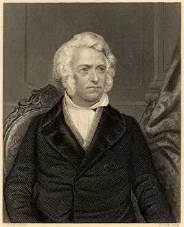
Background imagePhysiological Collection: Leopold Gmelin (1788-1853), German physiological chemist. Professor of chemistry

Leopold Gmelin (1788-1853), German physiological chemist. Professor of chemistry and medicine at Heidelberg University 1817-1852. From James Sheridan Muspratt Chemistry (London, c1860). Engraving

Background imagePhysiological Collection: Synaesthesia, conceptual artwork C017 / 7072

Synaesthesia, conceptual artwork C017 / 7072
Synaesthesia, conceptual artwork. Synaesthesia is a neurological condition where stimulation of one sensory or cognitive pathway leads to automatic

Background imagePhysiological Collection: Blood clot, SEM P260 / 0123

Blood clot, SEM P260 / 0123
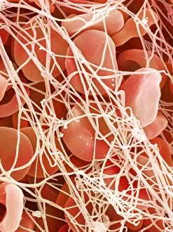
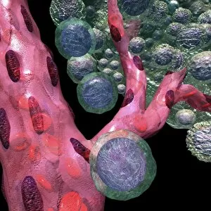
Blood clot. Coloured scanning electron micrograph (SEM) of a blood clot. The red blood cells (erythrocytes) are trapped in filaments of fibrin protein (pink)

Background imagePhysiological Collection: Physical fitness test C014 / 1103

Physical fitness test C014 / 1103
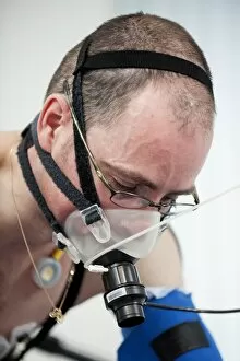
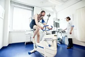
Physical fitness test. Athlete riding an exercise bike while his oxygen consumption is measured with a face mask. Electrodes on his chest monitor his heart rate

Background imagePhysiological Collection: Physical fitness test C014 / 1108

Physical fitness test C014 / 1108
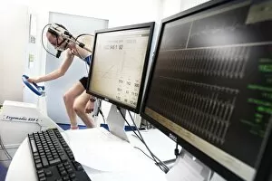
Physical fitness test. Athlete riding an exercise bike while his oxygen consumption is measured with a face mask. Electrodes on his chest monitor his heart rate (shown on screen)

Background imagePhysiological Collection: Muybridge motion study, 1870s C014 / 2071

Muybridge motion study, 1870s C014 / 2071
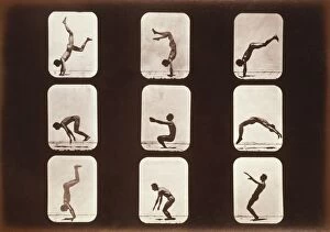
Muybridge motion study. Series of early photographs showing the motion of a man performing a back-flip or back handspring

Background imagePhysiological Collection: Blood vessel formation, artwork C016 / 9842

Blood vessel formation, artwork C016 / 9842
Blood vessel formation. Artwork showing malignant (cancerous) tumour cells (green) promoting the formation of new blood vessels (pink), a process known as angiogenesis

Background imagePhysiological Collection: Psychic brain, conceptual image

Psychic brain, conceptual image. Artwork showing the human brain and head, surrounded in flames, with the pineal gland highlighted (bright dot in centre of forehead)

Background imagePhysiological Collection: Senses within the brain, 16th century C017 / 6996

Senses within the brain, 16th century C017 / 6996
Senses within the brain. 16th-century woodcut of a mans head in profile, showing his brain, with various parts named, including memory, cognition, fantasia, sensus

Background imagePhysiological Collection: Physical fitness test C014 / 1105

Physical fitness test C014 / 1105
Physical fitness test. Athlete riding an exercise bike while his oxygen consumption is measured with a face mask. Electrodes on his chest monitor his heart rate

Background imagePhysiological Collection: Franz Joseph Gall, German physiologist

Franz Joseph Gall, German physiologist
Franz Joseph Gall (1758-1828), German physiologist. Gall believed that the brains shape was related to mental capacity and that different parts of the brain controlled specific parts of the human

Background imagePhysiological Collection: Sugar uptake in muscles, diagram

Sugar uptake in muscles, diagram
Sugar uptake in muscles. Diagram showing sugar from a drink (upper right) being taken up by the muscles of the human body

Background imagePhysiological Collection: Knee joint, x-ray, artwork

Knee joint, x-ray, artwork
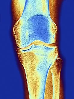
Knee joint. Coloured X-ray of a healthy knee joint, seen from the front. The knee is the largest joint in the body. It is formed by the articulation of the femur (thigh bone)

Background imagePhysiological Collection: Knee joint, x-ray, artwork F006 / 9187

Knee joint, x-ray, artwork F006 / 9187
Computer artwork of a coloured X-ray of a knee joint, depicting erosion of the bone structure and degeneration of the joint by arthrosis

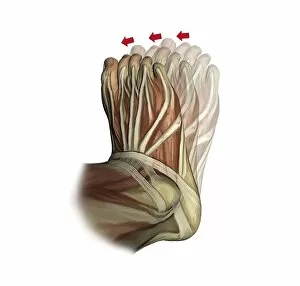
Background imagePhysiological Collection: Inversion of the foot, artwork C016 / 6802

Inversion of the foot, artwork C016 / 6802
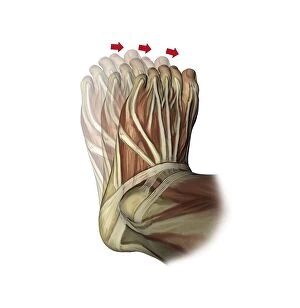
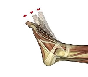
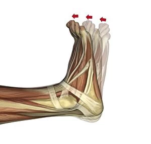
Inversion of the foot. Artwork of the muscles of the foot from above, with red arrows showing the direction of movement when inverting the foot

Background imagePhysiological Collection: Elbow flexion, artwork C016 / 6790

Elbow flexion, artwork C016 / 6790
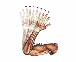
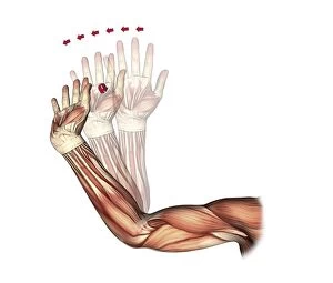
Elbow flexion. Artwork of the muscles of the arm from the front, with red arrows showing the direction of movement when flexing (bending) the elbow

Background imagePhysiological Collection: Plantarflexion of the foot, artwork C016 / 6800

Plantarflexion of the foot, artwork C016 / 6800
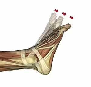
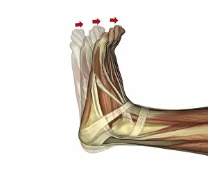
Plantarflexion of the foot. Artwork of the muscles of the foot from the side, with red arrows showing the direction of movement when flexing the foot in the direction of its lower (plantar)

Background imagePhysiological Collection: Ovulation, artwork C016 / 7537

Ovulation, artwork C016 / 7537
Ovulation, computer artwork

Background imagePhysiological Collection: Elbow flexion, artwork C016 / 6789

Elbow flexion, artwork C016 / 6789
Elbow flexion. Artwork of the muscles of the arm from the front, with red arrows showing the direction of movement when flexing (bending) the elbow

Background imagePhysiological Collection: Plantarflexion of the foot, artwork C016 / 6799

Plantarflexion of the foot, artwork C016 / 6799
Plantarflexion of the foot. Artwork of the muscles of the foot from the side, with red arrows showing the direction of movement when flexing the foot in the direction of its lower (plantar)

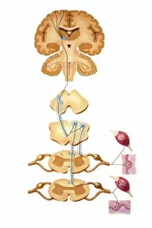
Background imagePhysiological Collection: Brain motor cortex pathways, artwork C016 / 6533

Brain motor cortex pathways, artwork C016 / 6533
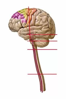
Brain motor cortex pathways. Artwork of a sectioned human brain, brainstem and spinal cord, showing neural pathways (black lines) from the motor cortex of the brain

Background imagePhysiological Collection: Elbow and wrist extension, artwork C016 / 6791

Elbow and wrist extension, artwork C016 / 6791
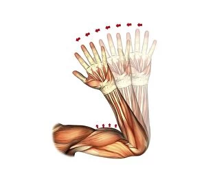
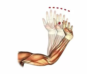
Elbow and wrist extension. Artwork of the muscles of the arm from the front, with red arrows showing the direction of movement when extending the elbow and wrist joints

Background imagePhysiological Collection: Little and ring finger flexion, artwork C016 / 6794

Little and ring finger flexion, artwork C016 / 6794
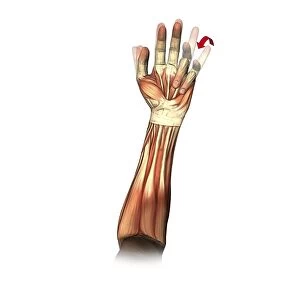
Little and ring finger flexion. Artwork of the muscles of the forearm and hand from the front, with red arrows showing the direction of movement of the little and ring (fourth and third)

Background imagePhysiological Collection: Runners anatomy, artwork

Runners anatomy, artwork
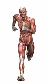
Runners anatomy. Artwork showing the musculature of a running human figure. The main muscles used when running are the quadriceps femoris (rectus femoris, vastus medialis, vastus lateralis)

Background imagePhysiological Collection: Elbow and wrist extension, artwork C016 / 6792

Elbow and wrist extension, artwork C016 / 6792
Elbow and wrist extension. Artwork of the muscles of the arm from the front, with red arrows showing the direction of movement when extending the elbow and wrist joints

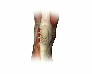
Background imagePhysiological Collection: Patella twitch, artwork C016 / 6816

Patella twitch, artwork C016 / 6816
Patella twitch. Artwork of the muscles of the knee from the front, with red arrows showing the direction of movement of the knee cap (patella) when the quadriceps muscles of the thigh contracts

Background imagePhysiological Collection: Galen, Ancient Greek physician C016 / 8854

Galen, Ancient Greek physician C016 / 8854
Galen (130-200 AD), Ancient Greek physician. After Galens death, his body of work became a medical authority among Europeans until his views on anatomy were overthrown in the 16th century by

Background imagePhysiological Collection: Inversion of the foot, artwork C016 / 6801

Inversion of the foot, artwork C016 / 6801
Inversion of the foot. Artwork of the muscles of the foot from above, with red arrows showing the direction of movement when inverting the foot

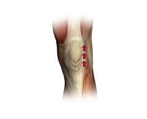
Background imagePhysiological Collection: Patella twitch, artwork C016 / 6803

Patella twitch, artwork C016 / 6803
Patella twitch. Artwork of the muscles of the knee from the front, with red arrows showing the direction of movement of the knee cap (patella) when the quadriceps muscles of the thigh contracts

Background imagePhysiological Collection: Shark heart-gill anatomy, artwork C016 / 6847

Shark heart-gill anatomy, artwork C016 / 6847
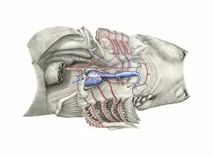
Shark heart-gill anatomy. Artwork of the heart and gill anatomy of a shark, seen from the underside, with the head at right (teeth at centre right)

Background imagePhysiological Collection: Duchennes physiognomy studies, 1860s

Duchennes physiognomy studies, 1860s
Duchennes physiognomy studies. Man with a facial expression induced by electrical probes applied by French neurologist Guillaume Duchenne (1806-1875)

Background imagePhysiological Collection: Brain motor cortex pathways, artwork C016 / 6531

Brain motor cortex pathways, artwork C016 / 6531
Brain motor cortex pathways. Artwork of a lateral (from the side) view of a human brain, showing neural pathways (pink lines) from the motor cortex of the brain to the brainstem and spinal cord

Background imagePhysiological Collection: Dorsiflexion of the foot, artwork C016 / 6797

Dorsiflexion of the foot, artwork C016 / 6797
Dorsiflexion of the foot. Artwork of the muscles of the foot from the side, with red arrows showing the direction of movement when flexing the foot in the direction of its upper (dorsal)

Background imagePhysiological Collection: Brain sensory neural pathways, artwork C016 / 6534

Brain sensory neural pathways, artwork C016 / 6534
Brain sensory neural pathways.. Artwork of a sectioned human brain, brainstem and spinal cord, showing neural pathways (black lines) from regions of the brain to receptors (lower right and lower left)

Background imagePhysiological Collection: Dorsiflexion of the foot, artwork C016 / 6798

Dorsiflexion of the foot, artwork C016 / 6798
Dorsiflexion of the foot. Artwork of the muscles of the foot from the side, with red arrows showing the direction of movement when flexing the foot in the direction of its upper (dorsal)


For sale as Licensed Images

Choose your image, Select your licence and Download the media

"Exploring the Intricacies of Physiology: From Airpumps to Blood Cells" Delving into the world of physiology, we uncover a fascinating array of scientific discoveries and visual wonders. Joseph Wright's masterpiece, "The Airpump, " captures the essence of early physiological experiments, showcasing humanity's thirst for knowledge. Intricate synapse nerve junctions depicted through Transmission Electron Microscopy (TEM) reveal the intricate communication network within our bodies. These microscopic connections enable seamless transmission of signals between neurons, shaping our thoughts and actions. Moving upwards to the head, we encounter an astonishing view of the muscular system. The complexity and precision required for even simple movements become apparent as ligaments intertwine with bones in perfect harmony. Venturing deeper into female physiology, a Scanning Electron Microscope (SEM) reveals the uterus lining during menstruation. This glimpse into nature's cyclical process sheds light on one aspect of womanhood that has both fascinated and perplexed scientists throughout history. Stepping away from biology but still within the realm of physiology, lie detector tests emerge as tools to decipher truth from deception. By monitoring physiological responses such as heart rate and perspiration levels, these tests attempt to unravel hidden truths buried within our bodies. Returning to cellular structures through TEM imaging once again unveils rough endoplasmic reticulum - a vital component responsible for protein synthesis in cells. Its intricate web-like structure showcases nature's elegance at work. Tracing back centuries ago brings us to Descartes' optics theory from the 17th century - an exploration into how vision is perceived by our eyes and interpreted by our brains. This groundbreaking theory laid foundations for understanding human perception today. Transitioning towards physicality itself leads us to skeletons intertwined with ligaments - providing structural support while allowing flexibility in movement. These silent heroes ensure smooth locomotion throughout life's journey.