Physiological Collection (#3)

"Exploring the Intricacies of Physiology: From Airpumps to Blood Cells" Delving into the world of physiology

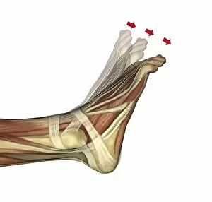
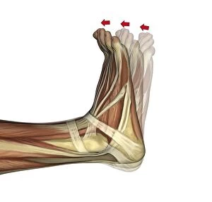
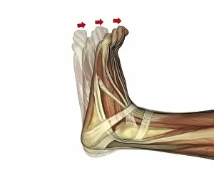
Background imagePhysiological Collection: Plantarflexion of the foot, artwork C016 / 6799

Plantarflexion of the foot, artwork C016 / 6799
Plantarflexion of the foot. Artwork of the muscles of the foot from the side, with red arrows showing the direction of movement when flexing the foot in the direction of its lower (plantar)

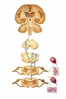
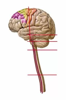
Background imagePhysiological Collection: Brain motor cortex pathways, artwork C016 / 6533

Brain motor cortex pathways, artwork C016 / 6533
Brain motor cortex pathways. Artwork of a sectioned human brain, brainstem and spinal cord, showing neural pathways (black lines) from the motor cortex of the brain

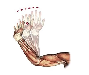
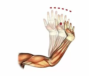
Background imagePhysiological Collection: Elbow and wrist extension, artwork C016 / 6791

Elbow and wrist extension, artwork C016 / 6791
Elbow and wrist extension. Artwork of the muscles of the arm from the front, with red arrows showing the direction of movement when extending the elbow and wrist joints

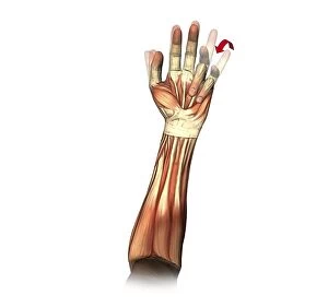
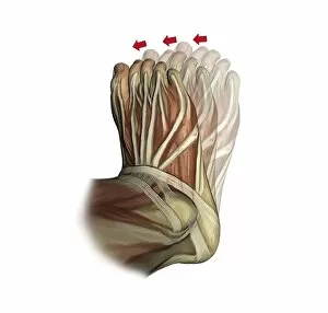
Background imagePhysiological Collection: Little and ring finger flexion, artwork C016 / 6794

Little and ring finger flexion, artwork C016 / 6794
Little and ring finger flexion. Artwork of the muscles of the forearm and hand from the front, with red arrows showing the direction of movement of the little and ring (fourth and third)

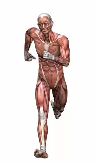
Background imagePhysiological Collection: Runners anatomy, artwork

Runners anatomy, artwork
Runners anatomy. Artwork showing the musculature of a running human figure. The main muscles used when running are the quadriceps femoris (rectus femoris, vastus medialis, vastus lateralis)
Background imagePhysiological Collection: Elbow and wrist extension, artwork C016 / 6792

Elbow and wrist extension, artwork C016 / 6792
Elbow and wrist extension. Artwork of the muscles of the arm from the front, with red arrows showing the direction of movement when extending the elbow and wrist joints

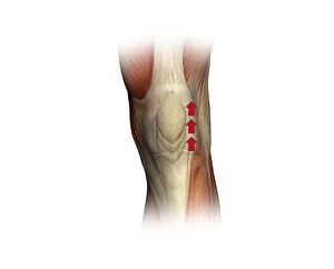
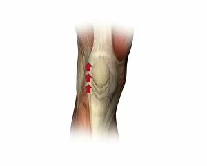
Background imagePhysiological Collection: Patella twitch, artwork C016 / 6816

Patella twitch, artwork C016 / 6816
Patella twitch. Artwork of the muscles of the knee from the front, with red arrows showing the direction of movement of the knee cap (patella) when the quadriceps muscles of the thigh contracts

Background imagePhysiological Collection: Galen, Ancient Greek physician C016 / 8854

Galen, Ancient Greek physician C016 / 8854
Galen (130-200 AD), Ancient Greek physician. After Galens death, his body of work became a medical authority among Europeans until his views on anatomy were overthrown in the 16th century by

Background imagePhysiological Collection: Inversion of the foot, artwork C016 / 6801

Inversion of the foot, artwork C016 / 6801
Inversion of the foot. Artwork of the muscles of the foot from above, with red arrows showing the direction of movement when inverting the foot

Background imagePhysiological Collection: Patella twitch, artwork C016 / 6803

Patella twitch, artwork C016 / 6803
Patella twitch. Artwork of the muscles of the knee from the front, with red arrows showing the direction of movement of the knee cap (patella) when the quadriceps muscles of the thigh contracts

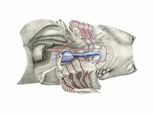
Background imagePhysiological Collection: Shark heart-gill anatomy, artwork C016 / 6847

Shark heart-gill anatomy, artwork C016 / 6847
Shark heart-gill anatomy. Artwork of the heart and gill anatomy of a shark, seen from the underside, with the head at right (teeth at centre right)

Background imagePhysiological Collection: Duchennes physiognomy studies, 1860s

Duchennes physiognomy studies, 1860s
Duchennes physiognomy studies. Man with a facial expression induced by electrical probes applied by French neurologist Guillaume Duchenne (1806-1875)

Background imagePhysiological Collection: Brain motor cortex pathways, artwork C016 / 6531

Brain motor cortex pathways, artwork C016 / 6531
Brain motor cortex pathways. Artwork of a lateral (from the side) view of a human brain, showing neural pathways (pink lines) from the motor cortex of the brain to the brainstem and spinal cord

Background imagePhysiological Collection: Dorsiflexion of the foot, artwork C016 / 6797

Dorsiflexion of the foot, artwork C016 / 6797
Dorsiflexion of the foot. Artwork of the muscles of the foot from the side, with red arrows showing the direction of movement when flexing the foot in the direction of its upper (dorsal)

Background imagePhysiological Collection: Brain sensory neural pathways, artwork C016 / 6534

Brain sensory neural pathways, artwork C016 / 6534
Brain sensory neural pathways.. Artwork of a sectioned human brain, brainstem and spinal cord, showing neural pathways (black lines) from regions of the brain to receptors (lower right and lower left)

Background imagePhysiological Collection: Dorsiflexion of the foot, artwork C016 / 6798

Dorsiflexion of the foot, artwork C016 / 6798
Dorsiflexion of the foot. Artwork of the muscles of the foot from the side, with red arrows showing the direction of movement when flexing the foot in the direction of its upper (dorsal)

Background imagePhysiological Collection: Transducin protein beta-gamma complex

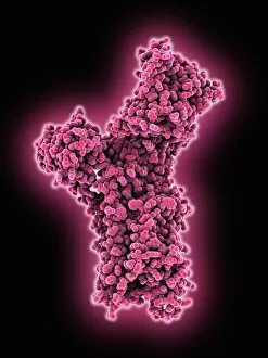
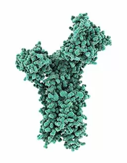
Transducin protein beta-gamma complex. Molecular model of the beta-gamma dimer of the heterotrimeric G protein transducin

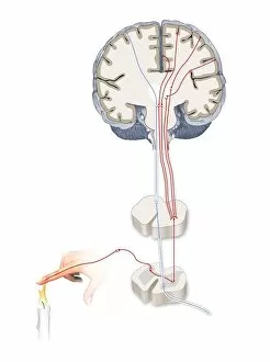
Background imagePhysiological Collection: Pain pathways, artwork

Pain pathways, artwork
Pain pathways, computer artwork. The heat of the candle is detected by nociceptors (special sensory nerve endings) in the finger

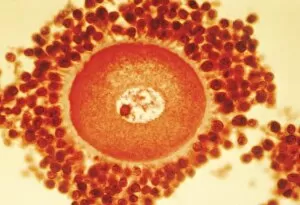
Background imagePhysiological Collection: Oocyte before ovulation, light micrograph

Oocyte before ovulation, light micrograph
Oocyte before ovulation. Light micrograph of a section through tissue from an ovary, showing an oocyte two days before ovulation

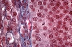
Background imagePhysiological Collection: Ovarian follicle tissue, light micrograph

Ovarian follicle tissue, light micrograph
Ovarian follicle tissue. Light micrograph of a section through tissue from an ovarian follicle, showing a boundary between two layers

Background imagePhysiological Collection: Suspended animation, artwork

Suspended animation, artwork
Suspended animation. Computer artwork of futuristic humans in suspended animation. This is a state where fundamental processes, such as breathing and heart rate, are slowed, which extends lifespan

Background imagePhysiological Collection: Genders of Nobel laureates, artwork

Genders of Nobel laureates, artwork
Genders of Nobel laureates. Artwork showing the gender divide between women (left) and men (right) who have won Nobel Prizes

Background imagePhysiological Collection: Anterior pituitary gland, TEM

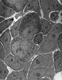
Anterior pituitary gland, TEM
Anterior pituitary gland. Transmission electron micrograph (TEM) of a section through cells of the anterior pituitary. The anterior pituitary, or adenohypophysis

Background imagePhysiological Collection: Olfactory bulb anatomy, artwork

Olfactory bulb anatomy, artwork
Olfactory bulb anatomy. Artwork of a human nose, showing the anatomy of the olfactory bulb, the organ of smell. This lies above the anterior aspect of the nasal cavity on the ethmoid bone

Background imagePhysiological Collection: Calcium ATPase ion pump C015 / 7143

Calcium ATPase ion pump C015 / 7143
Calcium ATPase ion pump, molecular model. This enzyme is found in muscle cell membranes, where it pumps calcium in and out of muscle cells and controls muscle contractions

Background imagePhysiological Collection: Liver, SEM C015 / 9924

Liver, SEM C015 / 9924
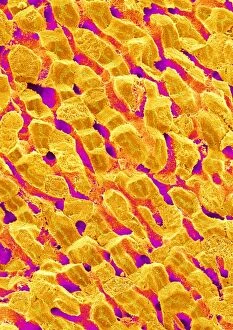
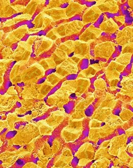
Liver vein. Coloured scanning electron micrograph (SEM) of liver tissue. The functions of the liver include destroying old red blood cells

Background imagePhysiological Collection: Liver, SEM C015 / 9922

Liver, SEM C015 / 9922
Liver vein. Coloured scanning electron micrograph (SEM) of liver tissue showing a large vein (pink, up centre). The functions of the liver include destroying old red blood cells

Background imagePhysiological Collection: Thrombin complexed with fibrinogen C015 / 7148

Thrombin complexed with fibrinogen C015 / 7148
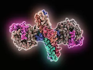
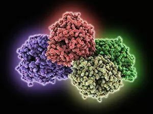
Thrombin complexed with fibrinogen, molecular model. The thrombin molecules (left and right, purple and green) are bound to the central part of the fibrinogen molecule (centre, multiple colours)

Background imagePhysiological Collection: Retinal-producing oxygenase enzyme C015 / 7150

Retinal-producing oxygenase enzyme C015 / 7150
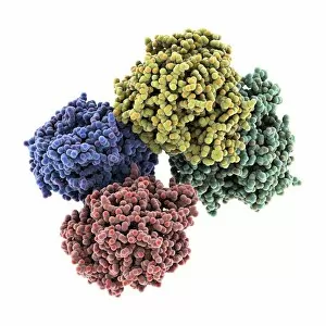
Retinal-producing oxygenase enzyme, molecular model. This enzyme, an oxygenase, plays a role in the production and metabolism of retinal and other apocarotenoids

Background imagePhysiological Collection: Liver, SEM C015 / 9923

Liver, SEM C015 / 9923
Liver vein. Coloured scanning electron micrograph (SEM) of liver tissue. The functions of the liver include destroying old red blood cells

Background imagePhysiological Collection: Thrombin complexed with fibrinogen C015 / 7149

Thrombin complexed with fibrinogen C015 / 7149
Thrombin complexed with fibrinogen, molecular model. The thrombin molecules (left and right, brown and pink) are bound to the central part of the fibrinogen molecule (centre, multiple colours)

Background imagePhysiological Collection: Calcium ATPase ion pump C015 / 7142

Calcium ATPase ion pump C015 / 7142
Calcium ATPase ion pump, molecular model. This enzyme is found in muscle cell membranes, where it pumps calcium in and out of muscle cells and controls muscle contractions

Background imagePhysiological Collection: Liver, SEM C015 / 9925

Liver, SEM C015 / 9925
Liver vein. Coloured scanning electron micrograph (SEM) of liver tissue. The functions of the liver include destroying old red blood cells

Background imagePhysiological Collection: Retinal-producing oxygenase enzyme C015 / 7151

Retinal-producing oxygenase enzyme C015 / 7151
Retinal-producing oxygenase enzyme, molecular model. This enzyme, an oxygenase, plays a role in the production and metabolism of retinal and other apocarotenoids

Background imagePhysiological Collection: Torn muscle, illustration C018 / 0806

Torn muscle, illustration C018 / 0806
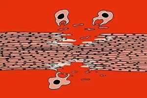
Torn muscle. Illustration of a tear in a skeletal muscle. Muscle tears can either result in muscle growth or injury depending on the circumstances

Background imagePhysiological Collection: Retina layers, SEM C018 / 0521

Retina layers, SEM C018 / 0521
Retina layers, coloured scanning electron micrograph (SEM). The retina of the human eye is found on the inside of the eyeball and is formed from a number of layers

Background imagePhysiological Collection: Physical fitness test C014 / 1104

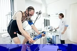
Physical fitness test C014 / 1104
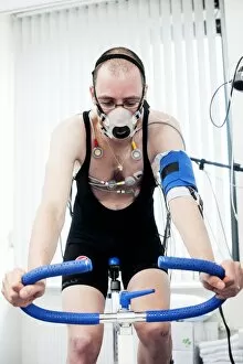
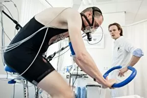
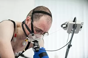
Physical fitness test. Athlete riding an exercise bike while his oxygen consumption is measured with a face mask. Electrodes on his chest monitor his heart rate

Background imagePhysiological Collection: Eye muscle, TEM C014 / 1467

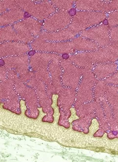
Eye muscle, TEM C014 / 1467
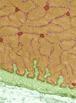
Eye muscle. Transmission electron micrograph (TEM) of a section through a striated muscle cell from the ciliary muscle of a human eye

Background imagePhysiological Collection: Heart and brain vascular system, artwork

Heart and brain vascular system, artwork
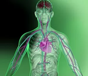
Heart and brain vascular system. Artwork showing the arteries (pink) and veins (blue) of the torso, arms, neck, head and brain. The brain is at top (red) and the heart at centre

Background imagePhysiological Collection: Eye muscle, TEM C014 / 1466

Eye muscle, TEM C014 / 1466
Eye muscle. Transmission electron micrograph (TEM) of a section through a striated muscle cell from the ciliary muscle of a human eye

Background imagePhysiological Collection: Psychic brain, conceptual image C014 / 1480

Psychic brain, conceptual image C014 / 1480
Psychic brain, conceptual image. Artwork showing the human brain from the side, surrounded by flames, with the pineal gland shown

Background imagePhysiological Collection: Physical fitness test C014 / 1101

Physical fitness test C014 / 1101
Physical fitness test. Athlete riding an exercise bike while his oxygen consumption is measured with a face mask. Electrodes on his chest monitor his heart rate

Background imagePhysiological Collection: Physical fitness test C014 / 1107

Physical fitness test C014 / 1107
Physical fitness test. Athlete riding an exercise bike while his oxygen consumption is measured with a face mask. Electrodes on his chest monitor his heart rate

Background imagePhysiological Collection: Physical fitness test C014 / 1102

Physical fitness test C014 / 1102
Physical fitness test. Athlete riding an exercise bike while his oxygen consumption is measured with a face mask. Electrodes on his chest monitor his heart rate

Background imagePhysiological Collection: Physical fitness test C014 / 1100

Physical fitness test C014 / 1100
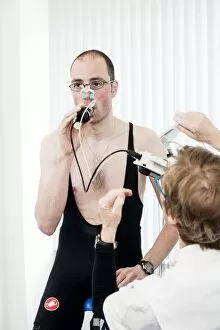
Physical fitness test. Athlete undergoing a physical examination. Here his lung capacity is being tested using a spirometer, which measures the volume and speed of air that he can exhale and inhale

Background imagePhysiological Collection: Nerve fibre node, TEM C014 / 1458

Nerve fibre node, TEM C014 / 1458
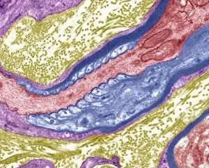
Nerve fibre node. Transmission electron micrograph (TEM) of a longitudinal section through a nerve fibre at a node of Ranvier, showing the nerve tissue (red) and the myelin sheath (dark blue)

Background imagePhysiological Collection: Psychic brain, conceptual image C014 / 1478

Psychic brain, conceptual image C014 / 1478
Psychic brain, conceptual image. Artwork of an anterior (frontal) view of the human brain. The cerebrum (the main visible area) is divided into two hemispheres

Background imagePhysiological Collection: Psychic brain, conceptual image C014 / 1479

Psychic brain, conceptual image C014 / 1479
Psychic brain, conceptual image. Artwork of an anterior (frontal) view of the human brain. The cerebrum (the main visible area) is divided into two hemispheres


For sale as Licensed Images

Choose your image, Select your licence and Download the media

"Exploring the Intricacies of Physiology: From Airpumps to Blood Cells" Delving into the world of physiology, we uncover a fascinating array of scientific discoveries and visual wonders. Joseph Wright's masterpiece, "The Airpump, " captures the essence of early physiological experiments, showcasing humanity's thirst for knowledge. Intricate synapse nerve junctions depicted through Transmission Electron Microscopy (TEM) reveal the intricate communication network within our bodies. These microscopic connections enable seamless transmission of signals between neurons, shaping our thoughts and actions. Moving upwards to the head, we encounter an astonishing view of the muscular system. The complexity and precision required for even simple movements become apparent as ligaments intertwine with bones in perfect harmony. Venturing deeper into female physiology, a Scanning Electron Microscope (SEM) reveals the uterus lining during menstruation. This glimpse into nature's cyclical process sheds light on one aspect of womanhood that has both fascinated and perplexed scientists throughout history. Stepping away from biology but still within the realm of physiology, lie detector tests emerge as tools to decipher truth from deception. By monitoring physiological responses such as heart rate and perspiration levels, these tests attempt to unravel hidden truths buried within our bodies. Returning to cellular structures through TEM imaging once again unveils rough endoplasmic reticulum - a vital component responsible for protein synthesis in cells. Its intricate web-like structure showcases nature's elegance at work. Tracing back centuries ago brings us to Descartes' optics theory from the 17th century - an exploration into how vision is perceived by our eyes and interpreted by our brains. This groundbreaking theory laid foundations for understanding human perception today. Transitioning towards physicality itself leads us to skeletons intertwined with ligaments - providing structural support while allowing flexibility in movement. These silent heroes ensure smooth locomotion throughout life's journey.