Physiological Collection (#5)

"Exploring the Intricacies of Physiology: From Airpumps to Blood Cells" Delving into the world of physiology

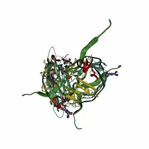
Background imagePhysiological Collection: Saliva chemicals, molecular model

Saliva chemicals, molecular model
Saliva chemicals. Molecular structure of a group of saliva molecules. These include the antibody immunoglobulin A (blue, double-y shape, see C014/5652)

Background imagePhysiological Collection: Human muscle fibres, diagram

Human muscle fibres, diagram
Human muscle fibres. Diagram showing the structure of human muscles from the cellular to the macroscopic level. At lower right, muscle cells contain many myofibrils

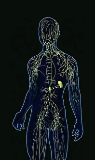
Background imagePhysiological Collection: Human lymphatic system, artwork

Human lymphatic system, artwork
Human lymphatic system. Artwork of a front view of a human, showing the lymphatic system (yellow). Also seen are associated organs including the spleen (centre right)

Background imagePhysiological Collection: Jean Fernel, French physician

Jean Fernel, French physician
Jean Francois Fernel (1497-1558), French physician. Fernel, the son of an innkeeper, lived in Paris and became personal physician to King Henry II of France

Background imagePhysiological Collection: Lining of the duodenum, endoscopic view C016 / 8321

Lining of the duodenum, endoscopic view C016 / 8321
Lining of the duodenum. Endoscopic view of the lining of the duodenum - the first section of the small intestine - showing lipid (fat, white) filled villi

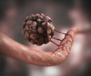
Background imagePhysiological Collection: Blood vessel formation, artwork

Blood vessel formation, artwork
Blood vessel formation. Artwork showing malignant (cancerous) tumour cells promoting the formation of new blood vessels, a process known as angiogenesis

Background imagePhysiological Collection: Lungs anatomy, artwork

Lungs anatomy, artwork
Lungs anatomy, computer artwork. At top left is the trachea (windpipe, white), which splits into two bronchi, one for each lung (blue)

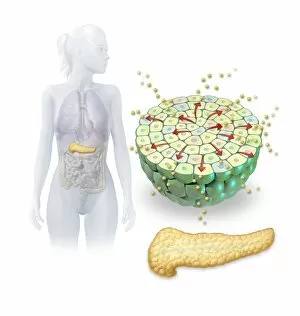
Background imagePhysiological Collection: Insulin production, artwork

Insulin production, artwork
Insulin production. Computer artwork of the human pancreas (yellow) and an islet of Langerhans (green) producing the hormone insulin (small spheres)

Background imagePhysiological Collection: Ovulation and fertilisation, artwork

Ovulation and fertilisation, artwork
Ovulation and fertilisation. Computer artwork of the female reproductive system, showing the development of an ovum (egg, small circle) inside an ovary (oval)

Background imagePhysiological Collection: Lactating breast tissue, light micrograph C015 / 7102

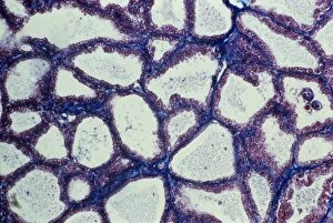
Lactating breast tissue, light micrograph C015 / 7102
Lactating breast tissue. Light micrograph of a section through healthy breast (mammary gland) tissue during lactation. Glandular tissue in the breast has proliferated to produce spaces (alveoli)

Background imagePhysiological Collection: Breathing and body temperature, artwork C013 / 5938

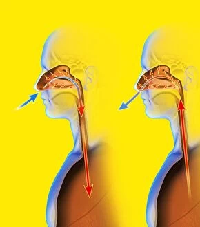
Breathing and body temperature, artwork C013 / 5938
Breathing and body temperature. Computer artwork showing how breathing through the nose helps to regulate body temperature

Background imagePhysiological Collection: Oxidoreductase enzyme complex C015 / 6554

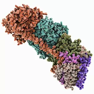
Oxidoreductase enzyme complex C015 / 6554
Oxidoreductase enzyme complex, molecular model. This is the membrane-bound domain formed from of a complex of NADH-quinone oxidoreductase subunits

Background imagePhysiological Collection: Oxidoreductase enzyme complex C015 / 6553

Oxidoreductase enzyme complex C015 / 6553
Oxidoreductase enzyme complex, molecular model. This is the membrane-bound domain formed from of a complex of NADH-quinone oxidoreductase subunits

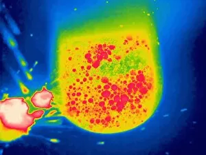
Background imagePhysiological Collection: Urine in a toilet, thermogram C016 / 7563

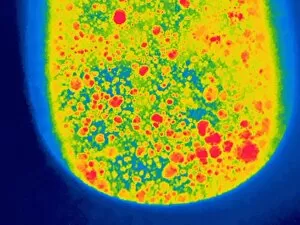
Urine in a toilet, thermogram C016 / 7563
Urine in a toilet. Thermogram of recently excreted urine mixing with the water in a toilet bowl. The colours show variation in temperature

Background imagePhysiological Collection: Urine in a toilet, thermogram C016 / 7561

Urine in a toilet, thermogram C016 / 7561
Urine in a toilet. Thermogram of recently excreted urine mixing with the water in a toilet bowl. The colours show variation in temperature

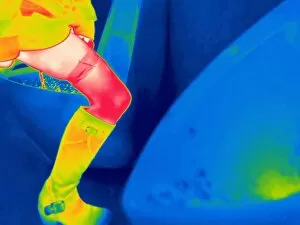
Background imagePhysiological Collection: Woman using a urinal, thermogram C016 / 7560

Woman using a urinal, thermogram C016 / 7560
Woman using a urinal. Thermogram of a woman urinating in a urinal. The colours show variation in temperature. The scale runs from black (coldest) through purple, pink, red, orange and yellow

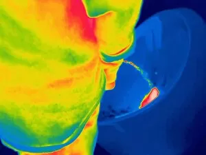
Background imagePhysiological Collection: Man urinating, thermogram C016 / 7554

Man urinating, thermogram C016 / 7554
Man urinating, thermogram. The colours show variation in temperature. The scale runs from black (coldest) through purple, pink, red, orange and yellow, to white (warmest)

Background imagePhysiological Collection: Man urinating, thermogram C016 / 7553

Man urinating, thermogram C016 / 7553
Man urinating. Thermogram of a man urinating into a urinal. The colours show variation in temperature. The scale runs from black (coldest) through purple, pink, red, orange and yellow

Background imagePhysiological Collection: Man urinating, thermogram C016 / 7551

Man urinating, thermogram C016 / 7551
Man urinating. Thermogram of a man urinating into a toilet. The colours show variation in temperature. The scale runs from black (coldest) through purple, pink, red, orange and yellow

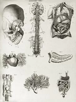
Background imagePhysiological Collection: Human anatomy, 1823 C017 / 8057

Human anatomy, 1823 C017 / 8057
Human anatomy. 19th-century illustrations showing the anatomy of a human skull (upper left), brain and spinal column (upper centre), chest and abdominal organs (lungs, heart, liver, stomach)

Background imagePhysiological Collection: Man urinating, thermogram C016 / 7548

Man urinating, thermogram C016 / 7548
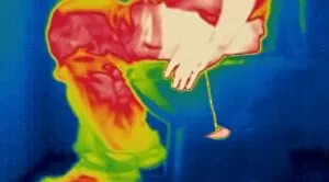
Man urinating. Thermogram of a man sitting on a toilet and urinating. The colours show variation in temperature. The scale runs from black (coldest) through purple, pink, red, orange and yellow

Background imagePhysiological Collection: Muybridge motion study, 1870s C016 / 4563

Muybridge motion study, 1870s C016 / 4563
Muybridge motion study. Series of early photographs showing the motion of a race horse harnessed to a sulky (racing cart) and demonstrating a trotting gait

Background imagePhysiological Collection: Muybridge motion study, 1907 C016 / 4564

Muybridge motion study, 1907 C016 / 4564
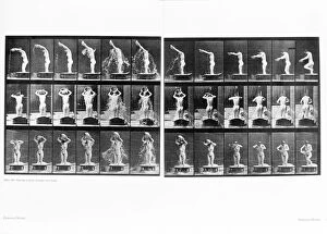
Muybridge motion study. Series of early photographs showing a nude woman pouring a basin of water over herself. These photographs were obtained by English photographer Eadweard J

Background imagePhysiological Collection: Muybridge motion study, 1870s C016 / 4562

Muybridge motion study, 1870s C016 / 4562
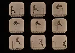
Muybridge motion study. Series of early photographs showing the motion of an athlete carrying out a walking leap over a hurdle

Background imagePhysiological Collection: Astronaut exercising on the ISS C016 / 4207

Astronaut exercising on the ISS C016 / 4207
Astronaut exercising on the ISS. NASA astronaut Sunita Williams (born 1965) exercising in a bungee harness on the Combined Operational Load Bearing External Resistance Treadmill (COLBERT)

Background imagePhysiological Collection: Luigi Galvani statue, Bologna, Italy

Luigi Galvani statue, Bologna, Italy
Luigi Galvani statue. Italian anatomist Luigi Galvani (1737-1798) noticed that dead frogs twitched when included in a circuit with metals (books and a dissected frog shown here)

Background imagePhysiological Collection: Hearing perception, conceptual artwork C013 / 9458

Hearing perception, conceptual artwork C013 / 9458
Hearing perception. Conceptual artwork of a piano key (right) blurring into two. This represents the phenomenon where a single auditory stimulus is heard as two separate sounds or tones

Background imagePhysiological Collection: Hearing protection, conceptual artwork

Hearing protection, conceptual artwork
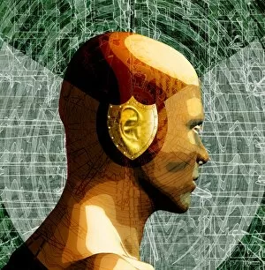
Hearing protection. Conceptual artwork of a shield covering the ear of a human head surrounded by loud noise (concentric circles)

Background imagePhysiological Collection: Hearing damage, conceptual artwork

Hearing damage, conceptual artwork
Hearing damage. Conceptual artwork of a man with distorted sound waves (green) entering his ears, with a volume-indicating dial (volume unit meter) in place of his mouth

Background imagePhysiological Collection: Erythropoietin hormone complex

Erythropoietin hormone complex. Computer model showing the secondary structure of a molecule of the human hormone Erythropoietin (EPO), complexed with an erythropoetin receptor molecule

Background imagePhysiological Collection: Preventing cancer spreading, artwork C013 / 8816

Preventing cancer spreading, artwork C013 / 8816
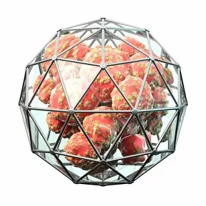
Preventing cancer spreading, conceptual artwork. Cancer cells inside a geodesic cage, representing the prevention of metastasis - spreading to other tissues of the body

Background imagePhysiological Collection: Angiogenesis inhibitors, conceptual image C013 / 7789

Angiogenesis inhibitors, conceptual image C013 / 7789
Angiogenesis inhibitors, conceptual image. Computer artwork showing a brick wall preventing new blood vessels from reaching a cancerous tumour

Background imagePhysiological Collection: Tomato (Solanum sp. ) close-up of fruit, with Catface physiological problem causing malformation

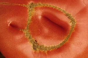
Tomato (Solanum sp. ) close-up of fruit, with Catface physiological problem causing malformation and scars, U. S. A
Tomato (Solanum sp.) close-up of fruit, with Catface physiological problem causing malformation and scars, U.S.A

Background imagePhysiological Collection: University Library, Nicholas Church and Physiological Institu

University Library, Nicholas Church and Physiological Institute, Griefswald (i.e. Greifswald), Pommeraina, Germany. Date between ca. 1890 and ca. 1900

Background imagePhysiological Collection: Fingerprint, computer artwork

Fingerprint, computer artwork
Fingerprint. Computer artwork of a fingerprint residue showing typical patterns for feature identification (whorl, ridge ending and bifurcation)

Background imagePhysiological Collection: Papillae on the tongue of a bat

Papillae on the tongue of a bat
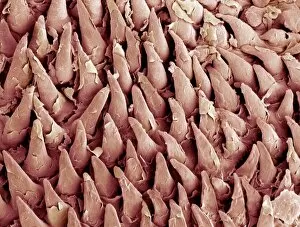
Papillae on the tongue of a pipistrelle bat. Coloured scanning electron micrograph (SEM) of the tongue of a pipistrelle bat (Pipistrellus pipistrellus)

Background imagePhysiological Collection: 4-cell embryo embryo, artwork

4-cell embryo embryo, artwork
4-cell embryo. Image 3 of 4. Computer model representing 4 daughter cells. The development of an embryo is called embryogenesis

Background imagePhysiological Collection: Womans legs, with knee X-ray

Womans legs, with knee X-ray
MODEL RELEASED. Womans legs superimposed on an X-ray of a knee joint. X-rays can be used to diagnose disorders of the skeletal system

Background imagePhysiological Collection: Multi-celled embryo, artwork

Multi-celled embryo, artwork
Multi-celled embryo. Image 4 of 4. Computer model representing a cluster of daughter cells. The development of an embryo is called embryogenesis

Background imagePhysiological Collection: Single-celled embryo, artwork

Single-celled embryo, artwork
Single-celled embryo. Image 1 of 4. Computer model representing a single cell. The development of an embryo is called embryogenesis

Background imagePhysiological Collection: Womans legs

Womans legs
MODEL RELEASED. Womans legs as she reclines. Her foot on tiptoes is at upper right, and in the background are historical anatomical drawings of the legs

Background imagePhysiological Collection: 2-cell embryo embryo, artwork

2-cell embryo embryo, artwork
2-cell embryo. Image 2 of 4. Computer model representing 2 daughter cells. The development of an embryo is called embryogenesis

Background imagePhysiological Collection: Blackhead, SEM

Blackhead, SEM
Black head. Coloured scanning electron micrograph (SEM) of hairs emerging from a blockage in the skin. The material which caused the obstruction

Background imagePhysiological Collection: Embryo in a fallopian tube, artwork

Embryo in a fallopian tube, artwork
Embryo inside a fallopian tube. Computer artwork representing an embryo (yellow) in the early stages of development inside a fallopian tube. Fallopian tubes lead from the ovaries to the uterus

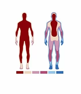
Background imagePhysiological Collection: Core body temperature maintenance

Core body temperature maintenance
Body temperature regulation. Artwork based on a thermogram of the human body under different temperature conditions. It is warm at left and cold at right

Background imagePhysiological Collection: Running man

Running man. Three shots of a man running, showing his muscular structure (centre) and his skeletal structure (right)

Background imagePhysiological Collection: Endocrine cell, TEM

Endocrine cell, TEM
Endocrine cell. Coloured transmission electron micrograph (TEM) of a cultured endocrine cell. Endocrine cells are part of the endocrine system

Background imagePhysiological Collection: Thyroid gland, scintigram

Thyroid gland, scintigram
Thyroid gland. Coloured scintigram of a healthy thyroid gland. This two-lobed endocrine gland is found in the neck, where it lies on top of the trachea (windpipe)


For sale as Licensed Images

Choose your image, Select your licence and Download the media

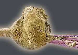
"Exploring the Intricacies of Physiology: From Airpumps to Blood Cells" Delving into the world of physiology, we uncover a fascinating array of scientific discoveries and visual wonders. Joseph Wright's masterpiece, "The Airpump, " captures the essence of early physiological experiments, showcasing humanity's thirst for knowledge. Intricate synapse nerve junctions depicted through Transmission Electron Microscopy (TEM) reveal the intricate communication network within our bodies. These microscopic connections enable seamless transmission of signals between neurons, shaping our thoughts and actions. Moving upwards to the head, we encounter an astonishing view of the muscular system. The complexity and precision required for even simple movements become apparent as ligaments intertwine with bones in perfect harmony. Venturing deeper into female physiology, a Scanning Electron Microscope (SEM) reveals the uterus lining during menstruation. This glimpse into nature's cyclical process sheds light on one aspect of womanhood that has both fascinated and perplexed scientists throughout history. Stepping away from biology but still within the realm of physiology, lie detector tests emerge as tools to decipher truth from deception. By monitoring physiological responses such as heart rate and perspiration levels, these tests attempt to unravel hidden truths buried within our bodies. Returning to cellular structures through TEM imaging once again unveils rough endoplasmic reticulum - a vital component responsible for protein synthesis in cells. Its intricate web-like structure showcases nature's elegance at work. Tracing back centuries ago brings us to Descartes' optics theory from the 17th century - an exploration into how vision is perceived by our eyes and interpreted by our brains. This groundbreaking theory laid foundations for understanding human perception today. Transitioning towards physicality itself leads us to skeletons intertwined with ligaments - providing structural support while allowing flexibility in movement. These silent heroes ensure smooth locomotion throughout life's journey.