Ribonucleic Acid Collection (page 2)

"Unraveling the Secrets of Ribonucleic Acid: The Double-Stranded RNA Molecule" In the intricate world of molecular biology

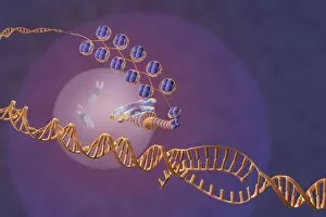
Background imageRibonucleic Acid Collection: DNA transcription, illustration C018 / 0900

DNA transcription, illustration C018 / 0900
DNA (deoxyribonucleic acid) transcription. Illustration of an RNA (ribonucelic acid) polymerase molecule (centre) synthesising an mRNA (messenger RNA) strand (bottom)

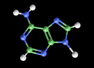
Background imageRibonucleic Acid Collection: Adenine molecule, artwork C017 / 7200

Adenine molecule, artwork C017 / 7200
Adenine molecule. Computer artwork showing the structure of a molecule of the nucleobase adenine. Atoms are colour-coded spheres: carbon (green), nitrogen (blue), and oxygen (white)

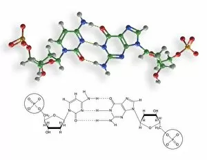
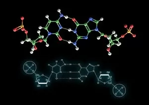
Background imageRibonucleic Acid Collection: Cytosine-guanine interaction, artwork C017 / 7215

Cytosine-guanine interaction, artwork C017 / 7215
Cytosine-guanine interaction. Computer artwork showing the structure of bound cytosine (left) and guanine molecules (right)

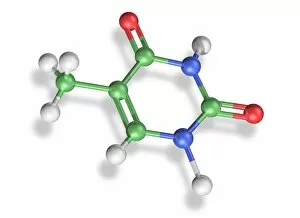
Background imageRibonucleic Acid Collection: Thymine molecule, artwork C017 / 7366

Thymine molecule, artwork C017 / 7366
Thymine molecule. Computer artwork showing the structure of a molecule of the nucleobase thymine. Atoms are colour-coded spheres: carbon (green), nitrogen (blue), oxygen (red), and hydrogen (white)

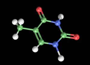
Background imageRibonucleic Acid Collection: Thymine molecule, artwork C017 / 7365

Thymine molecule, artwork C017 / 7365
Thymine molecule. Computer artwork showing the structure of a molecule of the nucleobase thymine. Atoms are colour-coded spheres: carbon (green), nitrogen (blue), oxygen (red), and hydrogen (white)

Background imageRibonucleic Acid Collection: Cytosine-guanine interaction, artwork C017 / 7216

Cytosine-guanine interaction, artwork C017 / 7216
Cytosine-guanine interaction. Computer artwork showing the structure of bound cytosine (left) and guanine molecules (right)

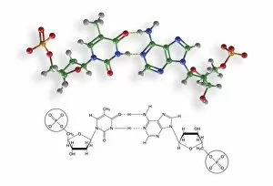
Background imageRibonucleic Acid Collection: Thymine-adenine interaction, artwork C017 / 7367

Thymine-adenine interaction, artwork C017 / 7367
Thymine-adenine interaction. Computer artwork showing the structure of bound thymine and adenine molecules. Atoms are shown as colour-coded spheres: carbon (green), hydrogen (white)

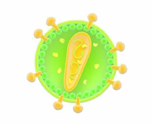
Background imageRibonucleic Acid Collection: Retrovirus, artwork F007 / 6437

Retrovirus, artwork F007 / 6437
Retrovirus, computer artwork. Retroviruses are viruses that have an RNA (ribonucleic acid) genome. They use reverse transcriptase to create a DNA (deoxyribonucleic acid)
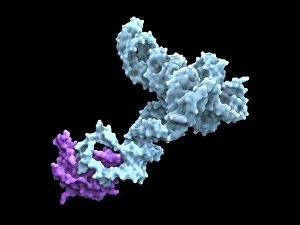
Background imageRibonucleic Acid Collection: Glycine riboswitch molecule F007 / 9921

Glycine riboswitch molecule F007 / 9921
Molecular model of the bacterial glycine riboswitch. This is an RNA element that can bind the amino acid glycine. Glycine riboswitches usually consist of two metabolite-binding aptamer domains tandem

Background imageRibonucleic Acid Collection: Glycine riboswitch molecule F007 / 9906

Glycine riboswitch molecule F007 / 9906
Molecular model of the bacterial glycine riboswitch. This is an RNA element that can bind the amino acid glycine. Glycine riboswitches usually consist of two metabolite-binding aptamer domains tandem

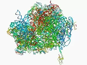
Background imageRibonucleic Acid Collection: Human 80S ribosome F007 / 9902

Human 80S ribosome F007 / 9902
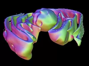
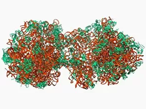
Ribosomal subunit. Computer model showing the structure of the RNA (ribonucleic acid) molecules in an 80S (large) ribosomal sub-unit. Ribosomes are composed of protein and RNA

Background imageRibonucleic Acid Collection: Human 80S ribosome F007 / 9898

Human 80S ribosome F007 / 9898
Ribosomal subunit. Computer model showing the structure of the RNA (ribonucleic acid) molecules in an 80S (large) ribosomal sub-unit. Ribosomes are composed of protein and RNA

Background imageRibonucleic Acid Collection: Microtubes of RNA samples F008 / 2041

Microtubes of RNA samples F008 / 2041
Microtubes of RNA samples

Background imageRibonucleic Acid Collection: tRNA molecule

tRNA molecule
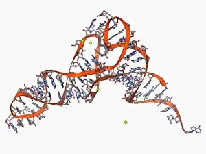
Transfer RNA (tRNA), molecular model. tRNA (transfer ribonucleic acid) translates messenger RNA (mRNA) into a protein product. Each tRNA molecule carries a specific amino acid, in this case tryptophan

Background imageRibonucleic Acid Collection: Gene expression, artwork

Gene expression, artwork
Gene expression. Computer artwork showing the process of transcription, the first stage or gene expression. Here, a chromosome (distance)

Background imageRibonucleic Acid Collection: Adenine molecule, artwork C017 / 7199

Adenine molecule, artwork C017 / 7199
Adenine molecule. Computer artwork showing the structure of a molecule of the nucleobase adenine. Atoms are colour-coded spheres: carbon (green), nitrogen (blue), and oxygen (white)

Background imageRibonucleic Acid Collection: Nucleus and endoplasmic reticulum F006 / 9196

Nucleus and endoplasmic reticulum F006 / 9196
Computer artwork showing part of a human or eukaryotic cell. In the middle the nucleus which has a membrane with nuclear pores. Inside the nucleus is the DNA

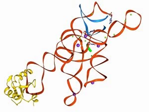
Background imageRibonucleic Acid Collection: Ribonuclease A molecule F006 / 9768

Ribonuclease A molecule F006 / 9768
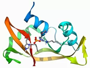
Ribonuclease A (RNAse A), molecular model. Ribonuclease (RNase) is a type of nuclease that catalyses the degradation of RNA (ribonucleic acid)

Background imageRibonucleic Acid Collection: tRNA molecule F006 / 9764

tRNA molecule F006 / 9764
Transfer RNA (tRNA), molecular model. tRNA (transfer ribonucleic acid) translates messenger RNA (mRNA) into a protein product

Background imageRibonucleic Acid Collection: Marburg viral protein 35 and RNA F006 / 9759

Marburg viral protein 35 and RNA F006 / 9759
Marburg viral protein 35 and RNA. Molecular model of the Marburg viral protein 35 (VP35) bound to a molecule of double stranded RNA (ribonucleic acid)

Background imageRibonucleic Acid Collection: RNA triplet repeat expansion F006 / 9749

RNA triplet repeat expansion F006 / 9749
RNA triplet repeat expansion. Molecular model of a CUG triplet repeat expansion in a molecule of double stranded RNA (ribonucleic acid)

Background imageRibonucleic Acid Collection: Iron-regulatory protein bound to RNA F006 / 9727

Iron-regulatory protein bound to RNA F006 / 9727
Iron-regulatory protein bound to RNA, molecular model. Iron regulatory protein 1 (IRP1, purple) bound to a short strand of RNA (ribonucleic acid, red) that includes iron-responsive elements (IREs)

Background imageRibonucleic Acid Collection: Ebola viral protein 35 and RNA F006 / 9697

Ebola viral protein 35 and RNA F006 / 9697
Ebola viral protein 35 and RNA. Molecular model of the Ebola viral protein 35 (VP35) bound to a molecule of double stranded RNA (ribonucleic acid)

Background imageRibonucleic Acid Collection: mRNA capping apparatus F006 / 9694

mRNA capping apparatus F006 / 9694
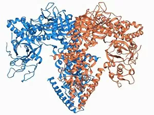
mRNA capping apparatus. Molecular model of the Cet-1-Ceg1 mRNA capping apparatus

Background imageRibonucleic Acid Collection: Toll-like receptor 3 and RNA F006 / 9666

Toll-like receptor 3 and RNA F006 / 9666
Toll-like receptor 3 and RNA. Molecular model of the toll-like receptor 3 (TLR3) protein (pink and blue) bound to a strand of RNA (ribonucleic acid, green and yellow)

Background imageRibonucleic Acid Collection: 70S ribosome, molecular model F006 / 9651

70S ribosome, molecular model F006 / 9651
70S ribosome, molecular model. Ribosomes are composed of protein and RNA (ribonucleic acid). In bacteria each ribosome consists of a small (30S) subunit and a large (50S) subunit

Background imageRibonucleic Acid Collection: SelB elongation factor bound to RNA F006 / 9648

SelB elongation factor bound to RNA F006 / 9648
SelB elongation factor bound to RNA. Molecular model of the SelB elongation factor bound to an mRNA (messenger ribonucleic acid) hairpin formed by the selenocysteine insertion sequence (SECIS)

Background imageRibonucleic Acid Collection: 70S ribosome, molecular model F006 / 9638

70S ribosome, molecular model F006 / 9638
70S ribosome. Molecular model of a 70S ribosome complex containing a Shine-Dalgarno helix, the point of mRNA (messenger ribonucleic acid) binding

Background imageRibonucleic Acid Collection: Metal-sensing RNA molecule F006 / 9636

Metal-sensing RNA molecule F006 / 9636
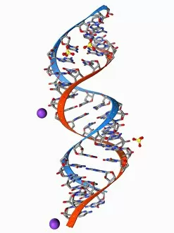
Metal-sensing RNA molecule. Molecular model of an M-box riboswitch, a length of RNA (ribonucleic acid) that regulates levels of metal ions in a cell

Background imageRibonucleic Acid Collection: PolyA polymerase and RNA F006 / 9635

PolyA polymerase and RNA F006 / 9635
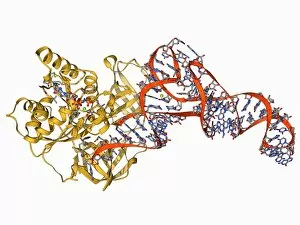
Poly(A) polymerase and RNA. Molecular model of poly(A) polymerase complexed with RNA (ribonucleic acid) and ATP (adenosine triphosphate)

Background imageRibonucleic Acid Collection: Internal ribosome entry site F006 / 9631

Internal ribosome entry site F006 / 9631
Internal ribosome entry site. Molecular model of an internal ribosome entry site nucleotide sequence from the hepatitis C virus. This sequence is essential for the initiation of viral translation

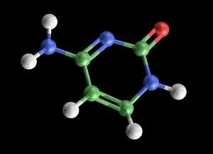
Background imageRibonucleic Acid Collection: Cytosine molecule, artwork C017 / 7214

Cytosine molecule, artwork C017 / 7214
Cytosine molecule. Computer artwork showing the structure of a molecule of the nucleobase cytosine (2-oxy-4-aminopyrimidine)

Background imageRibonucleic Acid Collection: RNA exosome complex, molecular model F006 / 9620

RNA exosome complex, molecular model F006 / 9620
RNA exosome complex, molecular model. This multi-protein complex functions to break up strands of RNA (ribonucleic acid, pink) during biochemical processes

Background imageRibonucleic Acid Collection: RNA editing enzyme F006 / 9615

RNA editing enzyme F006 / 9615
RNA editing enzyme, molecular model. This enzyme binds to double-stranded RNA (ribonucleic acid)

Background imageRibonucleic Acid Collection: RNA-dependent RNA polymerase molecule F006 / 9611

RNA-dependent RNA polymerase molecule F006 / 9611
RNA-dependent RNA polymerase, molecular model. This enzyme catalyses the replication of RNA (ribonucleic acid) from an RNA template

Background imageRibonucleic Acid Collection: Viral RNA packaging signal complex F006 / 9609

Viral RNA packaging signal complex F006 / 9609
Viral RNA packaging signal complex. Molecular model of the muPsi RNA packaging signal complex from the Rous sarcoma vuris

Background imageRibonucleic Acid Collection: RNA-editing enzyme, molecular model F006 / 9599

RNA-editing enzyme, molecular model F006 / 9599
RNA-editing enzyme. Molecular model of a left-handed, RNA double helix (Z-RNA, centre) bound by the Z alpha domain of the human RNA-editing enzyme ADAR1 (double-stranded RNA adenosine deaminase)

Background imageRibonucleic Acid Collection: Ribonuclease bound to transfer RNA F006 / 9591

Ribonuclease bound to transfer RNA F006 / 9591
Ribonuclease bound to transfer RNA, molecular model. This complex consists of the ribonuclease Z (RNase Z, green and pink) enzyme bound to a transfer RNA (tRNA) molecule (orange and blue)

Background imageRibonucleic Acid Collection: RNA-induced silencing complex F006 / 9587

RNA-induced silencing complex F006 / 9587
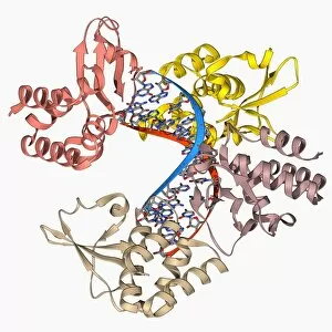
RNA-induced silencing complex (RISC), molecular model. This complex consists of a bacterial argonaute protein (top right) bound to a small interfering RNA (siRNA) molecule (red and blue)

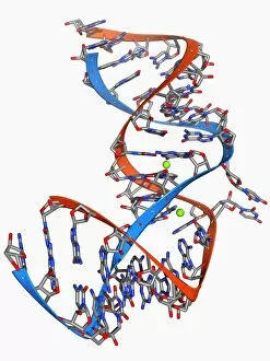
Background imageRibonucleic Acid Collection: Double-stranded RNA-ribonuclease III F006 / 9585

Double-stranded RNA-ribonuclease III F006 / 9585
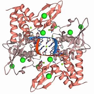
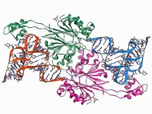
Double-stranded RNA-ribonuclease III. Molecular model of ribonuclease III (RNase III, D44N, pink and green) complexed with a double-stranded RNA (ribonucleic acid) strand (red and blue)

Background imageRibonucleic Acid Collection: RNA stem-loop motif, molecular model F006 / 9544

RNA stem-loop motif, molecular model F006 / 9544
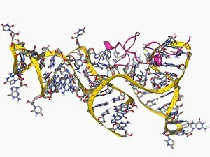
RNA stem-loop motif. Molecular model of the stem-loop II motif from the SARS (severe acute respiratory syndrome) coronavirus. This RNA (ribonucleic acid) element is a target for antiviral drugs

Background imageRibonucleic Acid Collection: Elongation factor Tu and tRNA F006 / 9522

Elongation factor Tu and tRNA F006 / 9522
Elongation factor Tu bound to tRNA (transfer ribonucleic acid), molecular model. This enzyme is involved in the elongation of polypeptide chains during translation

Background imageRibonucleic Acid Collection: Transcription factor and ribosomal RNA F006 / 9530

Transcription factor and ribosomal RNA F006 / 9530
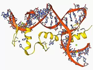
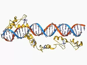
Transcription factor and ribosomal RNA (rRNA). Molecular model showing the 6 zinc fingers of transcription factor IIIA (yellow) bound to RNA (ribonucleic acid)

Background imageRibonucleic Acid Collection: Self-splicing RNA intron, molecular model F006 / 9527

Self-splicing RNA intron, molecular model F006 / 9527
Self-splicing RNA intron, molecular model. Splicing is the process where a non-coding fragment (intron) of a strand of nucleic acid (DNA, deoxyribonucleic acid; or RNA, ribonucleic acid) is removed

Background imageRibonucleic Acid Collection: Transcription factor and ribosomal RNA F006 / 9516

Transcription factor and ribosomal RNA F006 / 9516
Transcription factor and ribosomal RNA (rRNA). Molecular model showing the 6 zinc fingers of transcription factor IIIA (yellow) bound to RNA (ribonucleic acid)

Background imageRibonucleic Acid Collection: RNA-induced silencing complex F006 / 9502

RNA-induced silencing complex F006 / 9502
RNA-induced silencing complex (RISC), molecular model. This complex consists of a bacterial argonaute protein bound to a small interfering RNA (siRNA) molecule (red and blue)

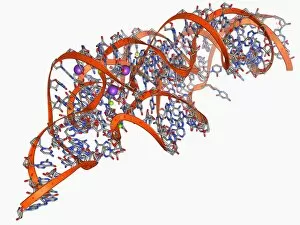
Background imageRibonucleic Acid Collection: Hammerhead ribozyme molecule F006 / 9492

Hammerhead ribozyme molecule F006 / 9492
Hammerhead ribozyme, molecular model. Ribozymes are RNA (ribonucleic acid) molecules that catalyse certain biochemical reactions

Background imageRibonucleic Acid Collection: RNA interference viral suppressor and RNA F006 / 9488

RNA interference viral suppressor and RNA F006 / 9488
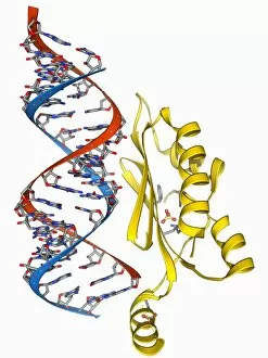
RNA interference viral suppressor and RNA. Molecular model of the p19 protein (yellow) from a Tombusvirus, suppressing a double-stranded, small interfering RNA (siRNA) molecule (red and blue)


For sale as Licensed Images

Choose your image, Select your licence and Download the media

"Unraveling the Secrets of Ribonucleic Acid: The Double-Stranded RNA Molecule" In the intricate world of molecular biology, ribonucleic acid (RNA) takes center stage as a vital player in various biological processes. This captivating molecule, often overshadowed by its famous cousin DNA, holds immense potential and complexity. DNA transcription sets the stage for RNA's crucial role. As a double-stranded RNA molecule unwinds, it serves as a template to synthesize single-stranded messenger RNA (mRNA), carrying genetic information from the nucleus to the cytoplasm. A mesmerizing molecular model showcases this elegant dance of transcription. Within bacterial ribosomes, another fascinating aspect unfolds. These cellular factories decode mRNA sequences into proteins through translation—a fundamental process that sustains life itself. Peering into their microscopic world reveals an awe-inspiring view of these tiny machines at work. But not all encounters with RNA are beneficial; some bring about disease-causing agents like human respiratory syncytial virus or paramyxovirus particles. Through electron microscopy, we witness their hauntingly beautiful structures—reminders of nature's delicate balance between beauty and danger. Electrophoresis techniques allow scientists to analyze and separate different types of RNAs based on size and charge—an invaluable tool in unraveling their mysteries. Such experiments reveal intriguing patterns under UV light that hint at hidden secrets within these molecules' structure and function. The realm of RNA extends beyond mere replication; it undergoes editing too. Molecular models showcase specialized enzymes responsible for altering specific nucleotides within an RNA sequence—a testament to nature's ingenuity in fine-tuning genetic information. Ribonucleases further highlight the multifaceted nature of RNAs—their ability to degrade both RNA-DNA hybrids and pure forms with precision is truly remarkable. Visualizing this interaction provides insights into how cells regulate gene expression through controlled degradation mechanisms.