Ribonucleic Acid Collection (page 5)

"Unraveling the Secrets of Ribonucleic Acid: The Double-Stranded RNA Molecule" In the intricate world of molecular biology

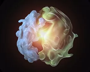
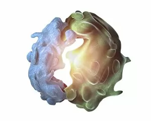
Background imageRibonucleic Acid Collection: Ribosome, artwork C015 / 6780

Ribosome, artwork C015 / 6780
Computer artwork of a ribosome. Ribosomes are protein particles that are found in cell cytoplasm. Each ribosome has a large and a small subunit

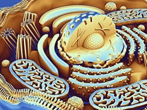
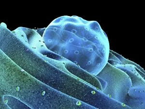
Background imageRibonucleic Acid Collection: Animal cell organelles, artwork C015 / 6775

Animal cell organelles, artwork C015 / 6775
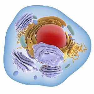
Animal cell organelles. Computer artwork showing the organelles in a eukaryotic cell. This is an animal cell. Structures include the nucleus (centre) which has a membrane with nuclear pores
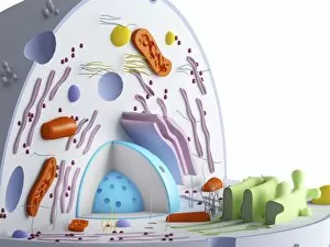
Background imageRibonucleic Acid Collection: Animal cell organelles, artwork C015 / 6778

Animal cell organelles, artwork C015 / 6778
Animal cell organelles. Computer artwork showing the organelles in a eukaryotic cell. This is an animal cell. Structures include the nucleus (centre) which has a membrane with nuclear pores

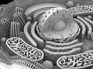
Background imageRibonucleic Acid Collection: Animal cell organelles, artwork C015 / 6777

Animal cell organelles, artwork C015 / 6777
Animal cell organelles. Computer artwork showing the organelles in a eukaryotic cell. This is an animal cell. Structures include the nucleus (centre) which has a membrane with nuclear pores

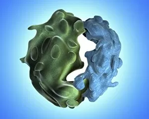
Background imageRibonucleic Acid Collection: Ribosome, artwork C015 / 6774

Ribosome, artwork C015 / 6774
Computer artwork of a ribosome. Ribosomes are protein particles that are found in cell cytoplasm. Each ribosome has a large and a small subunit

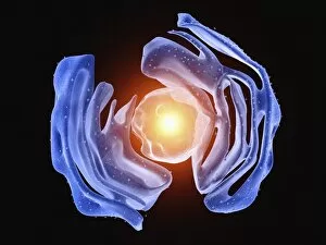
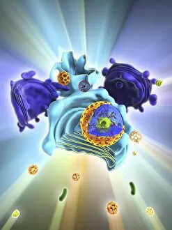
Background imageRibonucleic Acid Collection: Nucleus and endoplasmic reticulum C015 / 6772

Nucleus and endoplasmic reticulum C015 / 6772
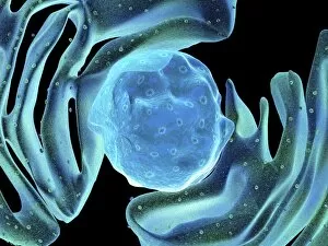
Computer artwork showing part of a human or eukaryotic cell. In the middle the nucleus which has a membrane with nuclear pores. Inside the nucleus is the DNA

Background imageRibonucleic Acid Collection: Ribosome, artwork C015 / 6769

Ribosome, artwork C015 / 6769
Computer artwork of a ribosome. Ribosomes are protein particles that are found in cell cytoplasm. Each ribosome has a large and a small subunit

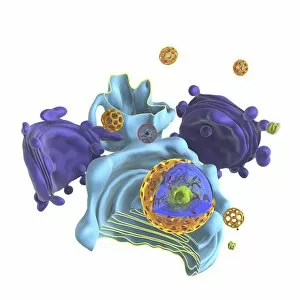
Background imageRibonucleic Acid Collection: Nucleus and endoplasmic reticulum C015 / 6768

Nucleus and endoplasmic reticulum C015 / 6768
Computer artwork showing part of a human or eukaryotic cell. In the middle the nucleus which has a membrane with nuclear pores. Inside the nucleus is the DNA

Background imageRibonucleic Acid Collection: Nucleus and endoplasmic reticulum C015 / 6767

Nucleus and endoplasmic reticulum C015 / 6767
Computer artwork showing part of a human or eukaryotic cell. In the middle the nucleus which has a membrane with nuclear pores. Inside the nucleus is the DNA

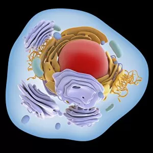
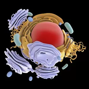
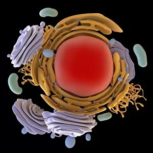
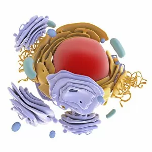
Background imageRibonucleic Acid Collection: Animal cell organelles, artwork C016 / 0619

Animal cell organelles, artwork C016 / 0619
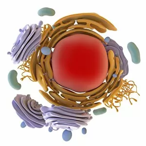
Computer artwork showing the organelles in a eukaryotic cell. This is an animal cell. Structures include the nucleus (centre, red) which has a membrane with nuclear pores

Background imageRibonucleic Acid Collection: Animal cell organelles, artwork C016 / 0621

Animal cell organelles, artwork C016 / 0621
Computer artwork showing the organelles in a eukaryotic cell. This is an animal cell. Structures include the nucleus (centre, red) which has a membrane with nuclear pores

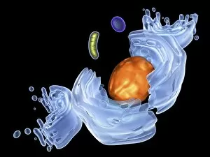
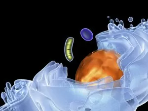
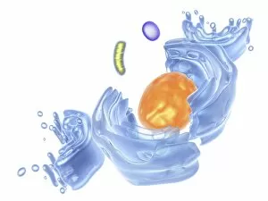
Background imageRibonucleic Acid Collection: Animal cell organelles, artwork C016 / 0620

Animal cell organelles, artwork C016 / 0620
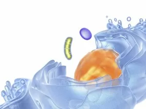
Computer artwork showing the organelles in a eukaryotic cell. This is an animal cell. Structures include the nucleus (centre, orange) which has a membrane with nuclear pores

Background imageRibonucleic Acid Collection: Animal cell organelles, artwork C016 / 0617

Animal cell organelles, artwork C016 / 0617
Computer artwork showing the organelles in a eukaryotic cell. This is an animal cell. Structures include the nucleus (centre, red) which has a membrane with nuclear pores

Background imageRibonucleic Acid Collection: Animal cell organelles, artwork C016 / 0618

Animal cell organelles, artwork C016 / 0618
Computer artwork showing the organelles in a eukaryotic cell. This is an animal cell. Structures include the nucleus (centre, orange) which has a membrane with nuclear pores

Background imageRibonucleic Acid Collection: Animal cell organelles, artwork C016 / 0615

Animal cell organelles, artwork C016 / 0615
Computer artwork showing the organelles in a eukaryotic cell. This is an animal cell. Structures include the nucleus (centre, orange) which has a membrane with nuclear pores

Background imageRibonucleic Acid Collection: Animal cell organelles, artwork C016 / 0616

Animal cell organelles, artwork C016 / 0616
Computer artwork showing the organelles in a eukaryotic cell. This is an animal cell. Structures include the nucleus (centre, orange) which has a membrane with nuclear pores

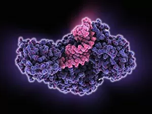
Background imageRibonucleic Acid Collection: Iron-regulatory protein bound to RNA C015 / 6691

Iron-regulatory protein bound to RNA C015 / 6691
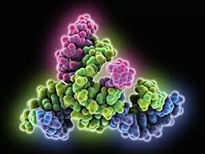
Iron-regulatory protein bound to RNA, molecular model. Iron regulatory protein 1 (IRP1, purple) bound to a short strand of RNA (ribonucleic acid, pink) that includes iron-responsive elements (IREs)

Background imageRibonucleic Acid Collection: Animal cell organelles, artwork C016 / 0611

Animal cell organelles, artwork C016 / 0611
Computer artwork showing the organelles in a eukaryotic cell. This is an animal cell. Structures include the nucleus (centre, red) which has a membrane with nuclear pores

Background imageRibonucleic Acid Collection: Iron-regulatory protein bound to RNA C015 / 6690

Iron-regulatory protein bound to RNA C015 / 6690
Iron-regulatory protein bound to RNA, molecular model. Iron regulatory protein 1 (IRP1, blue) bound to a short strand of RNA (ribonucleic acid, pink) that includes iron-responsive elements (IREs)

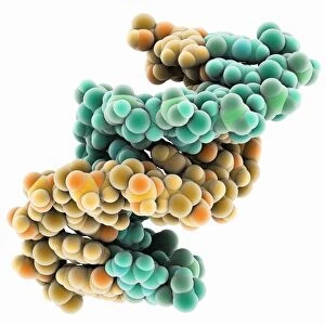
Background imageRibonucleic Acid Collection: DNA hybrid duplex, molecular model

DNA hybrid duplex, molecular model. This model shows a chimeric junction, where a DNA (deoxyribonucleic acid) strand changes from one form to another

Background imageRibonucleic Acid Collection: Animal cell organelles, artwork C016 / 0612

Animal cell organelles, artwork C016 / 0612
Computer artwork showing the organelles in a eukaryotic cell. This is an animal cell. Structures include the nucleus (centre, orange) which has a membrane with nuclear pores

Background imageRibonucleic Acid Collection: Animal cell organelles, artwork C016 / 0605

Animal cell organelles, artwork C016 / 0605
Computer artwork showing the organelles in a eukaryotic cell. This is an animal cell. Structures include the nucleus (centre, red) which has a membrane with nuclear pores

Background imageRibonucleic Acid Collection: Animal cell organelles, artwork C016 / 0610

Animal cell organelles, artwork C016 / 0610
Computer artwork showing the organelles in a eukaryotic cell. This is an animal cell. Structures include the nucleus (centre, orange) which has a membrane with nuclear pores

Background imageRibonucleic Acid Collection: Animal cell organelles, artwork C016 / 0604

Animal cell organelles, artwork C016 / 0604
Computer artwork showing the organelles in a eukaryotic cell. This is an animal cell. Structures include the nucleus (centre, red) which has a membrane with nuclear pores

Background imageRibonucleic Acid Collection: Animal cell organelles, artwork C016 / 0606

Animal cell organelles, artwork C016 / 0606
Computer artwork showing the organelles in a eukaryotic cell. This is an animal cell. Structures include the nucleus (centre, orange) which has a membrane with nuclear pores

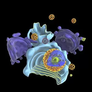
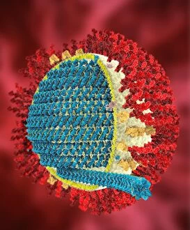
Background imageRibonucleic Acid Collection: Influenza virus, artwork C018 / 2894

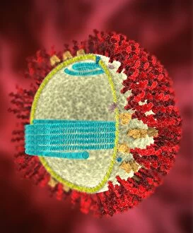
Influenza virus, artwork C018 / 2894
Influenza virus. Cut-away computer artwork of an influenza (flu) virus particle (virion). In each particles lipid envelope (blue) are two types of protein spike

Background imageRibonucleic Acid Collection: Influenza virus, artwork C018 / 2893

Influenza virus, artwork C018 / 2893
Influenza virus. Cut-away computer artwork of an influenza (flu) virus particle (virion). In each particles lipid envelope (blue) are two types of protein spike

Background imageRibonucleic Acid Collection: Influenza virus, artwork C018 / 2891

Influenza virus, artwork C018 / 2891
Influenza virus. Cut-away computer artwork of an influenza (flu) virus particle (virion). In each particles lipid envelope (blue) are two types of protein spike

Background imageRibonucleic Acid Collection: Influenza virus, artwork C018 / 2890

Influenza virus, artwork C018 / 2890
Influenza virus. Cut-away computer artwork of an influenza (flu) virus particle (virion). In each particles lipid envelope (green) are two types of protein spike

Background imageRibonucleic Acid Collection: Influenza virus, artwork C018 / 2892

Influenza virus, artwork C018 / 2892
Influenza virus. Cut-away computer artwork of an influenza (flu) virus particle (virion). In each particles lipid envelope (brown) are two types of protein spike

Background imageRibonucleic Acid Collection: VSIV virus protein complex C015 / 6423

VSIV virus protein complex C015 / 6423
VSIV virus protein complex, molecular model. This decameric (10-part) circular structure is a complex of nucleoproteins (nucleocapsid protein)

Background imageRibonucleic Acid Collection: VSIV virus protein complex C015 / 6422

VSIV virus protein complex C015 / 6422
VSIV virus protein complex, molecular model. This decameric (10-part) circular structure is a complex of nucleoproteins (nucleocapsid protein)

Background imageRibonucleic Acid Collection: Viral RNA interference complex C015 / 5356

Viral RNA interference complex C015 / 5356
Viral RNA interference complex, molecular model. Small interfering RNA (siRNA) complexed with tomato aspermy virus 2b protein (TAV2b)

Background imageRibonucleic Acid Collection: Viral RNA interference complex C015 / 5355

Viral RNA interference complex C015 / 5355
Viral RNA interference complex, molecular model. Small interfering RNA (siRNA) complexed with tomato aspermy virus 2b protein (TAV2b)

Background imageRibonucleic Acid Collection: Avian influenza virus, TEM C016 / 5843

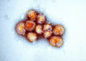
Avian influenza virus, TEM C016 / 5843
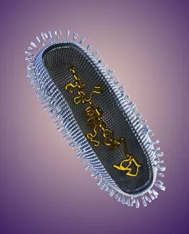
Avian influenza virus, type A strain H7N9, coloured transmission electron micrograph (TEM). This virus first emerged in the human population in China, in March 2013

Background imageRibonucleic Acid Collection: Avian influenza virus, TEM C016 / 5841

Avian influenza virus, TEM C016 / 5841
Avian influenza virus, type A strain H7N9, coloured transmission electron micrograph (TEM). This virus first emerged in the human population in China, in March 2013

Background imageRibonucleic Acid Collection: Avian influenza virus, TEM C016 / 5842

Avian influenza virus, TEM C016 / 5842
Avian influenza virus, type A strain H7N9, coloured transmission electron micrograph (TEM). This virus first emerged in the human population in China, in March 2013

Background imageRibonucleic Acid Collection: MERS coronavirus, artwork C016 / 3611

MERS coronavirus, artwork C016 / 3611
MERS coronavirus proteins. Cutaway computer model showing the protein structure of a MERS coronavirus particle (virion). MERS (Middle East respiratory syndrome)

Background imageRibonucleic Acid Collection: SARS coronavirus proteins, artwork C016 / 3056

SARS coronavirus proteins, artwork C016 / 3056
SARS coronavirus proteins. Computer model showing the spike proteins (red) of a SARS coronavirus particle (virion). SARS (severe acute respiratory syndrome)

Background imageRibonucleic Acid Collection: SARS coronavirus, artwork C016 / 3054

SARS coronavirus, artwork C016 / 3054
SARS coronavirus proteins. Cutaway computer model showing the protein structure of a SARS coronavirus particle (virion). SARS (severe acute respiratory syndrome)

Background imageRibonucleic Acid Collection: SARS coronavirus, artwork C016 / 3053

SARS coronavirus, artwork C016 / 3053
SARS coronavirus proteins. Cutaway computer model showing the protein structure of a SARS coronavirus particle (virion). SARS (severe acute respiratory syndrome)

Background imageRibonucleic Acid Collection: SARS coronavirus proteins, artwork C016 / 3052

SARS coronavirus proteins, artwork C016 / 3052
SARS coronavirus proteins. Computer model showing the proteins of a SARS coronavirus particle (virion). SARS (severe acute respiratory syndrome)

Background imageRibonucleic Acid Collection: Ribozyme enzyme and RNA C016 / 2829

Ribozyme enzyme and RNA C016 / 2829
Ribozyme enzyme and RNA, molecular model. Ribozymes are RNA (ribonucleic acid) molecules that catalyse certain biochemical reactions

Background imageRibonucleic Acid Collection: Ribozyme enzyme and RNA C016 / 2828

Ribozyme enzyme and RNA C016 / 2828
Ribozyme enzyme and RNA, molecular model. Ribozymes are RNA (ribonucleic acid) molecules that catalyse certain biochemical reactions

Background imageRibonucleic Acid Collection: RNA-silencing protein with RNA C016 / 2558

RNA-silencing protein with RNA C016 / 2558
RNA-silencing protein with RNA. Molecular model of RNA silencing taking place by human piwi-like protein (purple) acting on short loops (green) of RNA (ribonucleic acid)

Background imageRibonucleic Acid Collection: RNA-silencing protein with RNA C016 / 2557

RNA-silencing protein with RNA C016 / 2557
RNA-silencing protein with RNA. Molecular model of RNA silencing taking place by human piwi-like protein (green) acting on short loops (red) of RNA (ribonucleic acid)

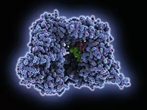
Background imageRibonucleic Acid Collection: RNA polymerase molecule C016 / 2391

RNA polymerase molecule C016 / 2391
RNA polymerase. Molecular model of RNA polymerase (blue and purple) transcribing a strand of mRNA (messenger ribonucleic acid, centre) from a DNA (deoxyribonucleic acid) template (pink and purple)

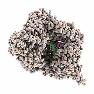
Background imageRibonucleic Acid Collection: RNA polymerase molecule C016 / 2390

RNA polymerase molecule C016 / 2390
RNA polymerase. Molecular model of RNA polymerase (beige and pink) transcribing a strand of mRNA (messenger ribonucleic acid, centre) from a DNA (deoxyribonucleic acid) template (pink and purple)


For sale as Licensed Images

Choose your image, Select your licence and Download the media

"Unraveling the Secrets of Ribonucleic Acid: The Double-Stranded RNA Molecule" In the intricate world of molecular biology, ribonucleic acid (RNA) takes center stage as a vital player in various biological processes. This captivating molecule, often overshadowed by its famous cousin DNA, holds immense potential and complexity. DNA transcription sets the stage for RNA's crucial role. As a double-stranded RNA molecule unwinds, it serves as a template to synthesize single-stranded messenger RNA (mRNA), carrying genetic information from the nucleus to the cytoplasm. A mesmerizing molecular model showcases this elegant dance of transcription. Within bacterial ribosomes, another fascinating aspect unfolds. These cellular factories decode mRNA sequences into proteins through translation—a fundamental process that sustains life itself. Peering into their microscopic world reveals an awe-inspiring view of these tiny machines at work. But not all encounters with RNA are beneficial; some bring about disease-causing agents like human respiratory syncytial virus or paramyxovirus particles. Through electron microscopy, we witness their hauntingly beautiful structures—reminders of nature's delicate balance between beauty and danger. Electrophoresis techniques allow scientists to analyze and separate different types of RNAs based on size and charge—an invaluable tool in unraveling their mysteries. Such experiments reveal intriguing patterns under UV light that hint at hidden secrets within these molecules' structure and function. The realm of RNA extends beyond mere replication; it undergoes editing too. Molecular models showcase specialized enzymes responsible for altering specific nucleotides within an RNA sequence—a testament to nature's ingenuity in fine-tuning genetic information. Ribonucleases further highlight the multifaceted nature of RNAs—their ability to degrade both RNA-DNA hybrids and pure forms with precision is truly remarkable. Visualizing this interaction provides insights into how cells regulate gene expression through controlled degradation mechanisms.