
Ribonucleic Acid Collection (page 4)
"Unraveling the Secrets of Ribonucleic Acid: The Double-Stranded RNA Molecule" In the intricate world of molecular biology


For sale as Licensed Images
Choose your image, Select your licence and Download the media
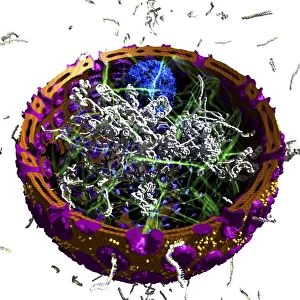
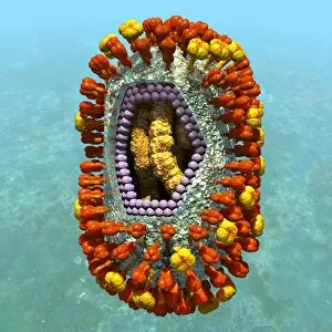
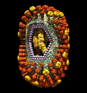
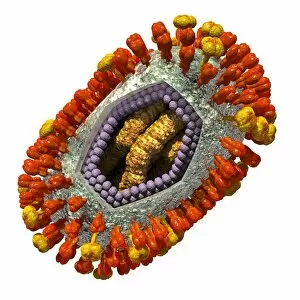
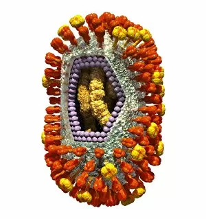
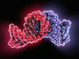
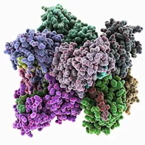
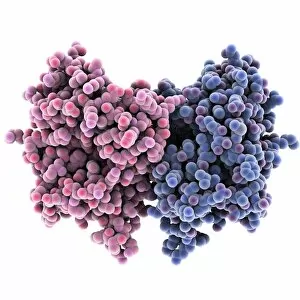
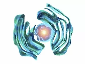
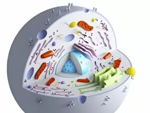
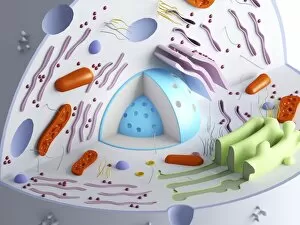
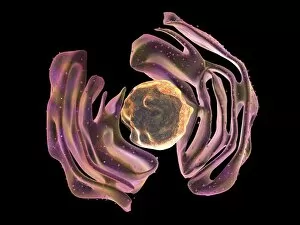
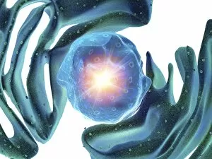
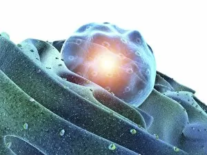
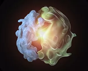
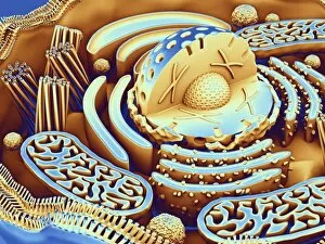
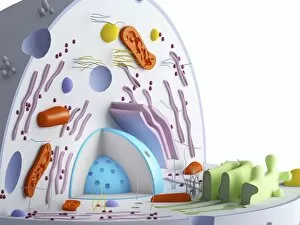
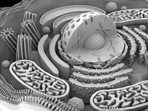
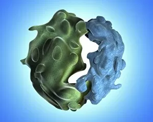
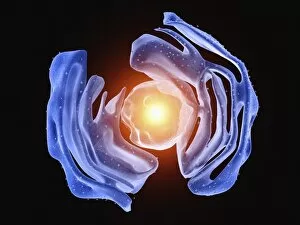
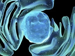
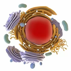
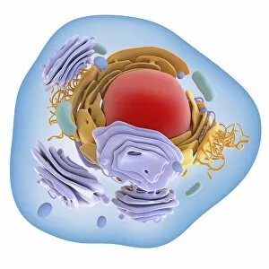
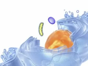
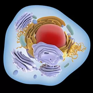
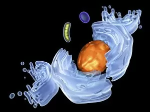
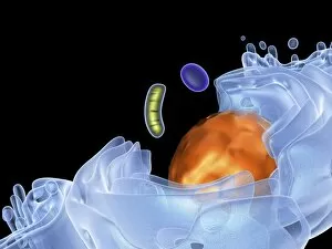
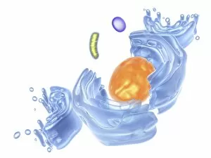
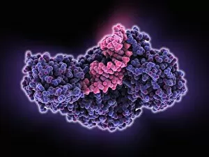
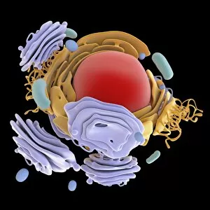
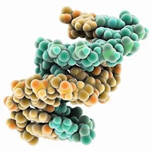
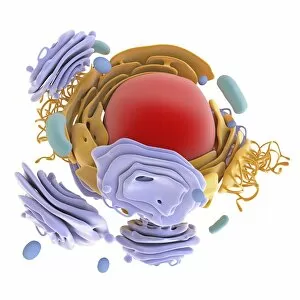
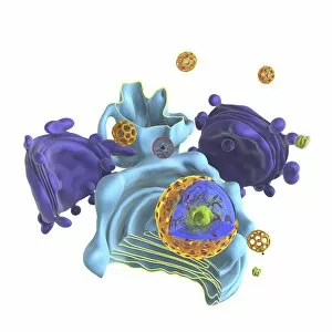
"Unraveling the Secrets of Ribonucleic Acid: The Double-Stranded RNA Molecule" In the intricate world of molecular biology, ribonucleic acid (RNA) takes center stage as a vital player in various biological processes. This captivating molecule, often overshadowed by its famous cousin DNA, holds immense potential and complexity. DNA transcription sets the stage for RNA's crucial role. As a double-stranded RNA molecule unwinds, it serves as a template to synthesize single-stranded messenger RNA (mRNA), carrying genetic information from the nucleus to the cytoplasm. A mesmerizing molecular model showcases this elegant dance of transcription. Within bacterial ribosomes, another fascinating aspect unfolds. These cellular factories decode mRNA sequences into proteins through translation—a fundamental process that sustains life itself. Peering into their microscopic world reveals an awe-inspiring view of these tiny machines at work. But not all encounters with RNA are beneficial; some bring about disease-causing agents like human respiratory syncytial virus or paramyxovirus particles. Through electron microscopy, we witness their hauntingly beautiful structures—reminders of nature's delicate balance between beauty and danger. Electrophoresis techniques allow scientists to analyze and separate different types of RNAs based on size and charge—an invaluable tool in unraveling their mysteries. Such experiments reveal intriguing patterns under UV light that hint at hidden secrets within these molecules' structure and function. The realm of RNA extends beyond mere replication; it undergoes editing too. Molecular models showcase specialized enzymes responsible for altering specific nucleotides within an RNA sequence—a testament to nature's ingenuity in fine-tuning genetic information. Ribonucleases further highlight the multifaceted nature of RNAs—their ability to degrade both RNA-DNA hybrids and pure forms with precision is truly remarkable. Visualizing this interaction provides insights into how cells regulate gene expression through controlled degradation mechanisms.