Secondary Structure Collection (page 6)

Secondary structure refers to the intricate folding patterns that proteins and nucleic acids adopt, playing a crucial role in their functionality

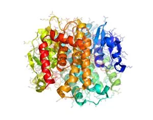
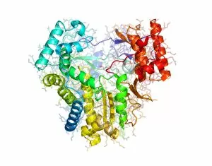
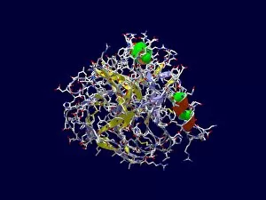
Background imageSecondary Structure Collection: GST enzyme conferring DDT resistance

GST enzyme conferring DDT resistance
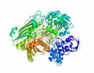
Glutathione S-transferase (GST) enzyme from a malaria-carrying strain of mosquito, Anopheles gambiae, molecular model. GSTs detoxify foreign substances in the body of the mosquito

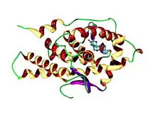
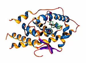
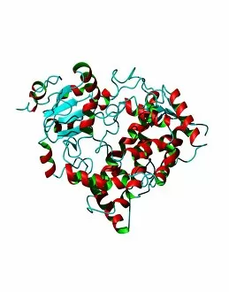
Background imageSecondary Structure Collection: Testosterone bound to receptor, artwork

Testosterone bound to receptor, artwork
Testosterone bound to receptor. Molecular model of a molecule of the male sex hormone testosterone (ball-and-stick primary structure) bound to a human androgen receptor (secondary structure)

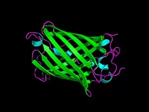
Background imageSecondary Structure Collection: Green fluorescent protein, computer model

Green fluorescent protein, computer model. This protein is found in the jellyfish Aequorea victoria. When ultraviolet or blue light shines on the protein, it emits green light

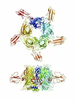
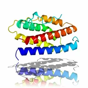
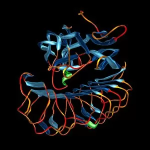
Background imageSecondary Structure Collection: Cobra venom action, molecular model

Cobra venom action, molecular model
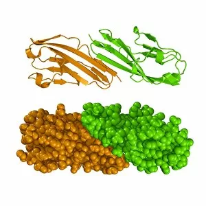
Cobra (Naja sp.) venom action. Molecular model showing top (upper centre) and side (lower centre) views of the secondary structure of an alpha-cobratoxin (snake venom protein)

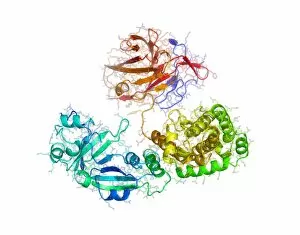
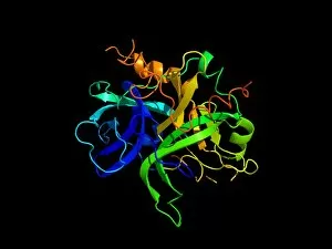
Background imageSecondary Structure Collection: RNA polymerase from Norwalk virus

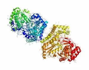
RNA polymerase from Norwalk virus
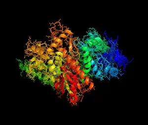
RNA polymerase enzyme from Norwalk virus, molecular model. This enzyme makes RNA (ribonucleic acid) from the RNA template within the virus

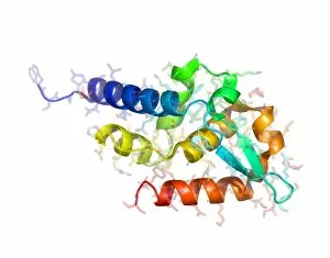
Background imageSecondary Structure Collection: RNA polymerase from rabies virus

RNA polymerase from rabies virus, molecular model. This is a single subunit from an enzyme, involved in the replication of the rabies viruss RNA after it has infected a host cell

Background imageSecondary Structure Collection: Interleukin-6, molecular model

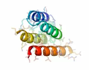
Interleukin-6, molecular model
Interleukin-6. Molecular model of the secondary structure of the cytokine protein interleukin-6. This protein is produced in the body and has a wide variety of functions in the immune system

Background imageSecondary Structure Collection: Phosphofructokinase bacterial enzyme

Phosphofructokinase bacterial enzyme
Phosphofructokinase enzyme, molecular model. This enzyme is from the spirochaete bacteria Borrelia burgdorferi, which causes Lyme disease

Background imageSecondary Structure Collection: Diphtheria toxin structure

Diphtheria toxin structure
Diphtheria toxin, molecular model. This model shows the toxin produced by the bacterium Corynebacterium diphtheriae, the cause of diphtheria

Background imageSecondary Structure Collection: Hepatitis C virus RNA polymerase enzyme

Hepatitis C virus RNA polymerase enzyme
Hepatitis C virus enzyme, molecular model. This protein, called NS5B, forms the active site of the viruss RNA-dependent RNA polymerase enzyme

Background imageSecondary Structure Collection: TGN1412 drug molecule

TGN1412 drug molecule. Computer model showing the secondary structure of the drug TGN1412. This drug was shown to have serious side effects during a clinical trial in the UK in 2006 when it caused

Background imageSecondary Structure Collection: Protein from measles virus

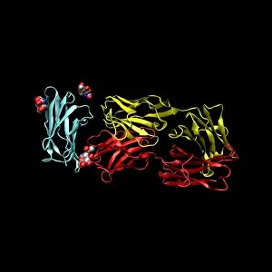
Protein from measles virus
Proteins from the measles virus, molecular model. A domain of the viruss P protein (upper, blue, green and yellow) is seen here in complex with part of the N protein (lower, red)

Background imageSecondary Structure Collection: Phenylbutazone anti-inflammatory molecule

Phenylbutazone anti-inflammatory molecule
Phenylbutazone anti-inflammatory drug. Molecular model showing the secondary structure of the non-steroidal anti-inflammatory drug (NSAID) phenylbutazone

Background imageSecondary Structure Collection: THG anabolic steroid and receptor

THG anabolic steroid and receptor
THG bound to receptor. Molecular model of a molecule of the anabolic steroid drug tetrahydrogestrinone (THG, ball-and-stick primary structure) bound to a human androgen receptor (secondary structure)

Background imageSecondary Structure Collection: HIV antibody therapy

HIV antibody therapy. Molecular model of the interaction of the HIV surface protein gp120 (green, lower right) as it interacts with a human white blood cell surface protein (CD4, blue)
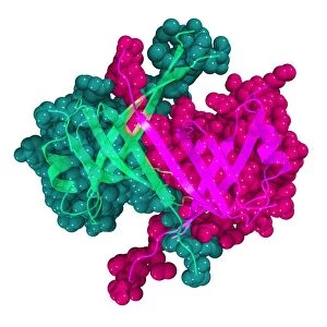
Background imageSecondary Structure Collection: Cholesterol enzyme affected by a drug

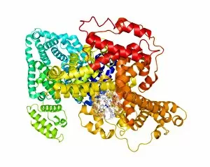
Cholesterol enzyme affected by a drug
Cholesterol enzyme being affected by a drug. Molecular model of the shape of the human enzyme Hmg-Coa reductase interacting (complexed) with the anti-cholesterol drug Fluvastatin (not seen)

Background imageSecondary Structure Collection: HIV enzyme being affected by a drug

HIV enzyme being affected by a drug. Molecular model of HIVs reverse transcriptase enzyme as it interacts with a drug (not seen)

Background imageSecondary Structure Collection: Follicle stimulating hormone molecule

Follicle stimulating hormone molecule. Computer artwork showing the secondary structure of a molecule of follicle stimulating hormone (FSH, bottom) and its receptor (FSHR, top)

Background imageSecondary Structure Collection: Erythropoietin molecule bound to receptor

Erythropoietin molecule bound to receptors. Computer model of a molecule of erythropoietin (EPO) (orange) bound to two extracellular EPO receptors (pink and purple)

Background imageSecondary Structure Collection: Insulin hormone, molecular model

Insulin hormone, molecular model
Insulin hormone. Molecular model of the bovine form of the hormone insulin, produced by the pancreas in mammals to aid the body in metabolising sugars

Background imageSecondary Structure Collection: Thrombin protein, secondary structure

Thrombin protein, secondary structure
Thrombin protein, computer model. Thrombin is a protein involved in the blood coagulation (clotting) process. It converts fibrinogen (a soluble plasma glycoprotein synthesised in the liver)

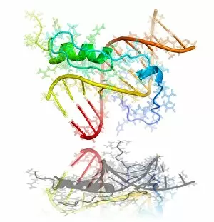
Background imageSecondary Structure Collection: GAGA transcription factor molecule

GAGA transcription factor molecule. Molecular model showing the primary (rods) and secondary (helices) structure of GAGA factor (green and blue)

Background imageSecondary Structure Collection: Chinese scorpion toxin structure

Chinese scorpion toxin structure. Molecular model showing the crystal structure of toxin BmBKTtx1, produced by the Chinese scorpion (Buthus martensi Karsch)

Background imageSecondary Structure Collection: NovoSeven clotting protein molecule

NovoSeven clotting protein molecule
NovoSeven clotting protein, molecular model. NovoSeven is the brand name for an artificial version of the natural blood protein Factor VII, or proconvertin

Background imageSecondary Structure Collection: Cytochrome P450 protein, molecular model

Cytochrome P450 protein, molecular model. This protein plays a crucial role in metabolism in animals (including humans), fungi, plants and bacteria

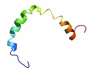
Background imageSecondary Structure Collection: Section of human apolipoprotein A-I

Section of human apolipoprotein A-I, molecular model. This is a fragment of a high-density lipoprotein (HDL) molecule known as an apolipoprotein

Background imageSecondary Structure Collection: Integration host factor and DNA

Integration host factor and DNA. Computer model of integration host factor (IHF, centre) bound to a molecule of DNA (deoxyribonucleic acid, semi- circle). The secondary structure of IHF is shown

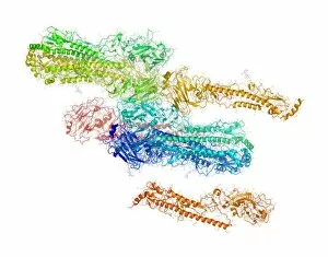
Background imageSecondary Structure Collection: C-reactive protein, molecular models

C-reactive protein, molecular models. Two molecules of the protein are seen. The protein is made up of five sub-units (monomers) arranged in a ring

Background imageSecondary Structure Collection: Human rhinovirus capsid proteins

Human rhinovirus capsid proteins, molecular model. These are proteins from the capsid (outer protein coat) of rhinovirus 14. Rhinoviruses are responsible for causing about 50% of common colds

Background imageSecondary Structure Collection: Haemagglutinin from bird flu virus

Haemagglutinin from bird flu virus, molecular model. This protein, H5, is found on the surface of the bird flu virus H5N1

Background imageSecondary Structure Collection: Lipase molecule

Lipase molecule. Computer model showing the secondary structure of lipase. Alpha-helices are blue and beta-sheets are purple

Background imageSecondary Structure Collection: DNA polymerase Klenow fragment

DNA polymerase Klenow fragment
Klenow fragment of DNA polymerase I. Computer model showing the secondary (alpha-helices and beta-sheets) and primary (ball-and-stick) structures of the Klenow, or large

Background imageSecondary Structure Collection: Protein tyrosine phosphatase molecule

Protein tyrosine phosphatase molecule. Computer model of the secondary structure of an intermediate form of protein tyrosine phosphatase. Beta-sheets are purple and alpha-helices are blue

Background imageSecondary Structure Collection: Enzyme from a sulphur-reducing bacterium

Enzyme from a sulphur-reducing bacterium. Molecular model of the enzyme aldehyde oxidoreductase from the Desulfovibrio gigas sulphur-reducing bacterium

Background imageSecondary Structure Collection: Yeast enzyme, molecular model

Yeast enzyme, molecular model
Yeast enzyme. Molecular model of an enzyme from Saccharomyces cerevisiae (Bakers yeast). This enzyme is 20S proteasome. A proteasome is a complex type of proteinase (protein-digesting enzyme)

Background imageSecondary Structure Collection: Fibroblast growth factor receptor 2

Fibroblast growth factor receptor 2 (FGFR2). Molecular models of the secondary structure (top) and the tertiary structure (bottom) of FGFR2

Background imageSecondary Structure Collection: Single stranded DNA-binding protein

Single stranded DNA-binding protein (SSBP). Molecular model showing the secondary and tertiary structures of a protein that binds to the single stranded DNA (deoxyribonucleic acid)

Background imageSecondary Structure Collection: Nerve growth factor, molecular model

Nerve growth factor, molecular model
Nerve growth factor. Molecular model showing the secondary structure of nerve growth factor (NGF). NGF is a small protein, which is involved in the growth

Background imageSecondary Structure Collection: Nitrogenase protein, molecular model

Nitrogenase protein, molecular model
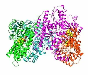
Nitrogenase protein. Molecular model of the MoFe protein, one of two proteins (MoFe and Fe) that combine to form the enzyme nitrogenase


For sale as Licensed Images

Choose your image, Select your licence and Download the media

Secondary structure refers to the intricate folding patterns that proteins and nucleic acids adopt, playing a crucial role in their functionality. In one captivating image, an anaesthetic inhibits an ion channel (C015/6718), highlighting how secondary structure impacts cellular processes. Another snapshot showcases DNA transcription, unveiling the molecular model of this essential process. Artwork depicting the secondary structure of proteins captivates our imagination as we marvel at the intricacy and beauty within each fold. The nucleosome molecule further emphasizes this complexity, showcasing how DNA wraps around histone proteins to form a compact structure. The bacterial ribosome stands tall as a testament to secondary structures' significance in protein synthesis. Meanwhile, the HIV reverse transcription enzyme reminds us of its vital role in converting viral RNA into DNA during infection. Molecular models provide insight into hepatitis C virus enzymes and interferon molecules—both critical players in disease progression and immune response modulation. Similarly, human growth hormone molecules hold immense importance for development and metabolism regulation. Exploring coagulation factor complex molecules (C014/0139) unravels mechanisms behind blood clotting—a process dependent on precise secondary structures working together seamlessly. Ghrelin hormone molecules intrigue us with their involvement in appetite regulation and energy balance maintenance. From anaesthetics influencing ion channels to hormones orchestrating bodily functions, understanding secondary structures unlocks countless mysteries within biological systems. These captivating images remind us of the intricate dance occurring at a microscopic level—the delicate folds dictating life's grand symphony.