Secondary Structure Collection (#2)

Secondary structure refers to the intricate folding patterns that proteins and nucleic acids adopt, playing a crucial role in their functionality

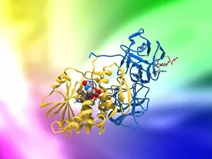
Background imageSecondary Structure Collection: Ricin molecule, artwork C017 / 3655

Ricin molecule, artwork C017 / 3655
Ricin molecule. Computer artwork showing the structure of a molecule of the toxic protein ricin. Ricin comprises two entwined amino acid chains; A (yellow) and B (blue)

Background imageSecondary Structure Collection: Blood clotting factor VIII molecule C014 / 0407

Blood clotting factor VIII molecule C014 / 0407
Blood clotting factor VIII, molecular model showing the secondary structure. Factor VIII, also known as anti-haemophilic factor, is an essential blood clotting protein in humans

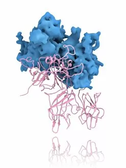
Background imageSecondary Structure Collection: Coagulation factor complex molecule C014 / 0409

Coagulation factor complex molecule C014 / 0409
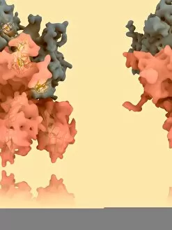
Coagulation factor complex molecule. Molecular model showing the interaction between coagulation factor VIII (FVIII, pink, blue and yellow), factor IXa (FIXa)

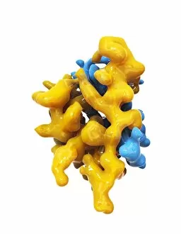
Background imageSecondary Structure Collection: Insulin molecule C014 / 2122

Insulin molecule C014 / 2122
Insulin, molecular module. Insulin is a hormone produced by the pancreas. It consists of two peptide chains, A (blue) and B (yellow), which are linked by disulphide bridges

Background imageSecondary Structure Collection: Blood clotting factor VIII molecule C014 / 0408

Blood clotting factor VIII molecule C014 / 0408
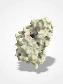
Blood clotting factor VIII, molecular model. Factor VIII, also known as anti-haemophilic factor, is an essential blood clotting protein in humans

Background imageSecondary Structure Collection: EcoRV restriction enzyme molecule C014 / 2113

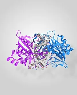
EcoRV restriction enzyme molecule C014 / 2113
EcoRV restriction enzyme. Molecular model of the type II restriction enzyme EcoRV (pink and blue) bound to a cleaved section of DNA (deoxyribonucleic acid, white)

Background imageSecondary Structure Collection: EcoRV restriction enzyme molecule C014 / 2118

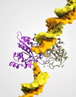
EcoRV restriction enzyme molecule C014 / 2118
EcoRV restriction enzyme. Molecular model of the type II restriction enzyme EcoRV (purple and beige) bound to a DNA molecule (deoxyribonucleic acid, yellow and orange)

Background imageSecondary Structure Collection: Coagulation factor complex molecule C014 / 0410

Coagulation factor complex molecule C014 / 0410
Coagulation factor complex molecule. Molecular model showing the interaction between coagulation factor VIII (FVIII, pink, blue and yellow), factor IXa (FIXa)

Background imageSecondary Structure Collection: Fibrinogen molecule C014 / 0472

Fibrinogen molecule C014 / 0472
Fibrinogen. Molecular model showing the secondary structure of the blood clotting glycoprotein fibrinogen (factor I). The molecule consists of two sets of alpha (grey)

Background imageSecondary Structure Collection: Stilbene synthase molecule C014 / 2292

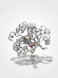
Stilbene synthase molecule C014 / 2292
Stilbene synthase, molecular model. This enzyme is produced by some plants (including grapes, peanuts and blueberries) in response to stress from either ultraviolet light or certain fungi

Background imageSecondary Structure Collection: Stilbene synthase molecule C014 / 2291

Stilbene synthase molecule C014 / 2291
Stilbene synthase, molecular model. This enzyme is produced by some plants (including grapes, peanuts and blueberries) in response to stress from either ultraviolet light or certain fungi

Background imageSecondary Structure Collection: UVR8 protein molecule C014 / 4913

UVR8 protein molecule C014 / 4913
UVR8 protein molecule. Computer model showing photoreception of UV-B (ultraviolet-B) light rays (white beam, left) by a UVR8 protein, whose secondary structure (purple ribbons) is shown

Background imageSecondary Structure Collection: Peptide YY obesity hormone molecule C014 / 4911

Peptide YY obesity hormone molecule C014 / 4911
Peptide YY obesity hormone molecule. Computer model showing the crystal structure of a molecule of the hormone peptide YY (PYY, or PYY3-36)

Background imageSecondary Structure Collection: Peptide YY obesity hormone molecule C014 / 4910

Peptide YY obesity hormone molecule C014 / 4910
Peptide YY obesity hormone molecule. Computer model showing the crystal structure of a molecule of the hormone peptide YY (PYY, or PYY3-36)

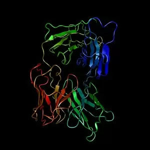
Background imageSecondary Structure Collection: Alemtuzumab antibody molecule

Alemtuzumab antibody molecule. Computer model showing the molecular structure of the monoclonal antibody and cancer drug alemtuzumab

Background imageSecondary Structure Collection: Activated ghrelin hormone molecule C014 / 4902

Activated ghrelin hormone molecule C014 / 4902
Activated ghrelin hormone molecule. Computer model showing the crystal structure of the human hormone ghrelin. The crystal structure consists of both the secondary structure

Background imageSecondary Structure Collection: Activated ghrelin hormone molecule C014 / 4903

Activated ghrelin hormone molecule C014 / 4903
Activated ghrelin hormone molecule. Computer model showing the structure of the human hormone ghrelin. Atoms are colour-coded spheres (carbon: grey, oxygen: red, nitrogen: blue)

Background imageSecondary Structure Collection: Obestatin molecule C014 / 4908

Obestatin molecule C014 / 4908
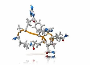
Obestatin molecule. Computer artwork showing the structure of a molecule of obestatin. Obestatin is thought to supress hunger and reduce food intake, thereby reducing weight gain

Background imageSecondary Structure Collection: Obestatin molecule C014 / 4909

Obestatin molecule C014 / 4909
Obestatin molecule. Computer artwork showing the structure of a molecule of obestatin. Obestatin is thought to supress hunger and reduce food intake, thereby reducing weight gain

Background imageSecondary Structure Collection: Bacterial outer membrane protein molecule C014 / 4949

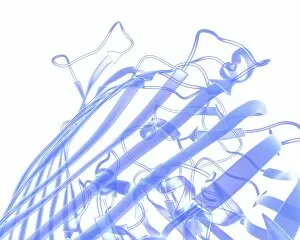
Bacterial outer membrane protein molecule C014 / 4949
Bacterial outer membrane protein molecule. Computer model showing the secondary structure of a molecule of outer membrane transporter FecA protein from Escherichia coli (E. coli) bacteria

Background imageSecondary Structure Collection: Bacterial outer membrane protein molecule

Bacterial outer membrane protein molecule. Computer model showing a part of the secondary structure of a molecule of outer membrane protein A from Escherichia coli (E. coli) bacteria

Background imageSecondary Structure Collection: UVR8 protein molecule C014 / 4912

UVR8 protein molecule C014 / 4912
UVR8 protein molecule. Computer model showing photoreception of UV-B (ultraviolet-B) light rays (top) by a UVR8 protein. The secondary structure (purple ribbons) of UVR8 is shown at bottom

Background imageSecondary Structure Collection: Activated ghrelin hormone molecule C014 / 4901

Activated ghrelin hormone molecule C014 / 4901
Activated ghrelin hormone molecule. Computer model showing the crystal structure of the human hormone ghrelin. The crystal structure consists of both the secondary structure

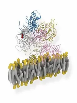
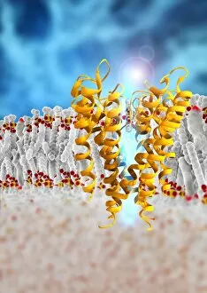
Background imageSecondary Structure Collection: Potassium ion channel

Potassium ion channel. Computer artwork of a KcsA potassium ion (K+) channel (ribbons) embedded in a phospholipid (spheres) cell membrane (horizontal, centre)

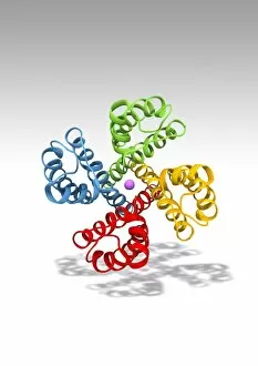
Background imageSecondary Structure Collection: Voltage-gated potassium channel

Voltage-gated potassium channel. Computer model showing the molecular structure of a voltage-gated potassium (Kv) ion channel

Background imageSecondary Structure Collection: Restriction enzyme cutting DNA

Restriction enzyme cutting DNA
Fragment of DNA bound by the restriction endonucleaseEcoRI. The protein is a dimer, with each subunitable to bind and cut one strand of DNA

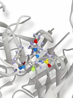
Background imageSecondary Structure Collection: Beta secretase inhibitor, molecular model

Beta secretase inhibitor, molecular model. The inhibitor molecule (centre, also shown in C015/1977) is bound to the beta secretase enzyme (partially seen)

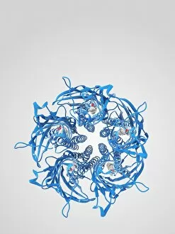
Background imageSecondary Structure Collection: Anaesthetic inhibiting an ion channel C015 / 6723

Anaesthetic inhibiting an ion channel C015 / 6723
Anaesthetic inhibiting an ion channel. Computer model showing the structure of propofol anaesthetic drug molecules (spheres) bound to a pentameric ligand-gated ion channel (pLGIC, blue ribbons)

Background imageSecondary Structure Collection: Coagulation factor complex molecule C014 / 0626

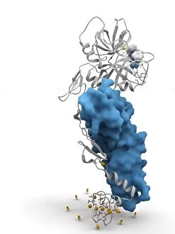
Coagulation factor complex molecule C014 / 0626
Coagulation factor complex molecule. Molecular model showing a blood clotting factor VIIa-tissue factor complex molecule (blue and white ribbons) with a tissue factor pathway inhibitor (TFPI)

Background imageSecondary Structure Collection: Anaesthetic inhibiting an ion channel C015 / 6722

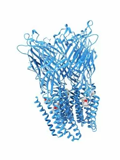
Anaesthetic inhibiting an ion channel C015 / 6722
Anaesthetic inhibiting an ion channel. Computer model showing the structure of propofol anaesthetic drug molecules (spheres) bound to a pentameric ligand-gated ion channel (pLGIC, blue ribbons)

Background imageSecondary Structure Collection: Anaesthetic inhibiting an ion channel C015 / 6720

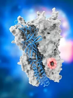
Anaesthetic inhibiting an ion channel C015 / 6720
Anaesthetic inhibiting an ion channel. Computer model showing the structure of propofol anaesthetic drug molecules (lower left and right) bound to a pentameric ligand-gated ion channel (pLGIC, grey)

Background imageSecondary Structure Collection: Coagulation factor complex molecule C014 / 0585

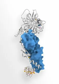
Coagulation factor complex molecule C014 / 0585
Coagulation factor complex molecule. Molecular model showing a blood clotting factor VIIa-tissue factor complex molecule (blue and white) with a tissue factor pathway inhibitor (TFPI)

Background imageSecondary Structure Collection: Coagulation factor complex molecule C014 / 0584

Coagulation factor complex molecule C014 / 0584
Coagulation factor complex molecule. Molecular model showing a blood clotting factor VIIa-tissue factor complex molecule (blue and white ribbons) with a tissue factor pathway inhibitor (TFPI)

Background imageSecondary Structure Collection: Oestrogen receptor, molecular model

Oestrogen receptor, molecular model
Oestrogen receptor. Molecular model of an oestrogen receptor, bound to a glucocorticoid receptor-interacting protein. Oestrogen receptors are cytoplasmic proteins that bind oestrogens

Background imageSecondary Structure Collection: Erythropoietin hormone complex C016 / 4461

Erythropoietin hormone complex C016 / 4461
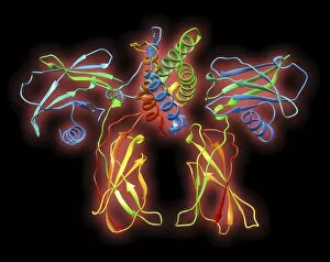
Erythropoietin hormone complex. Computer model showing the secondary structure of a molecule of the human hormone Erythropoietin (EPO), complexed with an erythropoetin receptor molecule

Background imageSecondary Structure Collection: Erythropoietin hormone complex C016 / 4458

Erythropoietin hormone complex C016 / 4458
Erythropoietin hormone complex. Computer model showing the secondary structure of a molecule of the human hormone Erythropoietin (EPO), complexed with an erythropoetin receptor molecule

Background imageSecondary Structure Collection: Erythropoietin hormone complex C016 / 4451

Erythropoietin hormone complex C016 / 4451
Erythropoietin hormone complex. Computer model showing the secondary structure of a molecule of the human hormone Erythropoietin (EPO), complexed with an erythropoetin receptor molecule

Background imageSecondary Structure Collection: Neuropeptide Y neurotransmitter molecule C014 / 0013

Neuropeptide Y neurotransmitter molecule C014 / 0013
Neuropeptide Y neurotransmitter molecule. Molecular model showing the structure of the neurotransmitter neuropeptide Y (NPY)

Background imageSecondary Structure Collection: B19 parvovirus capsid protein

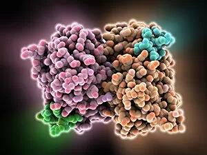
B19 parvovirus capsid protein, molecular model. This protein forms part of the capsid (protein shell) of parvovirus B19, one of only a few parvoviruses that infect humans

Background imageSecondary Structure Collection: Human growth hormone molecule C013 / 9006

Human growth hormone molecule C013 / 9006
Human growth hormone. Molecular model of human growth hormone (hGH, yellow) bound to the extracellular domain of the human growth hormone binding protein (hGHBP)

Background imageSecondary Structure Collection: RNA polymerase molecule C013 / 9005

RNA polymerase molecule C013 / 9005
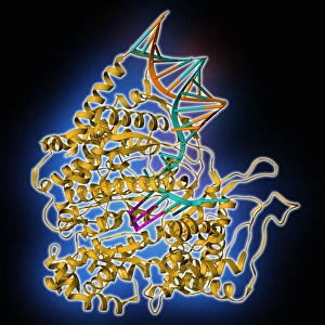
RNA polymerase. Molecular model of RNA polymerase (yellow) transcribing a strand of mRNA (messenger ribonucleic acid, pink) from a DNA (deoxyribonucleic acid) template (orange and turquoise)

Background imageSecondary Structure Collection: HIV reverse transcription enzyme C013 / 8998

HIV reverse transcription enzyme C013 / 8998
HIV reverse transcription enzyme. Molecular model of the reverse transcriptase enzyme (orange and blue) found in HIV (the human immunodeficiency virus)

Background imageSecondary Structure Collection: Erythropoietin molecule C013 / 8891

Erythropoietin molecule C013 / 8891
Erythropoietin molecule (EPO), molecular model. EPO is a glycoprotein that is produced by the kidneys in response to low blood oxygen levels

Background imageSecondary Structure Collection: Birch pollen allergen molecule C013 / 8889

Birch pollen allergen molecule C013 / 8889
Birch pollen allergen molecule. Computer model showing the secondary structure of a Bet v 1L molecule. This molecule is responsible for allergic reactions to pollen from birch (Betula sp.) trees

Background imageSecondary Structure Collection: Oxoguanine glycosylase complex C013 / 8886

Oxoguanine glycosylase complex C013 / 8886
Oxoguanine glycosylase complex. Computer model showing a molecule of human aG DNA repair glycosylase (right) bound to an DNA molecule (left)

Background imageSecondary Structure Collection: Oxoguanine glycosylase complex C013 / 8884

Oxoguanine glycosylase complex C013 / 8884
Oxoguanine glycosylase complex. Computer model showing an 8-Oxoguanine glycosylase (OGG1) molecule (green) bound to a section of DNA (deoxyribonucleic acid, pink and blue)

Background imageSecondary Structure Collection: Erythropoietin hormone complex

Erythropoietin hormone complex. Computer model showing the secondary structure of a molecule of the human hormone Erythropoietin (EPO), complexed with an erythropoetin receptor molecule

Background imageSecondary Structure Collection: TATA box-binding protein complex C013 / 8881

TATA box-binding protein complex C013 / 8881
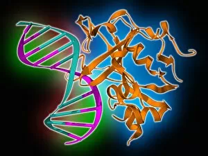
TATA box-binding protein complex. Molecular model showing a TATA box-binding protein (TBP) (purple) complexed with a strand of DNA (deoxyribonucleic acid, blue)


For sale as Licensed Images

Choose your image, Select your licence and Download the media

Secondary structure refers to the intricate folding patterns that proteins and nucleic acids adopt, playing a crucial role in their functionality. In one captivating image, an anaesthetic inhibits an ion channel (C015/6718), highlighting how secondary structure impacts cellular processes. Another snapshot showcases DNA transcription, unveiling the molecular model of this essential process. Artwork depicting the secondary structure of proteins captivates our imagination as we marvel at the intricacy and beauty within each fold. The nucleosome molecule further emphasizes this complexity, showcasing how DNA wraps around histone proteins to form a compact structure. The bacterial ribosome stands tall as a testament to secondary structures' significance in protein synthesis. Meanwhile, the HIV reverse transcription enzyme reminds us of its vital role in converting viral RNA into DNA during infection. Molecular models provide insight into hepatitis C virus enzymes and interferon molecules—both critical players in disease progression and immune response modulation. Similarly, human growth hormone molecules hold immense importance for development and metabolism regulation. Exploring coagulation factor complex molecules (C014/0139) unravels mechanisms behind blood clotting—a process dependent on precise secondary structures working together seamlessly. Ghrelin hormone molecules intrigue us with their involvement in appetite regulation and energy balance maintenance. From anaesthetics influencing ion channels to hormones orchestrating bodily functions, understanding secondary structures unlocks countless mysteries within biological systems. These captivating images remind us of the intricate dance occurring at a microscopic level—the delicate folds dictating life's grand symphony.