Sectioned Collection (page 3)

"Exploring the Intricacies: From Cerebellum Tissue to Fusion Research, Unveiling the World Sectioned" Delving into the depths of scientific exploration

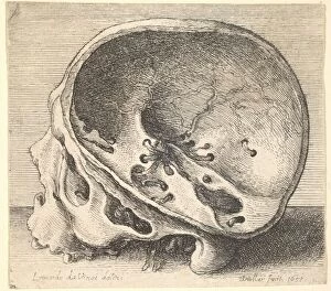
Background imageSectioned Collection: Sectioned skull in profile to left with the left side of the cranium removed, 1651

Sectioned skull in profile to left with the left side of the cranium removed, 1651

Background imageSectioned Collection: Close-up of puppy lying on sand

Close-up of puppy lying on sand

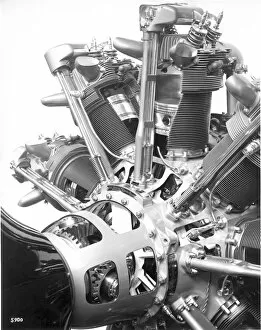
Background imageSectioned Collection: A sectioned Pratt & Whitney R-4360 Wasp Major radial

A sectioned Pratt & Whitney R-4360 Wasp Major radial

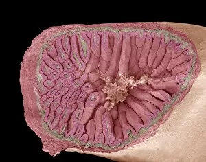
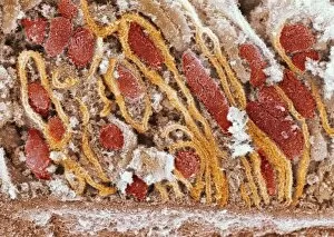
Background imageSectioned Collection: Intestinal villi, SEM

Intestinal villi, SEM
Intestinal villi. Coloured scanning electron micrograph (SEM) of a section through a part of the small intestine, showing the villi that cover its inner surface

Background imageSectioned Collection: Cervix, light micrograph F006 / 9805

Cervix, light micrograph F006 / 9805
Cervix. Light micrograph of a section though the mucosa of the cervix, the neck of the uterus. The purple-lined areas are cervical glands, which produce mucus (pink inside the glands)

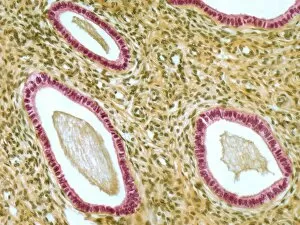
Background imageSectioned Collection: Fallopian tube, light micrograph F006 / 9799

Fallopian tube, light micrograph F006 / 9799
Fallopian tube. Light micrograph of a cross-section through a fallopian tube. The lumen (white) is seen, along with mucosal folds lined with simple columnar ciliated epithelium (purple)

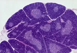
Background imageSectioned Collection: Thymus gland, light micrograph C015 / 4970

Thymus gland, light micrograph C015 / 4970
Thymus gland. Light micrograph of a section through tissue from the thymus gland, part of the lymphoid system. The dense areas are the thymic cortex, which produces lots of lymphocytes

Background imageSectioned Collection: Tuberculosis of the spine, MRI scan

Tuberculosis of the spine, MRI scan
Tuberculosis of the spine. Magnetic resonance imaging (MRI) scan of a sagittal section through the spine of a 74-year-old patient with tuberculosis (TB) of the spine, or Potts disease

Background imageSectioned Collection: Spondylolisthesis, 3D CT scan

Spondylolisthesis, 3D CT scan
Spondylolisthesis. Coloured 3D computed tomography (CT) scan of a section through the lumbar (lower back) spine of a 39-year-old patient with spondylolisthesis

Background imageSectioned Collection: Vascular system of the head, MRI scan

Vascular system of the head, MRI scan
Vascular system of the head. Magnetic resonance imaging (MRI) angiogram of a sagittal section through the head of a healthy 43-year-old, showing the blood vessels (white) of the brain and neck

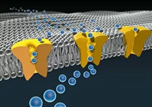
Background imageSectioned Collection: Cell membrane ion channels, artwork C016 / 7689

Cell membrane ion channels, artwork C016 / 7689
Cell membrane ion channels. Computer artwork of a section through the membrane of an animal cell, showing transmembrane ion channel proteins (yellow)

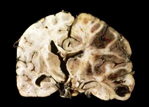
Background imageSectioned Collection: Brain injury C016 / 8920

Brain injury C016 / 8920
Brain injury. Gross specimen of a section through a brain showing the effects of a blunt trauma. The hemisphere at right is showing extensive haemorrhaging (bleeding)

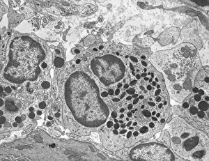
Background imageSectioned Collection: Eosinophil white blood cell, TEM

Eosinophil white blood cell, TEM
Eosinophil white blood cell. Transmission electron micrograph (TEM) of a section through an eosinophil white blood cell (leukocyte)

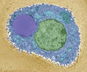
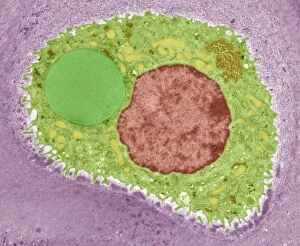
Background imageSectioned Collection: Pancreatic acinar cell

Pancreatic acinar cell. Transmission electron micrograph (TEM) of a section through an enzyme-secreting acinar cell in the human pancreas, showing part of the nucleus (round, far left)

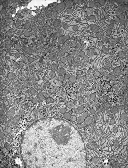
Background imageSectioned Collection: Hepatocyte liver cell, TEM

Hepatocyte liver cell, TEM
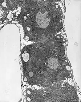
Hepatocyte liver cell. Transmission electron micrograph (TEM) of a section through an hepatocyte liver cell, showing rough and smooth endoplasmic reticulum (ER, dark lines)

Background imageSectioned Collection: Liver tissue, TEM

Liver tissue, TEM
Liver tissue. Transmission electron micrograph (TEM) of a section through the liver, showing part of a radial cord of hepatocyte liver cells (dark) and the vascular sinusoids (white)

Background imageSectioned Collection: Human head, X-ray artwork

Human head, X-ray artwork
Human head. Computer artwork of an X-ray through a human head, showing the brain (grey, top), cerebellum (blue), and brainstem and spinal cord (green)

Background imageSectioned Collection: Trachea lining, SEM C015 / 9936

Trachea lining, SEM C015 / 9936
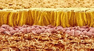
Trachea lining. Coloured scanning electron micrograph (SEM) of a section through the lining of the trachea (wind pipe). The trachea links the larynx (voice box) to the lungs

Background imageSectioned Collection: Vagus nerve anatomy, artwork C018 / 0300

Vagus nerve anatomy, artwork C018 / 0300
Vagus nerve anatomy. Artwork of a human body from the side, showing the internal anatomy and the location of the vagus nerve (yellow)

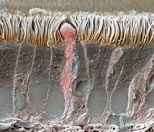
Background imageSectioned Collection: Retina layers, SEM C018 / 0520

Retina layers, SEM C018 / 0520
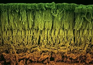
Retina layers, coloured scanning electron micrograph (SEM). The retina of the human eye is found on the inside of the eyeball and is formed from a number of layers

Background imageSectioned Collection: Dental filling polymerisation, artwork C014 / 2025

Dental filling polymerisation, artwork C014 / 2025
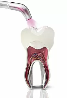
Dental filling polymerisation. Artwork of a cross-section of a human tooth, with a filling (white) being set using an ultraviolet light lamp tool

Background imageSectioned Collection: Cartilage cell, TEM C014 / 1433

Cartilage cell, TEM C014 / 1433
Cartilage cell. Transmission electron micrograph (TEM) of a section through a chondrocyte cell from hyaline cartilage of the trachea (windpipe)

Background imageSectioned Collection: Cartilage cell, TEM C014 / 1434

Cartilage cell, TEM C014 / 1434
Cartilage cell. Transmission electron micrograph (TEM) of a section through a chondrocyte cell from hyaline cartilage of the trachea (windpipe)

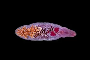
Background imageSectioned Collection: Lancet liver fluke C014 / 4846

Lancet liver fluke C014 / 4846
Lancet liver fluke (Dicrocoelium dendriticum). This parasitic trematode (flatworm) tends to live in cattle or other grazing mammals

Background imageSectioned Collection: Splenic infarct C015 / 6220

Splenic infarct C015 / 6220
Splenic infarct. Gross specimen of a sectioned spleen showing areas of dead tissue (pale). The tissue death results from a loss of blood flow to the area, known as an infarction

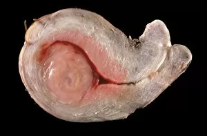
Background imageSectioned Collection: Uterine fibroid C015 / 6407

Uterine fibroid C015 / 6407
Uterine fibroid. Gross specimen of a sectioned uterine fibroid (left). A fibroid is a fibrous benign tumour originating from muscular tissue

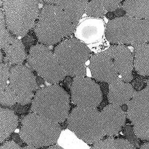
Background imageSectioned Collection: Skeletal muscle, TEM C016 / 5369

Skeletal muscle, TEM C016 / 5369
Skeletal muscle. Transmission electron micrograph (TEM) of a cross section through human skeletal (striated) muscle. Blocks of muscle (lighter grey) are surrounded by connective tissue (black)

Background imageSectioned Collection: Trachea lining, SEM C013 / 7122

Trachea lining, SEM C013 / 7122
Trachea lining. Coloured scanning electron micrograph (SEM) of a section through the wall of a trachea (wind pipe). The trachea links the larynx to the lungs

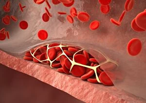
Background imageSectioned Collection: Thrombosed blood vessel, artwork C013 / 4649

Thrombosed blood vessel, artwork C013 / 4649
Thrombosed blood vessel. Computer artwork of a blood clot (thrombus) formed on the internal wall of a blood vessel

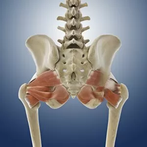
Background imageSectioned Collection: Buttock muscles, artwork C013 / 4414

Buttock muscles, artwork C013 / 4414
Buttock muscles. Computer artwork of a posterior view of the hips showing some of the muscles (red) of the buttocks. These are shown attached to the femurs (thigh bones) and the pelvis bones
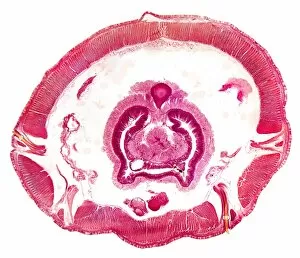
Background imageSectioned Collection: Earthworm, transverse section

Earthworm, transverse section
Earthworm. Light micrograph of a transverse section through the body of a round segmented earthworm (Lumbricus terrestris) in the intestinal region
Background imageSectioned Collection: De Laval steam turbine

De Laval steam turbine. Diagram of a cross-section through the steam turbine invented by the Swedish engineer Gustaf de Laval (1845-1913). De Laval built his first steam turbine in 1887

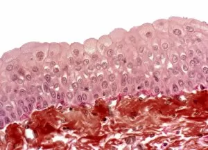
Background imageSectioned Collection: Bladder epithelium, light micrograph

Bladder epithelium, light micrograph
Bladder epithelium. Light micrograph of a vertical section through the wall of the urinary bladder. The inner surface is at top

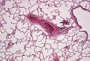
Background imageSectioned Collection: Lung alveoli

Lung alveoli. Light micrograph of a section through normal human lung tissue showing alveoli. These are tiny air sacs where most of the diffusion of gases to and from the blood occurs

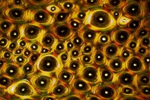
Background imageSectioned Collection: Whale bone tissue, light micrograph

Whale bone tissue, light micrograph

Background imageSectioned Collection: Hazel nut, light micrograph

Hazel nut, light micrograph
Hazel nut. Light micrograph of a section through a nut from a common hazel (Corylus avellana) tree, showing the large amount of fat (red) stored within endosperm tissue

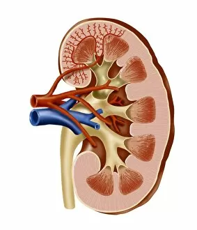
Background imageSectioned Collection: Kidney anatomy, artwork

Kidney anatomy, artwork
Kidney anatomy. Cutaway artwork showing the internal structure of a kidney. The kidney, one of a pair of such organs, is part of the human excretory system

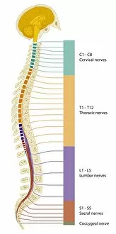
Background imageSectioned Collection: Spinal cord regions, diagram

Spinal cord regions, diagram
Spinal cord regions. Diagram of a sagittal section through the human skull and backbone, showing the five main regions of the spinal cord and the numbered nerves associated with them

Background imageSectioned Collection: Bacterial cell structure, artwork

Bacterial cell structure, artwork
Bacterial cell structure. Computer artwork showing the cell structure and components (organelles) of a typical rod-shaped bacteria (bacillus). Not all bacteria have a flagellum (long, tail-like)

Background imageSectioned Collection: Mitochondria, SEM

Mitochondria, SEM
Mitochondria. Coloured scanning electron micrograph (SEM) of mitochondria (red) in a kidney cell. Mitochondria are a type of organelle found in the cytoplasm of eukaryotic cells

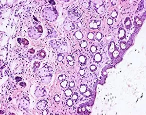
Background imageSectioned Collection: Skin and hair follicles, light micrograph

Skin and hair follicles, light micrograph
Skin and hair follicles. Light micrograph of a section through skin, showing hair follicles (dark purple circular structures). Haematoxylin and eosin stain

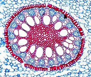
Background imageSectioned Collection: Iris root, light micrograph

Iris root, light micrograph
Iris root. Light micrograph of a section through the root of an Iris plant (Iris germanica) showing a vascular cylinder. The cylinder is comprised of a central cluster of parenchyma cells (red)

Background imageSectioned Collection: Prehistoric Hearths, Dundrum, Typical Section, Old Land Surf

Prehistoric Hearths, Dundrum, Typical Section, Old Land Surfaces(L), Exposed in the Aeolian Sands(S), Rich in Primitive Tools - a view of a mound in which the photographer has sectioned

Background imageSectioned Collection: India - Inner gate - Hoosimabad

India - Inner gate - Hoosimabad
A black and white lantern slide of an ornate five sectioned doorway. Slide number 305 of Box 221 Boswell Collection Date: circa 1890s

Background imageSectioned Collection: Scene in a car showroom

Scene in a car showroom, with a new Vauxhall 10 on display, stripped down to its engine. Prices start at 168

Background imageSectioned Collection: Design Circular Border Nine Female Figures Muses?

Design Circular Border Nine Female Figures Muses?
Artokoloro

Background imageSectioned Collection: Apple stem, light micrograph

Apple stem, light micrograph
Apple stem. Polarised light micrograph of a section through the stem of a young apple (Malus domestica) tree plant. Magnification: x100 when printed 10 centimetres wide

Background imageSectioned Collection: A sectioned Bristol Pegasus radial at the 1934 Paris Salon

A sectioned Bristol Pegasus radial at the 1934 Paris Salon. Date: 1934


For sale as Licensed Images

Choose your image, Select your licence and Download the media

"Exploring the Intricacies: From Cerebellum Tissue to Fusion Research, Unveiling the World Sectioned" Delving into the depths of scientific exploration, a light micrograph reveals the intricate structure of cerebellum tissue. Witnessing a Higgs boson event through the lens of ATLAS detector C013/6892, scientists capture an awe-inspiring moment in particle physics. A meticulously crafted model showcases the complexity and beauty of the human brain, inviting us to marvel at its wonders. Peering into hippocampus brain tissue under a microscope, we uncover secrets that may hold keys to memory formation and learning processes. In pursuit of limitless energy sources, fusion research takes center stage as we examine a tokamak device designed for controlled nuclear fusion reactions. The inner workings of our brains come alive through an MRI scan, revealing detailed brain anatomy like never before imagined. Illuminated by light microscopy, dicotyledon plant stem sections unveil nature's architectural masterpiece in stunning detail. EDTA crystals take on mesmerizing patterns when viewed under intense magnification - showcasing their unique molecular arrangement through light micrography. Journeying deep within our bodies, kidney tubules are sectioned to shed light on their vital role in maintaining fluid balance and waste removal systems. An X-ray image exposes every contour and intricacy of a human skull – offering glimpses into our skeletal framework with remarkable clarity. Through transmission electron microscopy (TEM), rough endoplasmic reticulum emerges as an interconnected network responsible for protein synthesis within cells' cytoplasmic matrix Computer artwork visualizes Alzheimer's-affected brain regions – reminding us why continued research is crucial in unraveling this devastating disease. In this captivating journey across various scientific disciplines and microscopic landscapes, "sectioned" unveils hidden worlds that expand our understanding of the universe and ourselves.