Sectioned Collection (#6)

"Exploring the Intricacies: From Cerebellum Tissue to Fusion Research, Unveiling the World Sectioned" Delving into the depths of scientific exploration

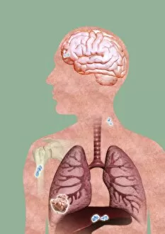
Background imageSectioned Collection: Metastatic cancer, artwork

Metastatic cancer, artwork
Metastatic cancer. Computer artwork of a section through a persons upper body and head, showing a primary cancer in the right liver (lower left) that has spread (metastasised)

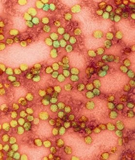
Background imageSectioned Collection: Yellow fever virus particles, TEM C016 / 9468

Yellow fever virus particles, TEM C016 / 9468
Yellow fever virus particles. Transmission electron micrograph (TEM) of yellow fever virus particles (virions, yellow). Yellow fever is endemic in tropical Africa and South America
Background imageSectioned Collection: Prosthetic hip joint and Gruen zones C016 / 6778

Prosthetic hip joint and Gruen zones C016 / 6778
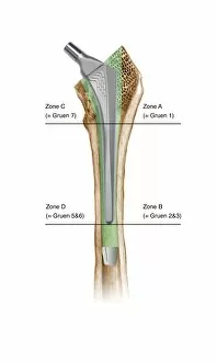
Prosthetic hip joint and Gruen zones. Cutaway diagram of a femur (thigh bone) showing a femoral component of a hip prosthesis and labels indicating Gruen zones

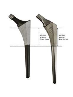
Background imageSectioned Collection: Prosthetic hip joints and Gruen zones C016 / 6781

Prosthetic hip joints and Gruen zones C016 / 6781
Prosthetic hip joints and Gruen zones. Diagram of the femoral components of two types of hip prosthesis with labels indicating standard and modified Gruen zones

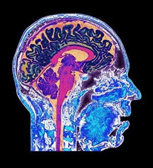
Background imageSectioned Collection: Normal human brain, MRI scan C016 / 8840

Normal human brain, MRI scan C016 / 8840
Brain. Coloured magnetic resonance imaging (MRI) scan of a sagittal section through a healthy human brain. The highly folded area (yellow) is the cerebrum, which is responsible for conscious thought

Background imageSectioned Collection: Oedema of larynx, artwork C016 / 6529

Oedema of larynx, artwork C016 / 6529
Oedema of larynx. Artwork of a transverse (axial) section through a larynx (seen from above) with and without swelling. The bottom image shows a healthy larynx

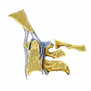
Background imageSectioned Collection: Atlanto-occipital joint, artwork C016 / 6554

Atlanto-occipital joint, artwork C016 / 6554
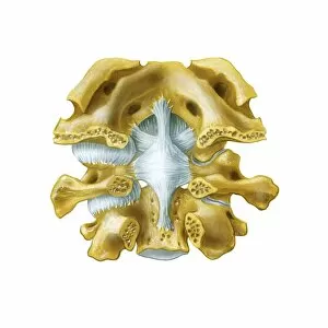
Atlanto-occipital joint. Artwork of the bones (yellow) and ligaments (blue) of the atlanto-occipital joint, as seen from behind (posterior view)

Background imageSectioned Collection: Normal human brain, MRI scan C016 / 8848

Normal human brain, MRI scan C016 / 8848
Brain. Coloured magnetic resonance imaging (MRI) scan of a sagittal section through a patients head showing a healthy human brain

Background imageSectioned Collection: Prosthetic hip joint, artwork C016 / 6782

Prosthetic hip joint, artwork C016 / 6782
Prosthetic hip joint. Cutaway artwork of a femur (thigh bone) showing a femoral component of a hip prosthesis. This component is implanted in the femur after the head of the femur has been surgically

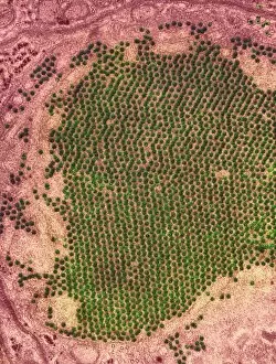
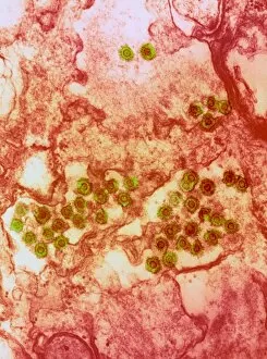
Background imageSectioned Collection: Yellow fever virus particles, TEM C016 / 9469

Yellow fever virus particles, TEM C016 / 9469
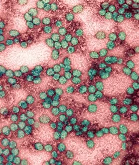
Yellow fever virus particles. Transmission electron micrograph (TEM) of yellow fever virus particles (virions, green). Yellow fever is endemic in tropical Africa and South America

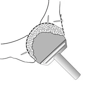
Background imageSectioned Collection: Bone drill and grafter, artwork C016 / 6845

Bone drill and grafter, artwork C016 / 6845
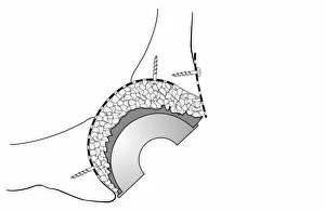
Bone drill and grafter. Artwork of a cross-section through a bone drill and grafter that is being used in a hip socket bone grafting operation

Background imageSectioned Collection: SARS virus particles, TEM C016 / 9445

SARS virus particles, TEM C016 / 9445
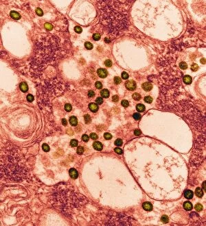
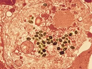
SARS virus particles. Coloured transmission electron micrograph (TEM) of a section through a tissue sample infected with numerous SARS coronavirus particles (virions, dark, round)

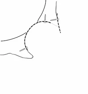
Background imageSectioned Collection: Hip socket bone grafting, diagram C016 / 6786

Hip socket bone grafting, diagram C016 / 6786
Hip socket bone grafting. Diagram showing completed hip socket bone grafting as part of a total hip replacement operation

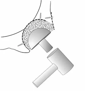
Background imageSectioned Collection: Hip socket bone grafting, diagram C016 / 6784

Hip socket bone grafting, diagram C016 / 6784
Hip socket bone grafting. Diagram showing the second stage in hip socket bone grafting as part of a total hip replacement operation

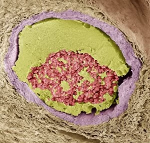
Background imageSectioned Collection: Blood clot, SEM C016 / 9088

Blood clot, SEM C016 / 9088
Blood clot. Coloured scanning electron micrograph (SEM) of a section through a blood clot (thrombus) in an arteriole (small blood vessel), showing red blood cells (red) in a fibrin mesh (green)

Background imageSectioned Collection: Normal human brain, MRI scan C016 / 8837

Normal human brain, MRI scan C016 / 8837
Brain. Coloured magnetic resonance imaging (MRI) scan of a sagittal section through a healthy human brain. The highly folded area (orange) is the cerebrum, which is responsible for conscious thought

Background imageSectioned Collection: Normal human brain, MRI scan C016 / 8841

Normal human brain, MRI scan C016 / 8841
Brain. Coloured magnetic resonance imaging (MRI) scan of a sagittal section through a healthy human brain. The highly folded area (red and yellow) is the cerebrum

Background imageSectioned Collection: Broken finger bone, SEM C016 / 9027

Broken finger bone, SEM C016 / 9027
Broken finger bone. Coloured scanning electron micrograph (SEM) showing the internal structure of a broken finger bone (phalanx)

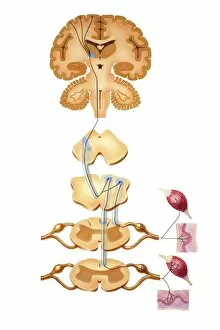
Background imageSectioned Collection: Brain motor cortex pathways, artwork C016 / 6533

Brain motor cortex pathways, artwork C016 / 6533
Brain motor cortex pathways. Artwork of a sectioned human brain, brainstem and spinal cord, showing neural pathways (black lines) from the motor cortex of the brain

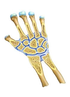
Background imageSectioned Collection: Wrist joint anatomy, artwork C016 / 6549

Wrist joint anatomy, artwork C016 / 6549
Wrist joint anatomy. Artwork of the bones (yellow) and ligaments (blue) of the wrist joint, in longitudinal section. The left hand has been sectioned in a palmar aspect to expose the synovial

Background imageSectioned Collection: Atlanto-occipital joint, artwork C016 / 6557

Atlanto-occipital joint, artwork C016 / 6557
Atlanto-occipital joint. Artwork of the bones (yellow) and ligaments (blue) of the atlanto-occipital joint, as seen from the side in a medial sagittal section. The rear of the spine is at right

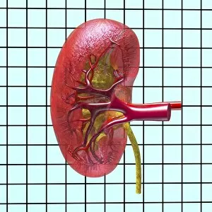
Background imageSectioned Collection: Kidney anatomy, artwork C016 / 8945

Kidney anatomy, artwork C016 / 8945
Kidney anatomy, artwork. The kidneys filter waste from the blood and excrete it as urine. The kidney consists of an outer cortex and an inner medulla

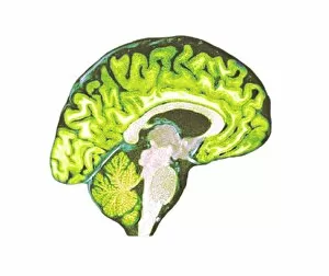
Background imageSectioned Collection: Normal human brain, MRI scan C016 / 8835

Normal human brain, MRI scan C016 / 8835
Brain. Coloured magnetic resonance imaging (MRI) scan of a sagittal section through a healthy human brain. The highly folded area across top is the cerebrum

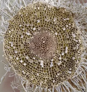
Background imageSectioned Collection: Dicotyledon root, SEM C016 / 9125

Dicotyledon root, SEM C016 / 9125
Dicotyledon root. Coloured scanning electron micrograph (SEM) of a transverse section through an immature root from a dicotyledonous plant

Background imageSectioned Collection: Hip socket bone grafting, diagram C016 / 6785

Hip socket bone grafting, diagram C016 / 6785
Hip socket bone grafting. Diagram showing the third stage in hip socket bone grafting as part of a total hip replacement operation

Background imageSectioned Collection: Hip socket bone grafting, diagram C016 / 6783

Hip socket bone grafting, diagram C016 / 6783
Hip socket bone grafting. Diagram showing the first stage in hip socket bone grafting as part of a total hip replacement operation

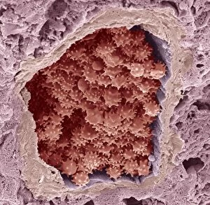
Background imageSectioned Collection: Crenated red blood cells, SEM C016 / 9029

Crenated red blood cells, SEM C016 / 9029
Crenated red blood cells. Coloured scanning electron micrograph (SEM) of a section through an arteriole (small blood vessel), showing crenated red blood cells inside

Background imageSectioned Collection: Normal human brain, MRI scan C016 / 8846

Normal human brain, MRI scan C016 / 8846
Brain. Coloured magnetic resonance imaging (MRI) scan of a sagittal section through a patients head showing a healthy human brain

Background imageSectioned Collection: Kidney stones, artwork C016 / 7531

Kidney stones, artwork C016 / 7531
Kidney stones. Computer artwork showing a section through a human kidney containing kidney stones (yellowish, right). The ureter (tube)

Background imageSectioned Collection: St. Louis encephalitis virus particles C016 / 9453

St. Louis encephalitis virus particles C016 / 9453
St. Louis encephalitis virus particles. Coloured transmission electron micrograph (TEM) of a section through a mosquito salivary gland containing numerous Saint Louis encephalitis virus particles

Background imageSectioned Collection: Prosthetic hip joint, diagram C016 / 6775

Prosthetic hip joint, diagram C016 / 6775
Prosthetic hip joint. Cutaway diagram of a femur (thigh bone) showing a femoral component of a hip prosthesis. This component is implanted in the femur after the head of the femur has been surgically

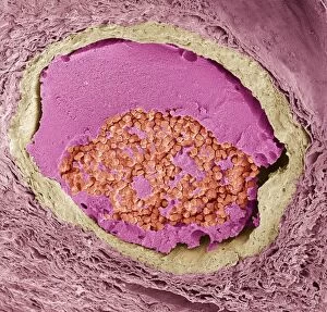
Background imageSectioned Collection: Blood clot, SEM C016 / 9089

Blood clot, SEM C016 / 9089
Blood clot. Coloured scanning electron micrograph (SEM) of a section through a blood clot (thrombus) in an arteriole (small blood vessel), showing red blood cells (red) in a fibrin mesh (bright pink)

Background imageSectioned Collection: Atherosclerosis in artery, artwork C016 / 6571

Atherosclerosis in artery, artwork C016 / 6571
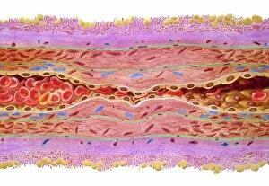
Atherosclerosis in artery. Artwork of a longitudinal section through an artery that has been narrowed by atherosclerosis. Narrowing is referred to as stenosis

Background imageSectioned Collection: Smallpox virus particles, TEM C016 / 9447

Smallpox virus particles, TEM C016 / 9447
Smallpox virus particles. Coloured transmission electron micrograph (TEM) of a section through a tissue sample infected with numerous Variola virus particles (virions, brown)

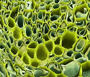
Background imageSectioned Collection: Dicotyledon vascular bundle, SEM C016 / 9603

Dicotyledon vascular bundle, SEM C016 / 9603
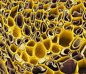
Dicotyledon vascular bundle. Coloured scanning electron micrograph (SEM) of a section through a vascular bundle from a dicotyledonous plant

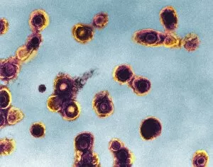
Background imageSectioned Collection: Spumavirus particles, TEM C016 / 9452

Spumavirus particles, TEM C016 / 9452
Spumavirus virus particles. Coloured transmission electron micrograph (TEM) of a section through a tissue sample infected with numerous spumavirus particles (virions, yellow)

Background imageSectioned Collection: Normal human brain, MRI scan C016 / 8834

Normal human brain, MRI scan C016 / 8834
Brain. Coloured magnetic resonance imaging (MRI) scan of a sagittal section through a healthy human brain. The highly folded area (orange) is the cerebrum, which is responsible for conscious thought

Background imageSectioned Collection: Normal human brain, MRI scan C016 / 8849

Normal human brain, MRI scan C016 / 8849
Brain. Coloured magnetic resonance imaging (MRI) scan of a sagittal section through a patients head showing a healthy human brain

Background imageSectioned Collection: Normal human brain, MRI scan C016 / 8836

Normal human brain, MRI scan C016 / 8836
Brain. Coloured magnetic resonance imaging (MRI) scan of a sagittal section through a healthy human brain. The highly folded area across top is the cerebrum

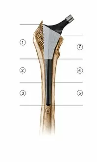
Background imageSectioned Collection: Prosthetic hip joint and Gruen zones C016 / 6777

Prosthetic hip joint and Gruen zones C016 / 6777
Prosthetic hip joint and Gruen zones. Cutaway diagram of a femur (thigh bone) showing a femoral component of a hip prosthesis and labels indicating Gruen zones

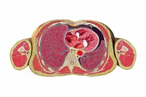
Background imageSectioned Collection: Pneumothorax cross-section, artwork C016 / 6569

Pneumothorax cross-section, artwork C016 / 6569
Pneumothorax cross-section. Artwork of an axial cross-section through a human body at the level of the heart and lungs, showing a pneumothorax (collapsed lung) during chest surgery

Background imageSectioned Collection: Normal human brain, MRI scan C016 / 8844

Normal human brain, MRI scan C016 / 8844
Brain. Coloured magnetic resonance imaging (MRI) scan of a sagittal section through a patients head showing a healthy human brain

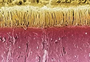
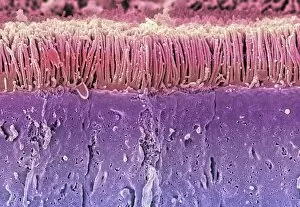
Background imageSectioned Collection: Intestinal microvilli, SEM C016 / 9066

Intestinal microvilli, SEM C016 / 9066
Intestinal microvilli. Coloured scanning electron micrograph (SEM) of a section through the lining of the small intestine, showing the densely packed microvilli (hair-like, top)

Background imageSectioned Collection: Dicotyledon root, SEM C016 / 9124

Dicotyledon root, SEM C016 / 9124
Dicotyledon root. Coloured scanning electron micrograph (SEM) of a transverse section through an immature root from a dicotyledonous plant

Background imageSectioned Collection: Oedema of larynx, artwork C016 / 6528

Oedema of larynx, artwork C016 / 6528
Oedema of larynx. Artwork of a vertical (coronal) section through a larynx, showing oedema (swelling caused by fluid accumulation)

Background imageSectioned Collection: Dicotyledon vascular bundle, SEM C016 / 9602

Dicotyledon vascular bundle, SEM C016 / 9602
Dicotyledon vascular bundle. Coloured scanning electron micrograph (SEM) of a section through a vascular bundle from a dicotyledonous plant

Background imageSectioned Collection: Varicella zoster virus particles, TEM C016 / 9466

Varicella zoster virus particles, TEM C016 / 9466
Varicella zoster virus particles. Transmission electron micrograph (TEM) of varicella zoster virus (VZV) particles (virions) from a patient with chickenpox

Background imageSectioned Collection: Intestinal microvilli, SEM C016 / 9067

Intestinal microvilli, SEM C016 / 9067
Intestinal microvilli. Coloured scanning electron micrograph (SEM) of a section through the lining of the small intestine, showing the densely packed microvilli (hair-like, top)


For sale as Licensed Images

Choose your image, Select your licence and Download the media

"Exploring the Intricacies: From Cerebellum Tissue to Fusion Research, Unveiling the World Sectioned" Delving into the depths of scientific exploration, a light micrograph reveals the intricate structure of cerebellum tissue. Witnessing a Higgs boson event through the lens of ATLAS detector C013/6892, scientists capture an awe-inspiring moment in particle physics. A meticulously crafted model showcases the complexity and beauty of the human brain, inviting us to marvel at its wonders. Peering into hippocampus brain tissue under a microscope, we uncover secrets that may hold keys to memory formation and learning processes. In pursuit of limitless energy sources, fusion research takes center stage as we examine a tokamak device designed for controlled nuclear fusion reactions. The inner workings of our brains come alive through an MRI scan, revealing detailed brain anatomy like never before imagined. Illuminated by light microscopy, dicotyledon plant stem sections unveil nature's architectural masterpiece in stunning detail. EDTA crystals take on mesmerizing patterns when viewed under intense magnification - showcasing their unique molecular arrangement through light micrography. Journeying deep within our bodies, kidney tubules are sectioned to shed light on their vital role in maintaining fluid balance and waste removal systems. An X-ray image exposes every contour and intricacy of a human skull – offering glimpses into our skeletal framework with remarkable clarity. Through transmission electron microscopy (TEM), rough endoplasmic reticulum emerges as an interconnected network responsible for protein synthesis within cells' cytoplasmic matrix Computer artwork visualizes Alzheimer's-affected brain regions – reminding us why continued research is crucial in unraveling this devastating disease. In this captivating journey across various scientific disciplines and microscopic landscapes, "sectioned" unveils hidden worlds that expand our understanding of the universe and ourselves.