Sectioned Collection (#4)

"Exploring the Intricacies: From Cerebellum Tissue to Fusion Research, Unveiling the World Sectioned" Delving into the depths of scientific exploration

Background imageSectioned Collection: Design Circular Border Nine Female Figures Muses?

Design Circular Border Nine Female Figures Muses?
Artokoloro

Background imageSectioned Collection: Apple stem, light micrograph

Apple stem, light micrograph
Apple stem. Polarised light micrograph of a section through the stem of a young apple (Malus domestica) tree plant. Magnification: x100 when printed 10 centimetres wide

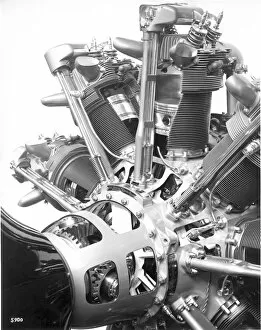
Background imageSectioned Collection: A sectioned Bristol Pegasus radial at the 1934 Paris Salon

A sectioned Bristol Pegasus radial at the 1934 Paris Salon. Date: 1934

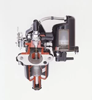
Background imageSectioned Collection: Sectioned view of S. U. Carburettor, 1965

Sectioned view of S. U. Carburettor, 1965
Sectioned view of S.U. Carburettor, 1965

Background imageSectioned Collection: Sectioned modern turbocharger from an car engine showing the inner workings

Sectioned modern turbocharger from an car engine showing the inner workings

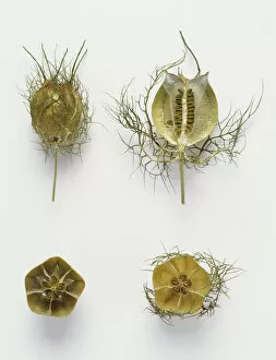
Background imageSectioned Collection: Love-in-a-mist, Nigella damascena, whole and sectioned fruit

Love-in-a-mist, Nigella damascena, whole and sectioned fruit

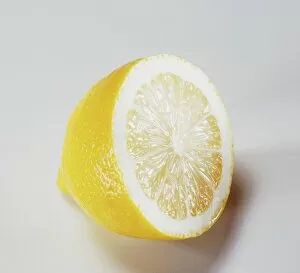
Background imageSectioned Collection: Halved Lemon

Halved Lemon

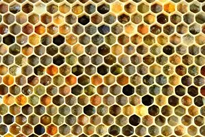
Background imageSectioned Collection: Honey bee comb C017 / 3713

Honey bee comb C017 / 3713
Honey bee comb. Close-up of a comb from the hive of a honey bee (Apis sp.). The cells of this comb are used to store pollen. The different colours are due to the different types of pollen being stored

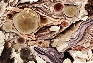
Background imageSectioned Collection: Freeze-fractured baby cream, SEM C017 / 7133

Freeze-fractured baby cream, SEM C017 / 7133
Freeze-fractured baby cream, coloured scanning electron micrograph (SEM). Mangification: x1680 when printed at 10 centimetres wide

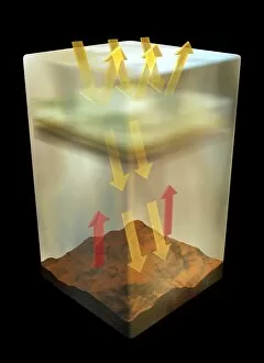
Background imageSectioned Collection: Venuss atmosphere, artwork

Venuss atmosphere, artwork
Venuss atmosphere, computer artwork. Venus has a thick, acidic atmosphere, that traps the heat of the Sun (arrows) causing a runaway green-house effect

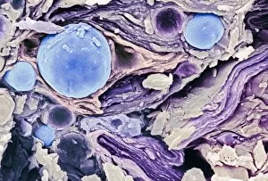
Background imageSectioned Collection: Freeze-fractured baby cream, SEM C017 / 7134

Freeze-fractured baby cream, SEM C017 / 7134
Freeze-fractured baby cream, coloured scanning electron micrograph (SEM). Mangification: x1680 when printed at 10 centimetres wide

Background imageSectioned Collection: Freeze-fractured baby cream, SEM C017 / 7135

Freeze-fractured baby cream, SEM C017 / 7135
Freeze-fractured baby cream, coloured scanning electron micrograph (SEM). Mangification: x1680 when printed at 10 centimetres wide

Background imageSectioned Collection: Polar bear insulating hair, SEM Z927 / 0132

Polar bear insulating hair, SEM Z927 / 0132
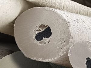
Polar bear insulating hair. Coloured scanning electron micrograph (SEM) of a cross-section through a shaft of an insulating hair from a polar bear (Ursus maritimus)

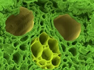
Background imageSectioned Collection: Synthetic cork, SEM

Synthetic cork, SEM
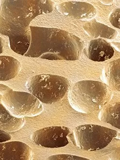
Synthetic cork. Coloured scanning electron micrograph (SEM) of a section through a plastic wine bottle cork. The air holes within the cork allow it to shrink and expand to fit the bottle neck

Background imageSectioned Collection: Compact bone

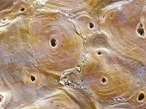
Compact bone. Coloured scanning electron micrograph (SEM) of Haversian canals in compact bone. Bundles of Haversian canals form the core of compact bone

Background imageSectioned Collection: Scalp skin showing hair follicles, LM

Scalp skin showing hair follicles, LM
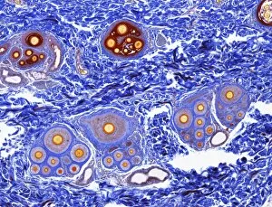
Scalp skin. Light micrograph of a section through skin from the scalp. Numerous hair shafts are seen (purple rings), which are surrounded by connective tissue (dark blue)

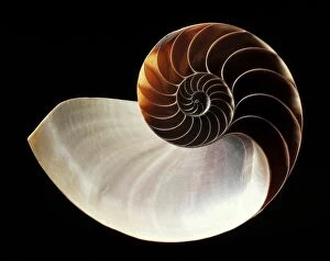
Background imageSectioned Collection: Chambered nautilus shell C019 / 1344

Chambered nautilus shell C019 / 1344
Chambered nautilus shell. Section through the shell of a chambered nautilus (Nautilus pompilius), showing the internal chambers

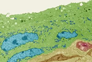
Background imageSectioned Collection: Bladder lining, TEM C014 / 1473

Bladder lining, TEM C014 / 1473
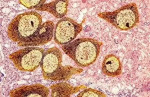
Bladder lining. Transmission electron micrograph (TEM) of a section through the epithelial cells that line of a human bladder, showing large flat vesicles (large, blue)

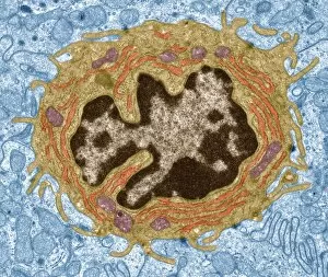
Background imageSectioned Collection: Intestinal gland cell, TEM C014 / 1441

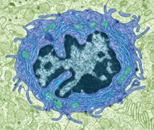
Intestinal gland cell, TEM C014 / 1441
Intestinal gland cell. Transmission electron micrograph (TEM) of a section through an undifferentiated cell from the intestinal tract, showing extensive rough endoplasmic reticulum (ER)

Background imageSectioned Collection: Cotton stem, confocal micrograph C014 / 4636

Cotton stem, confocal micrograph C014 / 4636
Cotton stem. Laser scanning confocal micrograph (LSCM) of a section through the stem of a cotton (Gossypium sp.) plant

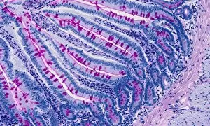
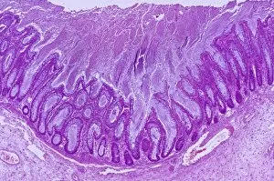
Background imageSectioned Collection: Small intestine, light micrograph

Small intestine, light micrograph
Small intestine. Light micrograph of a section through the small intestine. The finger-like projections at left are the villi. These increase the surface area for the absorption of food

Background imageSectioned Collection: Heart-lung anatomy, 1825 artwork

Heart-lung anatomy, 1825 artwork
Heart-lung anatomy. Axial section through the chest at the level of the fifth thoracic vertebra (T5). The front of the body is at top

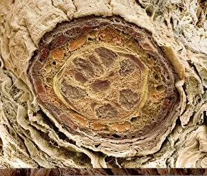
Background imageSectioned Collection: Hair follicle, SEM C014 / 0382

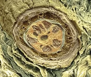
Hair follicle, SEM C014 / 0382
Hair follicle. Coloured scanning electron micrograph (SEM) of a section through a hair shaft and follicle (round structures at centre)

Background imageSectioned Collection: Intestinal gland cell, TEM C014 / 1440

Intestinal gland cell, TEM C014 / 1440
Intestinal gland cell. Transmission electron micrograph (TEM) of a section through an undifferentiated cell from the intestinal tract, showing extensive rough endoplasmic reticulum (ER)

Background imageSectioned Collection: Cartilage cell, TEM C014 / 1432

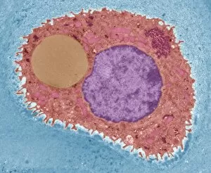
Cartilage cell, TEM C014 / 1432
Cartilage cell. Transmission electron micrograph (TEM) of a section through a chondrocyte cell from hyaline cartilage of the trachea (windpipe)

Background imageSectioned Collection: Kidney anatomy, 1825 artwork

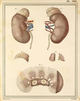
Kidney anatomy, 1825 artwork
Kidney anatomy. At top are the left and right kidneys (brown) with arteries (red), veins (blue), ureters (white) and adrenal glands (cream, on top of kidneys)

Background imageSectioned Collection: Cut root ginger C014 / 1251

Cut root ginger C014 / 1251
Cut root ginger. The rhizome (underground stem) of the ginger plant (Zingiber officinale) is used worldwide in a variety of forms as a cooking spice. It is also used in herbal remedies

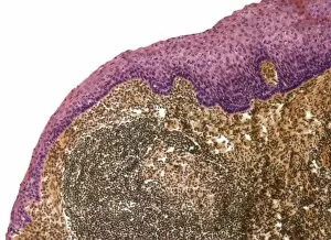
Background imageSectioned Collection: Tonsil, light micrograph F006 / 9818

Tonsil, light micrograph F006 / 9818
Tonsil. Light micrograph of a section through the surface of a tonsil showing the stratified squamous epithelium (pink) and underlying lymphoid tissue

Background imageSectioned Collection: Gall bladder lining, light micrograph F006 / 9814

Gall bladder lining, light micrograph F006 / 9814
Gall bladder lining. Light micrograph of a section through the gall bladder showing the folds of tall columnar epithelium (purple) overlying a connective tissue (brown)

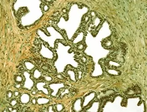
Background imageSectioned Collection: Seminal vesicle, light micrograph F006 / 9809

Seminal vesicle, light micrograph F006 / 9809
Seminal vesicle. Light micrograph of a section through the seminal vesicle showing the complex mucosal folding. The seminal vesicles are a pair of glands situated on each side of the male urinary

Background imageSectioned Collection: Seminal vesicle, light micrograph F006 / 9808

Seminal vesicle, light micrograph F006 / 9808
Seminal vesicle. Light micrograph of a section through the seminal vesicle showing the complex mucosal folding. The seminal vesicles are a pair of glands situated on each side of the male urinary

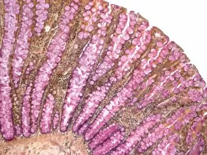
Background imageSectioned Collection: Colon, light micrograph F006 / 9806

Colon, light micrograph F006 / 9806
Colon, light micrograph. Goblet cells, which secrete mucous to lubricate the epithelium, are red. Magnification: x150 when printed at 10 centimetres wide

Background imageSectioned Collection: Acute bronchitis, light micrograph F006 / 9803

Acute bronchitis, light micrograph F006 / 9803
Acute bronchitis. Light micrograph of a section through a bronchus (airway) affected by acute bronchitis. The lumen is filled with pus (purple) consisting mainly of neutrophil white blood cells

Background imageSectioned Collection: Hair follicle, SEM C014 / 0383

Hair follicle, SEM C014 / 0383
Hair follicle. Coloured scanning electron micrograph (SEM) of a section through a hair shaft and follicle (round structures at centre)

Background imageSectioned Collection: Brain nerve cells, TEM C014 / 0357

Brain nerve cells, TEM C014 / 0357
Brain nerve cells. Transmission electron micrograph (TEM) of a section through brain tissue from the cerebral cortex, showing numerous neurons (nerve cells) surrounded by axons and dendrites

Background imageSectioned Collection: Trunk muscle anatomy, 1831 artwork

Trunk muscle anatomy, 1831 artwork
Trunk muscle anatomy. Coronal section of the trunk (anterior view) showing the anatomy of its muscles and bones. This anatomical artwork is plate 75 from volume 2 (1831)

Background imageSectioned Collection: Cell membrane, artwork C018 / 7905

Cell membrane, artwork C018 / 7905
Cell membrane. Computer artwork of a section though a cell membrane. This is a semi-permeable membrane that controls what substances leave and enter the cell

Background imageSectioned Collection: Bamboo stem, SEM C015 / 5073

Bamboo stem, SEM C015 / 5073
Bamboo stem. Coloured environmental scanning electron micrograph (ESEM) of a section through the stem of a bamboo plant (family Gramineae), showing a vascular bundle

Background imageSectioned Collection: Cancer cells spreading, artwork

Cancer cells spreading, artwork
Cancer cells spreading. Computer artwork showing cancerous cells (blue) migrating from a tumour (upper centre) into the blood stream (centre)

Background imageSectioned Collection: Oocyte, light micrograph

Oocyte, light micrograph
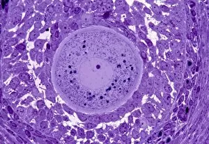
Oocyte. Light micrograph of a section through an oocyte within a early antral follicle in an ovary. Oocytes are immature ova, or egg cells

Background imageSectioned Collection: Cardiac muscle, light micrograph

Cardiac muscle, light micrograph
Cardiac muscle. Light micrograph of a section through muscle tissue from a heart, showing the cardiomyocyte cells. Heart muscle cells show central nuclei but the dominant feature is the abundance of

Background imageSectioned Collection: Spondylodiscitis, MRI scan

Spondylodiscitis, MRI scan
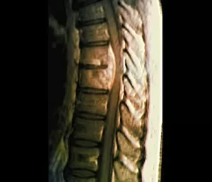
Spondylodiscitis. Coloured magnetic resonance imaging (MRI) scan of a sagittal section through the spine of a 69-year-old patient with spondylodiscitis at the level of the T7-T8 vertebra
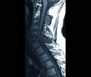
Background imageSectioned Collection: Abnormal bone growth, MRI scan

Abnormal bone growth, MRI scan
Abnormal bone growth. Magnetic resonance imaging (MRI) scan of a section through the cervical spine (neck) of a 28-year-old patient, showing abnormal growth (dysplasia) of the C6 vertebra

Background imageSectioned Collection: Thrombophlebitis in the brain, MRI scans

Thrombophlebitis in the brain, MRI scans
Thrombophlebitis in the brain. Coloured magnetic resonance imaging (MRI) angiograms comparing the blood vessels (white) of the brain and neck in a healthy 43-year-old (left)

Background imageSectioned Collection: Spinal cancer, MRI scan

Spinal cancer, MRI scan
Spinal cancer. Coloured magnetic resonance imaging (MRI) scan of a sagittal section through the cervical (neck) region of the spine of a 22-year-old patient

Background imageSectioned Collection: Colitis, light micrograph

Colitis, light micrograph
Colitis. Light micrograph of a section through colon (large intestine) tissue affected by bacterial infection, causing injury to the mucosal lining, which has resulted in inflammation (colitis)

Background imageSectioned Collection: Atrial extrasystole, anatomical artwork

Atrial extrasystole, anatomical artwork
Atrial extrasystole. Anatomical computer artwork of a transverse section through a human heart during atrial contraction in extrasystole

Background imageSectioned Collection: Hairy scalp skin, light micrograph

Hairy scalp skin, light micrograph
Hairy scalp skin. Light micrograph of a section through healthy skin from a human scalp, showing the numerous hair follicles. The scalp is an example of hairy skin


For sale as Licensed Images

Choose your image, Select your licence and Download the media

"Exploring the Intricacies: From Cerebellum Tissue to Fusion Research, Unveiling the World Sectioned" Delving into the depths of scientific exploration, a light micrograph reveals the intricate structure of cerebellum tissue. Witnessing a Higgs boson event through the lens of ATLAS detector C013/6892, scientists capture an awe-inspiring moment in particle physics. A meticulously crafted model showcases the complexity and beauty of the human brain, inviting us to marvel at its wonders. Peering into hippocampus brain tissue under a microscope, we uncover secrets that may hold keys to memory formation and learning processes. In pursuit of limitless energy sources, fusion research takes center stage as we examine a tokamak device designed for controlled nuclear fusion reactions. The inner workings of our brains come alive through an MRI scan, revealing detailed brain anatomy like never before imagined. Illuminated by light microscopy, dicotyledon plant stem sections unveil nature's architectural masterpiece in stunning detail. EDTA crystals take on mesmerizing patterns when viewed under intense magnification - showcasing their unique molecular arrangement through light micrography. Journeying deep within our bodies, kidney tubules are sectioned to shed light on their vital role in maintaining fluid balance and waste removal systems. An X-ray image exposes every contour and intricacy of a human skull – offering glimpses into our skeletal framework with remarkable clarity. Through transmission electron microscopy (TEM), rough endoplasmic reticulum emerges as an interconnected network responsible for protein synthesis within cells' cytoplasmic matrix Computer artwork visualizes Alzheimer's-affected brain regions – reminding us why continued research is crucial in unraveling this devastating disease. In this captivating journey across various scientific disciplines and microscopic landscapes, "sectioned" unveils hidden worlds that expand our understanding of the universe and ourselves.