Sectioned Collection (page 9)

"Exploring the Intricacies: From Cerebellum Tissue to Fusion Research, Unveiling the World Sectioned" Delving into the depths of scientific exploration

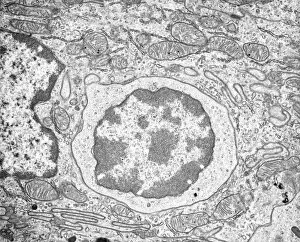
Background imageSectioned Collection: Lymphocyte white blood cell, TEM

Lymphocyte white blood cell, TEM
Lymphocyte white blood cell. Transmission electron micrograph (TEM) of a section through lymphocyte, showing the nucleus (large, round, centre), endoplasmic reticulum (ER)

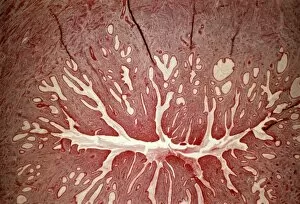
Background imageSectioned Collection: Cervix, light micrograph

Cervix, light micrograph
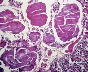
Cervix. Light micrograph of a section through the mucosa of the cervix, the neck of the uterus. This is the endocervix, which borders the uterus. It is lined with columnar epithelial cells

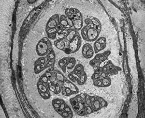
Background imageSectioned Collection: Unmyelinated nerve, TEM

Unmyelinated nerve, TEM
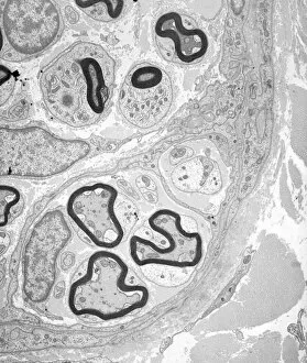
Unmyelinated nerve. Transmission electron micrograph (TEM) of a section through axon (nerve fibre) bundles of unmyelinated nerves

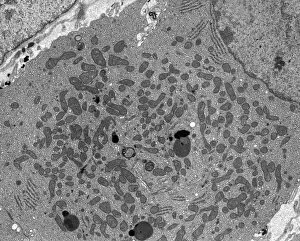
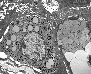
Background imageSectioned Collection: Steroid secreting cell, TEM

Steroid secreting cell, TEM
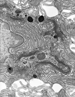
Steroid secreting cell. Transmission electron micrograph (TEM) of a section through a cell involved with steroid synthesis and secretion (steroidogenesis)

Background imageSectioned Collection: Enterocyte, TEM

Enterocyte, TEM
Enterocyte. Transmission electron micrograph (TEM) of a section through the cytoplasm and part of the nucleus of an enterocyte cell located in a crypt of Lieberkuhn of the small intestine

Background imageSectioned Collection: Islet of Langerhans, TEM

Islet of Langerhans, TEM
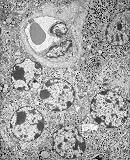
Islet of Langerhans. Transmission electron micrograph (TEM) of a section through cells clustered in an islet of Langerhans in the pancreas

Background imageSectioned Collection: Capillary, TEM

Capillary, TEM
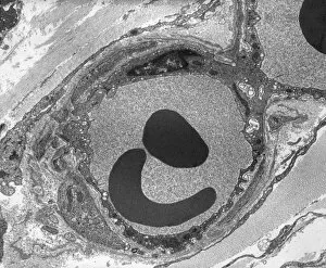
Capillary. Transmission electron micrograph (TEM) of a section through a capillary, showing two red blood cells (erythrocytes, black) in its interior

Background imageSectioned Collection: Kidney mitochondria, TEM

Kidney mitochondria, TEM
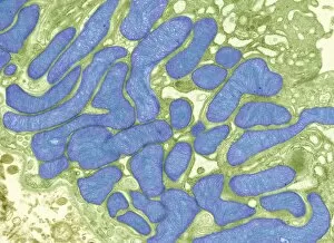
Kidney mitochondria. Transmission electron micrograph (TEM) of a section through a kidney tubule, showing numerous mitochondria (blue)

Background imageSectioned Collection: Berry aneurysm, MRI scan

Berry aneurysm, MRI scan
Stroke. Coloured computed tomography (CT) scan of an axial section through the brain of a 58-year-old patient, showing the damage (dark) caused 24 hours after a cerebral infarction

Background imageSectioned Collection: Creutzfeldt-Jakob disease, MRI scan

Creutzfeldt-Jakob disease, MRI scan
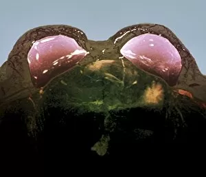
Creutzfeldt-Jakob disease. Coloured magnetic resonance imaging (MRI) scan of an axial section through the brain of a 35-year-old patient

Background imageSectioned Collection: Pineal gland, light micrograph

Pineal gland, light micrograph. The pineal gland is a small endocrine gland located near to the centre of the brain, between the two hemispheres

Background imageSectioned Collection: Stroke, CT scan

Stroke, CT scan
Stroke. Coloured computed tomography (CT) scan of an axial section through the brain of a 58-year-old patient, showing the damage (purple) caused 24 hours after a cerebral infarction

Background imageSectioned Collection: Double mastectomy breast implants, MRI

Double mastectomy breast implants, MRI
Double mastectomy breast implants. Post-operative coloured magnetic resonance imaging (MRI) scan of a section through the chest of a 61-year-old patient after a double mastectomy (breast removal)

Background imageSectioned Collection: Myelinated nerve tissue, TEM

Myelinated nerve tissue, TEM
Myelinated nerve tissue. Transmission electron micrograph (TEM) of a section through myelinated nerve fibres (axons). Each axon is coated with many layers of myelin

Background imageSectioned Collection: Kidney tumour, CT scan

Kidney tumour, CT scan
Kidney tumour. Coloured computed tomography (CT) scan of a section through the abdomen of a 62-year-old patient, showing necrosis (death) of tissue in the right kidney

Background imageSectioned Collection: Calcified heart valve, CT scans

Calcified heart valve, CT scans
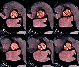
Calcified heart valve. Coloured computed tomography (CT) scans of a section through the heart of a 67-year-old patient showing calcification of the aortic valve (round)

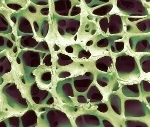
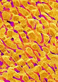
Background imageSectioned Collection: Bone tissue, SEM

Bone tissue, SEM
Bone tissue. Coloured scanning electron micrograph (SEM) of cancellous (spongy) bone. Bone tissue can be either cortical (compact) or cancellous

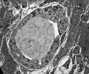
Background imageSectioned Collection: Ovarian follicle, TEM

Ovarian follicle, TEM
Ovarian follicle. Transmission electron micrograph (TEM) of a section through an ovary, showing a primary follicle. Primary follicles contain a central oocyte (female germ cell)

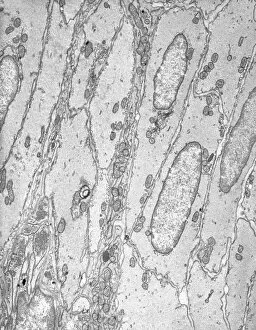
Background imageSectioned Collection: Smooth muscle tissue, TEM

Smooth muscle tissue, TEM
Smooth muscle tissue. Transmission electron micrograph (TEM) of a longitudinal section through smooth muscle cells. Smooth muscle cells are spindle-shaped and often closely packed together

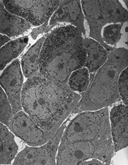
Background imageSectioned Collection: Anterior pituitary gland, TEM

Anterior pituitary gland, TEM
Anterior pituitary gland. Transmission electron micrograph (TEM) of a section through cells of the anterior pituitary. The anterior pituitary, or adenohypophysis

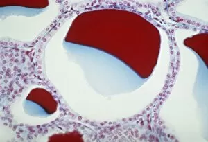
Background imageSectioned Collection: Thyroid follicles, light micrograph

Thyroid follicles, light micrograph
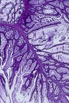
Thyroid follicles. Light micrograph of a section through a thyroid gland showing the follicles. The follicles are lined by a single layer of cuboidal epithelial cells
Background imageSectioned Collection: Intestinal brush border, TEM

Intestinal brush border, TEM
Intestinal brush border. Transmission electron micrograph (TEM) of intestinal absorptive cells sectioned horizontally at their apex to show the surface microvilli (round)

Background imageSectioned Collection: Enteroendocrine cell, TEM

Enteroendocrine cell, TEM
Enteroendocrine cell. Transmission electron micrograph (TEM) of a section through an enteroendocrine cell within a gland of the stomach

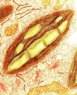
Background imageSectioned Collection: Chloroplast, TEM C016 / 6297

Chloroplast, TEM C016 / 6297
Chloroplast. Coloured transmission electron micrograph (TEM) of chloroplast from the moss Physcomitrella patens. Chloroplasts are the sites of photosynthesis

Background imageSectioned Collection: Polished coral C016 / 5994

Polished coral C016 / 5994
Polished coral (Isastraea oblonga). Specimen from the Jurassic period, found in upper Portlandian beds in Tisbury, Wiltshire, UK

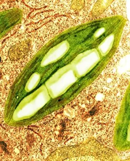
Background imageSectioned Collection: Chloroplast, TEM C016 / 6298

Chloroplast, TEM C016 / 6298
Chloroplast. Coloured transmission electron micrograph (TEM) of chloroplast from the moss Physcomitrella patens. Chloroplasts are the sites of photosynthesis

Background imageSectioned Collection: Trachea lining, SEM C015 / 9939

Trachea lining, SEM C015 / 9939
Trachea lining. Coloured scanning electron micrograph (SEM) of a section through the lining of the trachea (wind pipe). The trachea links the larynx (voice box) to the lungs

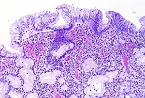
Background imageSectioned Collection: Gastritis, light micrograph C016 / 0508

Gastritis, light micrograph C016 / 0508
Gastritis. Light micrograph of a section through an inflamed human stomach (gastritis), showing an abnormal accumulation of white blood cells (leucocytes)

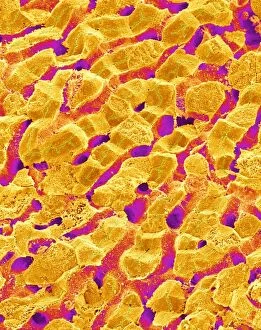
Background imageSectioned Collection: Liver, SEM C015 / 9924

Liver, SEM C015 / 9924
Liver vein. Coloured scanning electron micrograph (SEM) of liver tissue. The functions of the liver include destroying old red blood cells

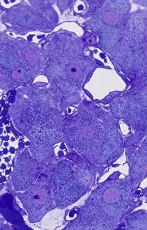
Background imageSectioned Collection: Corpus luteum of ovary, light micrograph C016 / 0527

Corpus luteum of ovary, light micrograph C016 / 0527
Corpus luteum of ovary. Light micrograph of a section through steroid-secreting luteal cells in the corpus luteum of an ovary. The corpus luteum develops from an ovulated follicle

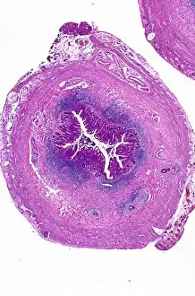
Background imageSectioned Collection: Appendix, light micrograph C016 / 0510

Appendix, light micrograph C016 / 0510
Appendix. Light micrograph of a section through a human appendix. The appendix is a blind-ending small diameter tubular extension of the gut, arising from the caecum of the large bowel

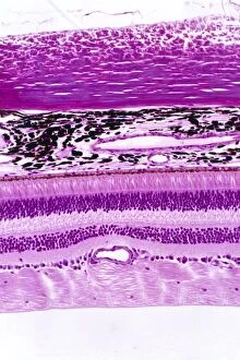
Background imageSectioned Collection: Retina of the eye, light micrograph C016 / 0528

Retina of the eye, light micrograph C016 / 0528
Retina of the eye. Light micrograph of a section through the retina from a human eye. From top down: nerve fibres of the optic nerve and a blood vessel; several layers of neurons (nerve cells)

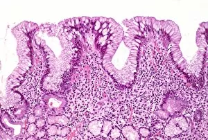
Background imageSectioned Collection: Gastritis, light micrograph C016 / 0507

Gastritis, light micrograph C016 / 0507
Gastritis. Light micrograph of a section through an inflamed human stomach (gastritis), showing an abnormal accumulation of white blood cells (leucocytes)

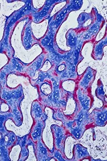
Background imageSectioned Collection: Cancellous bone, light micrograph C016 / 0509

Cancellous bone, light micrograph C016 / 0509
Cancellous bone. Light micrograph of a section through cancellous, or spongy, bone. Constructed in a honeycomb-type architecture

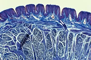
Background imageSectioned Collection: Tongue, light micrograph C015 / 7105

Tongue, light micrograph C015 / 7105
Tongue. Light micrograph of a section through healthy tongue tissue. The surface of the tongue (across top) consists of stratified squamous epithelium (dark purple) with a rough keratinised surface

Background imageSectioned Collection: Liver, SEM C015 / 9922

Liver, SEM C015 / 9922
Liver vein. Coloured scanning electron micrograph (SEM) of liver tissue showing a large vein (pink, up centre). The functions of the liver include destroying old red blood cells

Background imageSectioned Collection: Colon polyp, light micrograph C016 / 0512

Colon polyp, light micrograph C016 / 0512
Colon polyp. Light micrograph of a section through part of a tubulovillous adenoma of the mucosa of the large bowel (colon)

Background imageSectioned Collection: Lens of the eye, light micrograph C016 / 0526

Lens of the eye, light micrograph C016 / 0526
Lens of the eye. Light micrograph of a section through the lens from a human eye. The lens consists of thousands of precisely aligned stacks of fibres seen here end-on

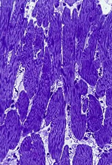
Background imageSectioned Collection: Heart muscle, light micrograph C016 / 0517

Heart muscle, light micrograph C016 / 0517
Heart muscle. Light micrograph of a section through heart (cardiac) muscle tissue, showing cardiomyocyte muscle cells. These cells have a centrally located nucleus and are branched

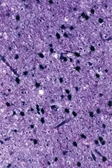
Background imageSectioned Collection: Brain glial cells, light micrograph C016 / 0524

Brain glial cells, light micrograph C016 / 0524
Brain glial cells. Light micrograph of a section through brain tissue, showing numerous glial cells (dark). Glial cells, or neuroglia, are non-neural cells of the central nervous system (CNS)

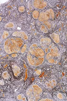
Background imageSectioned Collection: Cirrhosis of liver, light micrograph C016 / 0530

Cirrhosis of liver, light micrograph C016 / 0530
Cirrhosis of liver. Light micrograph of a section through liver tissue, damaged by cirrhosis. Cirrhosis occurs as a result of a range of factors causing damage to liver function

Background imageSectioned Collection: Liver, SEM C015 / 9923

Liver, SEM C015 / 9923
Liver vein. Coloured scanning electron micrograph (SEM) of liver tissue. The functions of the liver include destroying old red blood cells


For sale as Licensed Images

Choose your image, Select your licence and Download the media

"Exploring the Intricacies: From Cerebellum Tissue to Fusion Research, Unveiling the World Sectioned" Delving into the depths of scientific exploration, a light micrograph reveals the intricate structure of cerebellum tissue. Witnessing a Higgs boson event through the lens of ATLAS detector C013/6892, scientists capture an awe-inspiring moment in particle physics. A meticulously crafted model showcases the complexity and beauty of the human brain, inviting us to marvel at its wonders. Peering into hippocampus brain tissue under a microscope, we uncover secrets that may hold keys to memory formation and learning processes. In pursuit of limitless energy sources, fusion research takes center stage as we examine a tokamak device designed for controlled nuclear fusion reactions. The inner workings of our brains come alive through an MRI scan, revealing detailed brain anatomy like never before imagined. Illuminated by light microscopy, dicotyledon plant stem sections unveil nature's architectural masterpiece in stunning detail. EDTA crystals take on mesmerizing patterns when viewed under intense magnification - showcasing their unique molecular arrangement through light micrography. Journeying deep within our bodies, kidney tubules are sectioned to shed light on their vital role in maintaining fluid balance and waste removal systems. An X-ray image exposes every contour and intricacy of a human skull – offering glimpses into our skeletal framework with remarkable clarity. Through transmission electron microscopy (TEM), rough endoplasmic reticulum emerges as an interconnected network responsible for protein synthesis within cells' cytoplasmic matrix Computer artwork visualizes Alzheimer's-affected brain regions – reminding us why continued research is crucial in unraveling this devastating disease. In this captivating journey across various scientific disciplines and microscopic landscapes, "sectioned" unveils hidden worlds that expand our understanding of the universe and ourselves.