Transmission Electron Collection (#5)

"Unlocking the Intricacies of Life

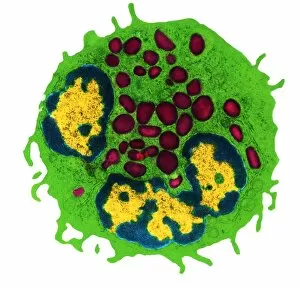
Background imageTransmission Electron Collection: Coloured TEM of a basophil white blood cell

Coloured TEM of a basophil white blood cell
Polynuclear basophil. Coloured Transmission Electron Micrograph (TEM) of a basophil white blood cell. This basophil has one bi-lobed nucleus (yellow, lower centre) and a second nucleus at upper left

Background imageTransmission Electron Collection: Coronary artery, TEM

Coronary artery, TEM
Coronary artery. Coloured transmission electron micrograph (TEM) of a cross-section through the wall of a coronary artery

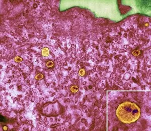
Background imageTransmission Electron Collection: HIV particles in infected cell, TEM

HIV particles in infected cell, TEM
HIV particles. Coloured transmission electron micrograph (TEM) of human immunodeficiency virus (HIV) particles (orange) in a host cell. HIV causes the disease AIDS

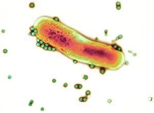
Background imageTransmission Electron Collection: Adenovirus particles and bacterium, TEM

Adenovirus particles and bacterium, TEM
Adenovirus particles and bacterium. Coloured transmission electron micrograph (TEM) of adenovirus particles (green) with a gut bacterium (orange)

Background imageTransmission Electron Collection: Colour TEM of a cluster of five influenza viruses

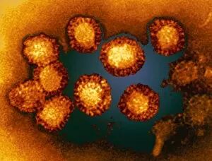
Colour TEM of a cluster of five influenza viruses
Influenza virus. Coloured transmission electron micrograph of a cluster of five spherical-shaped influenza (flu) viruses. Each virus is made up of a core of ribonucleic acid (RNA, not seen here)

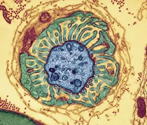
Background imageTransmission Electron Collection: Nerve fibre node, TEM

Nerve fibre node, TEM
Nerve fibre node. Coloured transmission electron micrograph (TEM) of a cross-section through a nerve fibre (axon) at a node of Ranvier

Background imageTransmission Electron Collection: Neutrophil and trapped bacteria, TEM

Neutrophil and trapped bacteria, TEM
Neutrophil and trapped bacteria. Coloured transmission electron micrograph (TEM) of Neisseria gonorrhoeae bacteria (blue) trapped by a neutrophil (orange), a type of white blood cell

Background imageTransmission Electron Collection: Immunoglobulin G antibodies, TEM

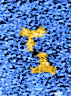
Immunoglobulin G antibodies, TEM
Immunoglobulin G antibody molecules, coloured transmission electron micrograph (TEM). IgG is the most abundant human immunoglobulin, and is found in all body fluids

Background imageTransmission Electron Collection: Blood platelet, TEM

Blood platelet, TEM

Background imageTransmission Electron Collection: Colour TEM of red blood cells, rouleau formation

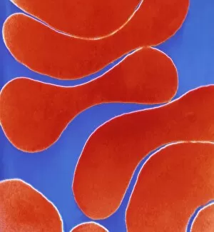
Colour TEM of red blood cells, rouleau formation
Red blood cells. Coloured Transmission Electron Micrograph (TEM) of sectioned human red blood cells (erythrocytes) in a rouleau formation

Background imageTransmission Electron Collection: AIDS viruses budding from T-cell

AIDS viruses budding from T-cell
AIDS viruses. Coloured transmission electron micrograph (TEM) of rounded Human Immunodeficiency viruses (HIV) budding from the surface of an infected T-lymphocyte blood cell (T-cell)

Background imageTransmission Electron Collection: Polyoma viruses

Polyoma viruses. Coloured transmission electron micrograph (TEM) of polyoma viruses. These simian (monkey) viruses (SV40) are a type of papovavirus

Background imageTransmission Electron Collection: West Nile viruses

West Nile viruses. Coloured transmission electron micrograph (TEM) of a group of West Nile viruses (WNV). This virus is known to cause encephalitis in humans

Background imageTransmission Electron Collection: Hydronephrosis

Hydronephrosis. Coloured transmission electron micrograph (TEM) of a section through a human kidney, showing a renal tubule in hydronephrosis

Background imageTransmission Electron Collection: Nerve demyelination, TEM

Nerve demyelination, TEM
Nerve demyelination. Coloured transmission electron micrograph (TEM) of a section through a Schwann cell and a nerve fibre, showing the early collapse of its myelin sheath

Background imageTransmission Electron Collection: Alzheimers disease brain cell, TEM

Alzheimers disease brain cell, TEM
Alzheimers disease brain cell. Coloured transmission electron micrograph (TEM) of a neurofibrillary tangle in a nerve cell from the brain of a patient with Alzheimers disease

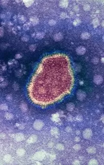
Background imageTransmission Electron Collection: Influenza virus, TEM

Influenza virus, TEM
Influenza virus. Coloured transmission electron micrograph (TEM) of an influenza ( flu) virus particle. Influenza belongs to the orthomyxovirus group of viruses

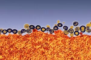
Background imageTransmission Electron Collection: Budding HIV particles, TEM

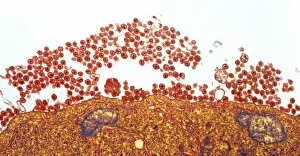
Budding HIV particles, TEM
Budding HIV particles. Coloured transmission electron micrograph (TEM) of HIV particles (blue and yellow) budding from the membrane of the host cell (orange)

Background imageTransmission Electron Collection: Reovirus particles in a cell, TEM

Reovirus particles in a cell, TEM
Reovirus particles. Coloured transmission electron micrograph (TEM) of hundreds of particles (blue) of a reovirus infecting a host cell. The nucleus of the cell (green) is at centre

Background imageTransmission Electron Collection: West Nile viruses, TEM

West Nile viruses, TEM

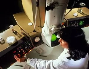
Background imageTransmission Electron Collection: Operator using a transmission electron microscope

Operator using a transmission electron microscope (TEM). The operating console is seen with a TV monitor and magnified image

Background imageTransmission Electron Collection: Cell nucleolus, TEM

Cell nucleolus, TEM
Nucleolus. Coloured transmission electron micrograph of the nucleolus (blue), a component of the nucleus (orange) of a cell. The cell cytoplasm is green

Background imageTransmission Electron Collection: Fallopian tube, TEM

Fallopian tube, TEM
Fallopian tube, coloured transmission electron micrograph (TEM). Section through non-ciliated columnar epithelium from a fallopian tube. The ciliated cells are not shown here

Background imageTransmission Electron Collection: Artery, TEM

Artery, TEM
Artery. Coloured transmission electron micrograph (TEM) of a transverse section through an artery. Red blood cells (erythrocytes, red) and white blood cells (leucocytes)

Background imageTransmission Electron Collection: Nasal lining, TEM

Nasal lining, TEM
Nasal lining. Transmission electron micrograph (TEM) of a section through cilia (green) covering the epithelial lining of the nasopharyx, the part of the throat behind the nasal cavity

Background imageTransmission Electron Collection: Smell receptors, TEM

Smell receptors, TEM
Smell receptors. Coloured transmission electron micrograph (TEM) of a section through smell receptors (cilia) projecting from an olfactory neurone (blue)

Background imageTransmission Electron Collection: Mast cell, TEM

Mast cell, TEM
Mast cell, coloured transmission electron micrograph (TEM). Mast cells are a type of white blood cell found in connective tissue

Background imageTransmission Electron Collection: Influenza viruses, TEM

Influenza viruses, TEM
Influenza viruses. Coloured transmission electron micrograph (TEM) of influenza (flu) viruses (yellow) budding from a host cell. The host cell nucleus is black

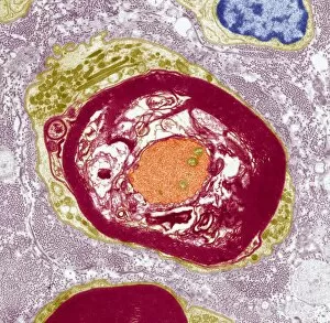
Background imageTransmission Electron Collection: Capillary and red blood cell, TEM

Capillary and red blood cell, TEM
Capillary and red blood cell, coloured transmission electron micrograph (TEM). This cross-section shows a single red blood cell almost filling the lumen of the capillary


For sale as Licensed Images

Choose your image, Select your licence and Download the media

"Unlocking the Intricacies of Life: Exploring the Microscopic World through Transmission Electron Microscopy (TEM)" Delving into the depths of cellular structures and biological processes, transmission electron microscopy (TEM) has revolutionized our understanding of various aspects of life. With its high-resolution imaging capabilities, TEM allows us to witness intricate details that were once hidden from our sight. At synapse nerve junctions, TEM reveals a mesmerizing dance between neurotransmitters, enabling communication between neurons with remarkable precision. Norovirus particles come alive under TEM's lens, showcasing their unique shape and arrangement – a crucial insight in combating these notorious pathogens. E. Coli bacteria appear as tiny rods when observed through TEM, reminding us of their omnipresence in nature and sometimes unfortunate encounters in human health. Fat cells take on an unexpected beauty when magnified by TEM; their delicate structure resembling a web-like network that stores energy for our bodies. The myelination process is brought to life through TEM images capturing nerve fibers coated with protective sheaths. These stunning visuals help unravel the mysteries behind efficient neural signaling and hold promise for treating demyelinating diseases such as multiple sclerosis. TEM exposes the menacing presence of MRSA-resistant Staphylococcus bacteria – formidable adversaries in healthcare settings worldwide. Another glimpse at E. coli bacterium showcases its intricate internal machinery responsible for vital functions within this single-celled organism. Nerve cells reveal their complexity under TEM's scrutiny – branching dendrites reaching out like tree branches while axons transmit electrical signals across vast distances. Mitochondria steal the spotlight as they power these nerve cells' activities, appearing as dynamic organelles teeming with energy-producing potential. Plasma cells burst forth with vibrant colors when examined using TEM; their role in producing antibodies becomes even more awe-inspiring upon closer inspection. Once again, myelinated nerve fibers captivate us with their elegant architecture – a testament to nature's ingenuity in optimizing neural communication.