Transmission Electron Collection (#8)

"Unlocking the Intricacies of Life

Background imageTransmission Electron Collection: Influenza C virus, TEM

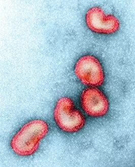
Influenza C virus, TEM
Influenza C virus. Coloured transmission electron micrograph (TEM) of the influenza C virus, which is one of the causes of influenza (flu) in humans and pigs

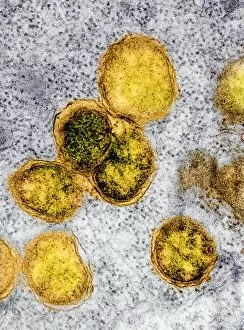
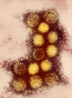
Background imageTransmission Electron Collection: Adenovirus particles, TEM

Adenovirus particles, TEM
Adenovirus particles. Coloured transmission electron micrograph (TEM) of adenovirus particles. Each particle consists of an icosahedral protein coat (capsid)

Background imageTransmission Electron Collection: Monkeypox virus particle, TEM

Monkeypox virus particle, TEM
Monkeypox virus particle, coloured transmission electron micrograph (TEM). Monkeypox is a poxvirus that was identified in Cynomolgus monkeys in 1958 and then in humans in 1970

Background imageTransmission Electron Collection: Mosquito testis infected with bacteria

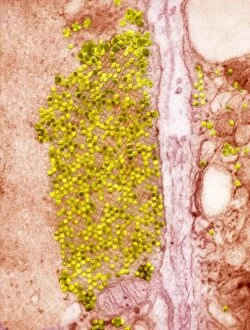
Mosquito testis infected with bacteria. Coloured transmission electron micrograph (TEM) of a testis from a Culex pipiens mosquito infected with Walbachia sp. bacteria (yellow)

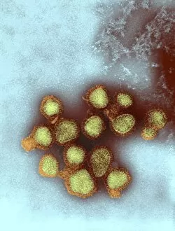
Background imageTransmission Electron Collection: H5N3 influenza A virus particles, TEM

H5N3 influenza A virus particles, TEM
H5N3 influenza A virus particles. Coloured transmission electron micrograph (TEM) of the H5N3 strain of influenza virus type A. All five H5 strains (H5N1, H5N2, H5N3, H5N8 and H5N9) cause bird flu

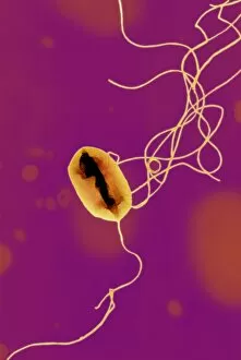
Background imageTransmission Electron Collection: E. coli bacterium strain O157: H7, TEM

E. coli bacterium strain O157: H7, TEM
E. coli bacterium strain O157:H7, coloured transmission electron micrograph (TEM). E. coli are Gram-negative rod-shaped bacteria that are part of the normal flora of the human gut

Background imageTransmission Electron Collection: Listeria bacteria, TEM

Listeria bacteria, TEM
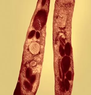
Listeria sp. bacteria, coloured transmission electron micrograph (TEM). Listeria are Gram-positive rod-shaped bacteria. The species L

Background imageTransmission Electron Collection: Human coronavirus, TEM

Human coronavirus, TEM
Human coronavirus. Coloured transmission electron micrograph (TEM) of Human coronavirus particles. Coronaviruses primarily infect the upper respiratory and gastrointestinal tract

Background imageTransmission Electron Collection: Nerve fibre, TEM

Nerve fibre, TEM
Nerve fibre. Coloured transmission electron micrograph (TEM) of a section through a non-myelinated peripheral nerve fibre

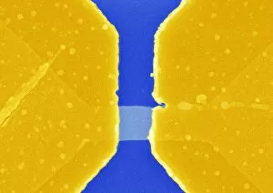
Background imageTransmission Electron Collection: Graphene transistor, SEM

Graphene transistor, SEM
Graphene transistor. Coloured scanning electron micrograph (SEM) of a transistor composed of a graphene wire (centre), gold electrodes (dark yellow) and silicon (blue)

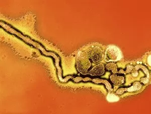
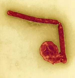
Background imageTransmission Electron Collection: Marburg virus, TEM

Marburg virus, TEM
Marburg virus, coloured transmission electron micrograph (TEM). This tubular RNA (ribonucleic acid) virus causes Marburg haemorrhagic fever in humans and non-human primates

Background imageTransmission Electron Collection: Macrophage cells, TEM

Macrophage cells, TEM
Macrophage cells, coloured transmission electron micrograph (TEM). The cell nuclei are purple. Mitochondria (dark pink ovals) in the cytoplasm produce energy for the cell

Background imageTransmission Electron Collection: La Crosse encephalitis virus, TEM

La Crosse encephalitis virus, TEM
La Crosse encephalitis virus. Coloured transmission electron micrograph (TEM) of La Cross (LAC) encephalitis virus particles (virions)

Background imageTransmission Electron Collection: Mosquito ovary infected with bacteria TEM

Mosquito ovary infected with bacteria TEM
Mosquito ovary infected with bacteria. Coloured transmission electron micrograph (TEM) of an ovary from a Culex pipiens mosquito infected with Walbachia sp. bacteria (yellow)

Background imageTransmission Electron Collection: Ebola virus particles, TEM

Ebola virus particles, TEM
Ebola virus particles, coloured transmission electron micrograph (TEM). Each particle consists of a ribonucleic acid (RNA) genome within a nucleocapsid (purple)

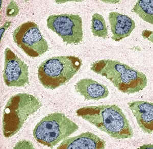
Background imageTransmission Electron Collection: Cartilage cells, TEM

Cartilage cells, TEM
Cartilage cells. Coloured transmission electron micrograph (TEM) of a section through chondrocytes from nasal hyaline cartilage

Background imageTransmission Electron Collection: E. coli bacteria strain O157: H7, TEM

E. coli bacteria strain O157: H7, TEM
E. coli bacteria strain O157:H7, coloured transmission electron micrograph (TEM). E. coli are Gram-negative rod-shaped bacteria that are part of the normal flora of the human gut

Background imageTransmission Electron Collection: Graphene, SEM

Graphene, SEM
Graphene. Coloured scanning electron micrograph (SEM) of a crumpled layer of graphene. Graphene is composed of a single layer of carbon atoms arranged in a honeycomb crystal lattice

Background imageTransmission Electron Collection: California encephalitis virus, TEM

California encephalitis virus, TEM
California encephalitis virus. Coloured transmission electron micrograph of California encephalitis virus particles (virions)

Background imageTransmission Electron Collection: Changuinola virus, TEM

Changuinola virus, TEM
Changuinola virus. Coloured transmission electron micrograph of Changuinola virus particles (virions). Each particle consists of a protein coat (capsid)

Background imageTransmission Electron Collection: Avian influenza virus particles, TEM

Avian influenza virus particles, TEM
Avian influenza virus particles. Coloured transmission electron micrograph (TEM) of a H5 strain of influenza virus type A. All five H5 strains (H5N1, H5N2, H5N3, H5N8 and H5N9) cause bird flu

Background imageTransmission Electron Collection: Unmyelinated nerve bundle, TEM

Unmyelinated nerve bundle, TEM
Unmyelinated nerve bundle. Coloured transmission electron micrograph (TEM) of a section through a group of unmylinated nerve fibres

Background imageTransmission Electron Collection: Rhabdovirus, TEM

Rhabdovirus, TEM
Rhabdovirus. Transmission electron micrograph (TEM) of particles of the rhabdovirus vesicular stomatitis virus (VSV). These particles bullet shape is characteristic of rhabdoviruses

Background imageTransmission Electron Collection: H1N1 Influenza A virus particles, TEM

H1N1 Influenza A virus particles, TEM
H1N1 Influenza A virus particles, coloured transmission electron micrograph (TEM). This influenza virus can infect multiple species

Background imageTransmission Electron Collection: Anthrax bacteria, TEM

Anthrax bacteria, TEM
Anthrax bacteria. Coloured transmission electron micrograph of Bacillus anthracis bacteria. B. anthracis is a Gram-positive, rod-shaped, spore- forming bacterium that causes the disease anthrax

Background imageTransmission Electron Collection: Legionella bacterium, TEM

Legionella bacterium, TEM
Legionella bacteria. Coloured transmission electron micrograph of a Legionella pneumophila bacterium, the cause of Legionnaires disease

Background imageTransmission Electron Collection: H5N1 avian influenza virus particles, TEM

H5N1 avian influenza virus particles, TEM
H5N1 avian influenza virus particles, coloured transmission electron micrograph (TEM). Each virus particle consists of ribonucleic acid (RNA)

Background imageTransmission Electron Collection: Coxsackie B3 virus particles, TEM

Coxsackie B3 virus particles, TEM
Coxsackie B3 virus particles, coloured transmission electron micrograph (TEM). Each Coxsackie B3 virus particle (yellow) consists of a non-enveloped icosahedral (20-sided) protein capsid (coat)

Background imageTransmission Electron Collection: Syphilis bacterium, TEM

Syphilis bacterium, TEM
Syphilis bacterium. Coloured transmission electron micrograph (TEM) showing the spirochaete bacterium Treponema pallidum, which causes syphilis

Background imageTransmission Electron Collection: Spleen cell, TEM

Spleen cell, TEM
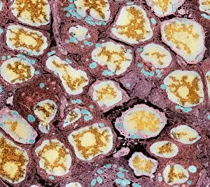
Spleen cell. Coloured transmission electron micrograph (TEM) of a section through a cell in the spleen, an organ located in the upper left side of the abdomen in vertebrate animals

Background imageTransmission Electron Collection: Spleen cells, TEM

Spleen cells, TEM
Spleen cells. Coloured transmission electron micrograph (TEM) of a section through cells in the spleen, an organ located in the upper left side of the abdomen in vertebrate animals

Background imageTransmission Electron Collection: Lung alveoli and red blood cells, TEM

Lung alveoli and red blood cells, TEM
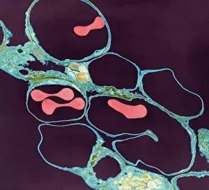
Lung alveoli and red blood cells. Coloured transmission electron micrograph (TEM) of a section through lung alveoli (blue) and red blood cells (red)

Background imageTransmission Electron Collection: Bacteria contamination, TEM

Bacteria contamination, TEM
Bacteria contamination. Coloured transmission electron micrograph (TEM) of a group of bacteria that have contaminated the growth medium in a cell culture plate

Background imageTransmission Electron Collection: Lung alveoli and blood cells, TEM

Lung alveoli and blood cells, TEM
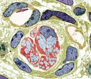
Lung alveoli and blood cells. Coloured transmission electron micrograph (TEM) of a section through lung alveoli and blood cells


For sale as Licensed Images

Choose your image, Select your licence and Download the media

"Unlocking the Intricacies of Life: Exploring the Microscopic World through Transmission Electron Microscopy (TEM)" Delving into the depths of cellular structures and biological processes, transmission electron microscopy (TEM) has revolutionized our understanding of various aspects of life. With its high-resolution imaging capabilities, TEM allows us to witness intricate details that were once hidden from our sight. At synapse nerve junctions, TEM reveals a mesmerizing dance between neurotransmitters, enabling communication between neurons with remarkable precision. Norovirus particles come alive under TEM's lens, showcasing their unique shape and arrangement – a crucial insight in combating these notorious pathogens. E. Coli bacteria appear as tiny rods when observed through TEM, reminding us of their omnipresence in nature and sometimes unfortunate encounters in human health. Fat cells take on an unexpected beauty when magnified by TEM; their delicate structure resembling a web-like network that stores energy for our bodies. The myelination process is brought to life through TEM images capturing nerve fibers coated with protective sheaths. These stunning visuals help unravel the mysteries behind efficient neural signaling and hold promise for treating demyelinating diseases such as multiple sclerosis. TEM exposes the menacing presence of MRSA-resistant Staphylococcus bacteria – formidable adversaries in healthcare settings worldwide. Another glimpse at E. coli bacterium showcases its intricate internal machinery responsible for vital functions within this single-celled organism. Nerve cells reveal their complexity under TEM's scrutiny – branching dendrites reaching out like tree branches while axons transmit electrical signals across vast distances. Mitochondria steal the spotlight as they power these nerve cells' activities, appearing as dynamic organelles teeming with energy-producing potential. Plasma cells burst forth with vibrant colors when examined using TEM; their role in producing antibodies becomes even more awe-inspiring upon closer inspection. Once again, myelinated nerve fibers captivate us with their elegant architecture – a testament to nature's ingenuity in optimizing neural communication.