
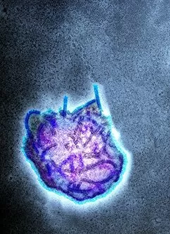
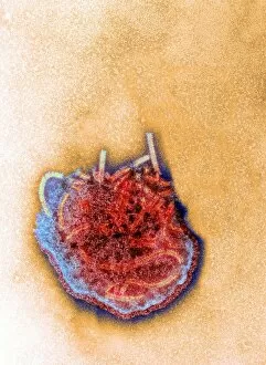
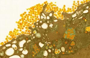
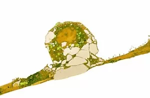
Transmission Electron Collection (#2)

"Unlocking the Intricacies of Life

Background imageTransmission Electron Collection: Activated plasma cell, TEM

Activated plasma cell, TEM
Activated plasma cell, coloured transmission electron micrograph (TEM). Plasma cells are derived from stimulated B lymphocytes (white blood cells)

Background imageTransmission Electron Collection: MERS coronavirus, TEM C015 / 1772

MERS coronavirus, TEM C015 / 1772
MERS coronavirus, coloured transmission electron micrograph (TEM). This virus (originally novel coronavirus 2012) has been named Middle East respiratory syndrome (MERS) coronavirus

Background imageTransmission Electron Collection: MERS coronavirus, TEM C015 / 1774

MERS coronavirus, TEM C015 / 1774
MERS coronavirus, coloured transmission electron micrograph (TEM). This virus (originally novel coronavirus 2012) has been named Middle East respiratory syndrome (MERS) coronavirus

Background imageTransmission Electron Collection: Granulocyte white blood cell, TEM

Granulocyte white blood cell, TEM
Granulocyte white blood cell. Coloured transmission electron micrograph (TEM) of a neutrophil granulocyte, a type of white blood cell

Background imageTransmission Electron Collection: MERS coronavirus, TEM C015 / 1776

MERS coronavirus, TEM C015 / 1776
MERS coronavirus, coloured transmission electron micrograph (TEM). This virus (originally novel coronavirus 2012) has been named Middle East respiratory syndrome (MERS) coronavirus

Background imageTransmission Electron Collection: MERS coronavirus, TEM C015 / 1773

MERS coronavirus, TEM C015 / 1773
MERS coronavirus, coloured transmission electron micrograph (TEM). This virus (originally novel coronavirus 2012) has been named Middle East respiratory syndrome (MERS) coronavirus

Background imageTransmission Electron Collection: Chloroplast, TEM C016 / 6297

Chloroplast, TEM C016 / 6297
Chloroplast. Coloured transmission electron micrograph (TEM) of chloroplast from the moss Physcomitrella patens. Chloroplasts are the sites of photosynthesis

Background imageTransmission Electron Collection: Chloroplast, TEM C016 / 6298

Chloroplast, TEM C016 / 6298
Chloroplast. Coloured transmission electron micrograph (TEM) of chloroplast from the moss Physcomitrella patens. Chloroplasts are the sites of photosynthesis

Background imageTransmission Electron Collection: Measles virus particle, TEM C015 / 7160

Measles virus particle, TEM C015 / 7160
Measles virus particle, coloured transmission electron micrograph (TEM). This virus, from the Morbillivirus group of viruses, consists of an RNA (ribonucleic acid)

Background imageTransmission Electron Collection: Measles virus particle, TEM C015 / 7161

Measles virus particle, TEM C015 / 7161
Measles virus particle, coloured transmission electron micrograph (TEM). This virus, from the Morbillivirus group of viruses, consists of an RNA (ribonucleic acid)

Background imageTransmission Electron Collection: MERS coronavirus, TEM C015 / 7155

MERS coronavirus, TEM C015 / 7155
MERS coronavirus. Coloured transmission electron micrograph (TEM) of MERS coronavirus particles (orange) budding from a host cell (brown)

Background imageTransmission Electron Collection: MERS coronavirus, TEM C015 / 7157

MERS coronavirus, TEM C015 / 7157
MERS coronavirus. Coloured transmission electron micrograph (TEM) of MERS coronavirus particles (small green dots) budding from a host cell (brown)

Background imageTransmission Electron Collection: MERS coronavirus, TEM C015 / 7158

MERS coronavirus, TEM C015 / 7158
MERS coronavirus. Transmission electron micrograph (TEM) of MERS coronavirus particles (small dots) budding from a host cell (centre)

Background imageTransmission Electron Collection: Avian influenza virus, TEM C015 / 8800

Avian influenza virus, TEM C015 / 8800
Avian influenza virus, type A strain H7N9, coloured transmission electron micrograph (TEM). This virus first emerged in the human population in China, in March 2013

Background imageTransmission Electron Collection: Measles virus particle, TEM C015 / 7162

Measles virus particle, TEM C015 / 7162
Measles virus particle, coloured transmission electron micrograph (TEM). This virus, from the Morbillivirus group of viruses

Background imageTransmission Electron Collection: Measles virus particle, TEM C015 / 7159

Measles virus particle, TEM C015 / 7159
Measles virus particle, coloured transmission electron micrograph (TEM). This virus, from the Morbillivirus group of viruses, consists of an RNA (ribonucleic acid)

Background imageTransmission Electron Collection: Avian influenza virus, TEM C015 / 8799

Avian influenza virus, TEM C015 / 8799
Avian influenza virus, type A strain H7N9, coloured transmission electron micrograph (TEM). This virus first emerged in the human population in China, in March 2013

Background imageTransmission Electron Collection: Avian influenza virus, TEM C015 / 8797

Avian influenza virus, TEM C015 / 8797
Avian influenza virus, type A strain H7N9, coloured transmission electron micrograph (TEM). This virus first emerged in the human population in China, in March 2013

Background imageTransmission Electron Collection: Measles virus particle, TEM C015 / 7164

Measles virus particle, TEM C015 / 7164
Measles virus particle, coloured transmission electron micrograph (TEM). This virus, from the Morbillivirus group of viruses

Background imageTransmission Electron Collection: Measles virus particle, TEM C015 / 7163

Measles virus particle, TEM C015 / 7163
Measles virus particle, coloured transmission electron micrograph (TEM). This virus, from the Morbillivirus group of viruses

Background imageTransmission Electron Collection: MERS coronavirus, TEM C015 / 7156

MERS coronavirus, TEM C015 / 7156
MERS coronavirus. Coloured transmission electron micrograph (TEM) of MERS coronavirus particles (small orange dots) budding from a host cell (red)

Background imageTransmission Electron Collection: Budding HIV particles, TEM C018 / 0125

Budding HIV particles, TEM C018 / 0125
Budding HIV particles. Coloured transmision electron micrograph (TEM) of human immunodeficiency virus (HIV) particles (green) budding from the surface of a T lymphocyte (orange)

Background imageTransmission Electron Collection: Budding HIV particles, TEM C018 / 0124

Budding HIV particles, TEM C018 / 0124
Budding HIV particles. Coloured transmision electron micrograph (TEM) of human immunodeficiency virus (HIV) particles (gren) budding from the surface of a T lymphocyte (orange) from the H9 cell line

Background imageTransmission Electron Collection: Budding HIV particles, TEM C018 / 0123

Budding HIV particles, TEM C018 / 0123
Budding HIV particles. Coloured transmision electron micrograph (TEM) of human immunodeficiency virus (HIV) particles (blue) budding from the surface of a T lymphocyte (red) from the H9 cell line

Background imageTransmission Electron Collection: Budding HIV particles, TEM C018 / 0127

Budding HIV particles, TEM C018 / 0127
Budding HIV particles. Coloured transmision electron micrograph (TEM) of human immunodeficiency virus (HIV) particles (orange) budding from the surface of a T lymphocyte (brown)

Background imageTransmission Electron Collection: Budding HIV particles, TEM C018 / 0126

Budding HIV particles, TEM C018 / 0126
Budding HIV particles. Coloured transmision electron micrograph (TEM) of human immunodeficiency virus (HIV) particles (pink) budding from the surface of a T lymphocyte (blue) from the H9 cell line

Background imageTransmission Electron Collection: Brain nerve cells, TEM C014 / 0356

Brain nerve cells, TEM C014 / 0356
Brain nerve cells. Transmission electron micrograph (TEM) of a section through brain tissue from the cerebral cortex, showing numerous neurons (nerve cells) surrounded by axons and dendrites

Background imageTransmission Electron Collection: Schmallenberg virus, TEM

Schmallenberg virus, TEM
Schmallenberg virus. Transmission electron micrograph (TEM) of a virus particle (virion) of the Schmallenberg virus. This is an informal name for a viral disease of livestock that was first reported

Background imageTransmission Electron Collection: Monkeypox virus particles, TEM C016 / 7386

Monkeypox virus particles, TEM C016 / 7386
Monkeypox virus (MPV) particles, coloured transmission electron micrograph (TEM). Each particle is composed of a DNA (deoxyribonucleic acid) genome surrounded by a protein coat and lipid envelope

Background imageTransmission Electron Collection: Monkeypox virus particles, TEM C016 / 7387

Monkeypox virus particles, TEM C016 / 7387
Monkeypox virus (MPV) particles, coloured transmission electron micrograph (TEM). Each particle is composed of a DNA (deoxyribonucleic acid) genome surrounded by a protein coat and lipid envelope

Background imageTransmission Electron Collection: Monkeypox virus particles, TEM C016 / 7385

Monkeypox virus particles, TEM C016 / 7385
Monkeypox virus (MPV) particles, coloured transmission electron micrograph (TEM). Each particle is composed of a DNA (deoxyribonucleic acid) genome surrounded by a protein coat and lipid envelope

Background imageTransmission Electron Collection: Monkeypox virus particles, TEM C016 / 7384

Monkeypox virus particles, TEM C016 / 7384
Monkeypox virus (MPV) particles, coloured transmission electron micrograph (TEM). Each particle is composed of a DNA (deoxyribonucleic acid) genome surrounded by a protein coat and lipid envelope

Background imageTransmission Electron Collection: Monkeypox virus particle, TEM C016 / 7383

Monkeypox virus particle, TEM C016 / 7383
Monkeypox virus (MPV) particle, coloured transmission electron micrograph (TEM). Each particle is composed of a DNA (deoxyribonucleic acid) genome surrounded by a protein coat and lipid envelope

Background imageTransmission Electron Collection: Monkeypox virus particle, TEM C016 / 7380

Monkeypox virus particle, TEM C016 / 7380
Monkeypox virus (MPV) particle, coloured transmission electron micrograph (TEM). Each particle is composed of a DNA (deoxyribonucleic acid) genome surrounded by a protein coat and lipid envelope

Background imageTransmission Electron Collection: Influenza virus particle, TEM

Influenza virus particle, TEM
Influenza virus particle, coloured transmission electron micrograph (TEM). The virus consists of ribonucleic acid (RNA), surrounded by a nucleocapsid (light green) and a lipid envelope (dark green)

Background imageTransmission Electron Collection: MERS coronavirus, TEM C017 / 8300

MERS coronavirus, TEM C017 / 8300
MERS coronavirus. Coloured transmission electron micrograph (TEM) of a MERS coronavirus particle (yellow) budding from a host cell (orange)

Background imageTransmission Electron Collection: Chloroplast, TEM C017 / 8233

Chloroplast, TEM C017 / 8233
Chloroplast. Coloured transmission electron micrograph (TEM) of chloroplast from the leaf of a Coleus blumei plant. Chloroplasts are the sites of photosynthesis

Background imageTransmission Electron Collection: Islet of Langerhans, TEM C015 / 6418

Islet of Langerhans, TEM C015 / 6418
Islet of Langerhans. Transmission electron micrograph (TEM) of a section through cells clustered in an islet of Langerhans in the pancreas

Background imageTransmission Electron Collection: Avian influenza virus, TEM C016 / 5843

Avian influenza virus, TEM C016 / 5843
Avian influenza virus, type A strain H7N9, coloured transmission electron micrograph (TEM). This virus first emerged in the human population in China, in March 2013

Background imageTransmission Electron Collection: Avian influenza virus, TEM C016 / 5841

Avian influenza virus, TEM C016 / 5841
Avian influenza virus, type A strain H7N9, coloured transmission electron micrograph (TEM). This virus first emerged in the human population in China, in March 2013

Background imageTransmission Electron Collection: Myelinated nerve, TEM C016 / 5840

Myelinated nerve, TEM C016 / 5840
Myelinated nerve. Coloured transmission electron micrograph (TEM) of a section through a myelinated nerve fibre and Schwann cell

Background imageTransmission Electron Collection: Myelinated nerve, TEM C016 / 5839

Myelinated nerve, TEM C016 / 5839
Myelinated nerve. Coloured transmission electron micrograph (TEM) of a section through a myelinated nerve fibre and Schwann cell

Background imageTransmission Electron Collection: Avian influenza virus, TEM C016 / 5842

Avian influenza virus, TEM C016 / 5842
Avian influenza virus, type A strain H7N9, coloured transmission electron micrograph (TEM). This virus first emerged in the human population in China, in March 2013

Background imageTransmission Electron Collection: Myelinated nerve, TEM C016 / 5838

Myelinated nerve, TEM C016 / 5838
Myelinated nerve. Coloured transmission electron micrograph (TEM) of a section through a myelinated nerve fibre and Schwann cell

Background imageTransmission Electron Collection: Intraglomerular mesangial cells, TEM C016 / 5837

Intraglomerular mesangial cells, TEM C016 / 5837
Intraglomerular mesangial cells. Transmission electron micrograph (TEM) of a section through intraglomerular mesangial cells from the glomerulus of a kidney

Background imageTransmission Electron Collection: Glomerulosclerosis, TEM C016 / 5836

Glomerulosclerosis, TEM C016 / 5836
Glomerulosclerosis. Transmission electron micrograph (TEM) of a section through through the glomerulus of a kidney affected by glomerulosclerosis

Background imageTransmission Electron Collection: Kidney glomerulus, TEM C016 / 5831

Kidney glomerulus, TEM C016 / 5831
Kidney glomerulus. Transmission electron micrograph (TEM) of a section through a renal glomerulus. At centre left is a capillary with a red blood cell (black) in its lumen

Background imageTransmission Electron Collection: Alveolar soft part sarcoma, TEM C016 / 5826

Alveolar soft part sarcoma, TEM C016 / 5826
Alveolar soft part sarcoma. Transmission electron micrograph (TEM) of a section through an alveolar soft part sarcoma (ASPS) showing a characteristic rhomboid crystal (black, centre)


For sale as Licensed Images

Choose your image, Select your licence and Download the media

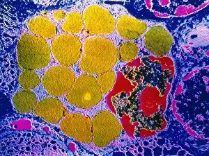
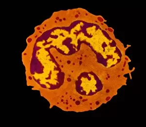
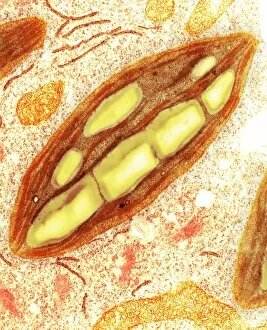
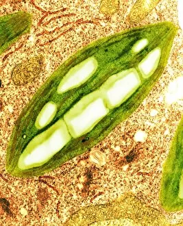
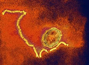
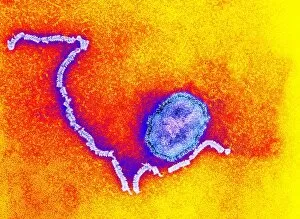
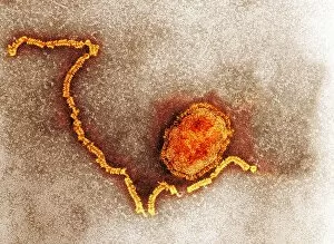
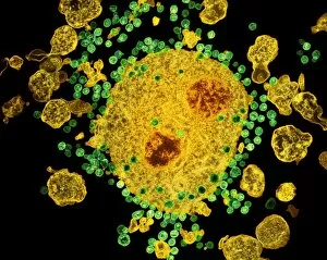
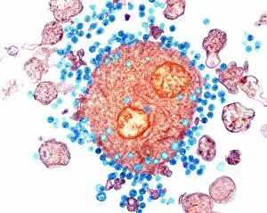
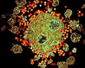
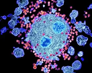
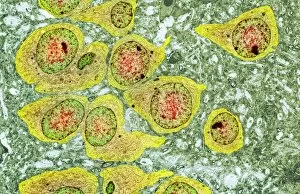
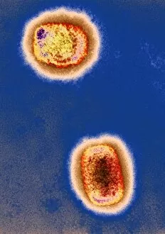
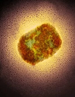
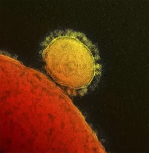
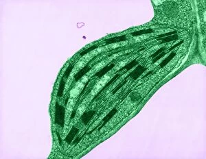
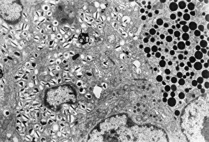
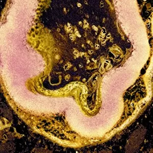
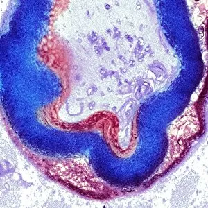
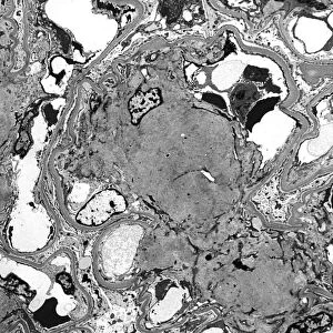
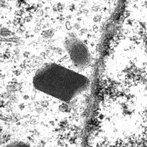
"Unlocking the Intricacies of Life: Exploring the Microscopic World through Transmission Electron Microscopy (TEM)" Delving into the depths of cellular structures and biological processes, transmission electron microscopy (TEM) has revolutionized our understanding of various aspects of life. With its high-resolution imaging capabilities, TEM allows us to witness intricate details that were once hidden from our sight. At synapse nerve junctions, TEM reveals a mesmerizing dance between neurotransmitters, enabling communication between neurons with remarkable precision. Norovirus particles come alive under TEM's lens, showcasing their unique shape and arrangement – a crucial insight in combating these notorious pathogens. E. Coli bacteria appear as tiny rods when observed through TEM, reminding us of their omnipresence in nature and sometimes unfortunate encounters in human health. Fat cells take on an unexpected beauty when magnified by TEM; their delicate structure resembling a web-like network that stores energy for our bodies. The myelination process is brought to life through TEM images capturing nerve fibers coated with protective sheaths. These stunning visuals help unravel the mysteries behind efficient neural signaling and hold promise for treating demyelinating diseases such as multiple sclerosis. TEM exposes the menacing presence of MRSA-resistant Staphylococcus bacteria – formidable adversaries in healthcare settings worldwide. Another glimpse at E. coli bacterium showcases its intricate internal machinery responsible for vital functions within this single-celled organism. Nerve cells reveal their complexity under TEM's scrutiny – branching dendrites reaching out like tree branches while axons transmit electrical signals across vast distances. Mitochondria steal the spotlight as they power these nerve cells' activities, appearing as dynamic organelles teeming with energy-producing potential. Plasma cells burst forth with vibrant colors when examined using TEM; their role in producing antibodies becomes even more awe-inspiring upon closer inspection. Once again, myelinated nerve fibers captivate us with their elegant architecture – a testament to nature's ingenuity in optimizing neural communication.