Metal Print > Popular Themes > Human Body
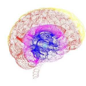
Metal Print : Brain tumour, fMRI and tractography C017 / 7102


![]()
Metal Prints from Science Photo Library
Brain tumour, fMRI and tractography C017 / 7102
Brain tumour, fMRI and tractography. Combined functional magnetic resonance imaging (fMRI, blue and green) and tractography (yellow and red) imaging of a brain with a tumour (upper left). The front of the brain is at right. The tumour (solid red) has been imaged using tractography, also known as 3D diffusion tensor imaging (DTI) magnetic resonance imaging (MRI). It shows the nerve pathways (red and yellow) affected by the tumour. Brain tumours can be benign or malignant (cancerous). They can cause seizures, headaches, and memory and personality changes. Hot spots on the fMRI areas are activated functional areas
Science Photo Library features Science and Medical images including photos and illustrations
Media ID 9341939
© SHERBROOKE CONNECTIVITY IMAGING LAB/SCIENCE PHOTO LIBRARY
Brain Imaging Brain Scan Cancer Cancerous Central Nervous System Cerebral Diffusion Tensor Imaging Dti Scan Fiber Fibers Fibre Fibres Imaging Technique Magnetic Resonance Imaging Malignancy Malignant Mri Scan Mri Scanner Nerve Nerve Fibre Nerves Neural Pathway Neural Tract Neuropathology Oncology Paths Pathway Pathways Structural Tractogram Tractography Tumor Tumour Brain Condition Disorder Neurological Neurology
16"x20" (51x41cm) Metal Print
Discover the depths of the human brain with Media Storehouse's Metal Prints featuring the captivating image "Brain tumour, fMRI and tractography C017 / 7102" by SHERBROOKE CONNECTIVITY IMAGING LAB/SCIENCE PHOTO LIBRARY. This striking image showcases a brain scan combining functional magnetic resonance imaging (fMRI) in blue and green, and tractography in yellow and red. Witness the intricate details of the brain's neural connections, highlighted against the contrast of the tumour. Our high-quality Metal Prints bring this scientific masterpiece to life, making it a unique and thought-provoking addition to any space.
Made with durable metal and luxurious printing techniques, our metal photo prints go beyond traditional canvases, adding a cool, modern touch to your space. Wall mount on back. Eco-friendly 100% post-consumer recycled ChromaLuxe aluminum surface. The thickness of the print is 0.045". Featuring a Scratch-resistant surface and Rounded corners. Backing hangers are attached to the back of the print and float the print 1/2-inch off the wall when hung, the choice of hanger may vary depending on size and International orders will come with Float Mount hangers only. Finished with a brilliant white high gloss surface for unsurpassed detail and vibrance. Printed using Dye-Sublimation and for best care we recommend a non-ammonia glass cleaner, water, or isopropyl (rubbing) alcohol to prevent harming the print surface. We recommend using a clean, lint-free cloth to wipe off the print. The ultra-hard surface is scratch-resistant, waterproof and weatherproof. Avoid direct sunlight exposure.
Made with durable metal and luxurious printing techniques, metal prints bring images to life and add a modern touch to any space
Estimated Image Size (if not cropped) is 50.8cm x 40.6cm (20" x 16")
Estimated Product Size is 51.4cm x 41.2cm (20.2" x 16.2")
These are individually made so all sizes are approximate
Artwork printed orientated as per the preview above, with landscape (horizontal) orientation to match the source image.
EDITORS COMMENTS
This print showcases the intricate details of a brain affected by a tumour, using advanced imaging techniques. The combination of functional magnetic resonance imaging (fMRI) and tractography provides a comprehensive view of the brain's structure and disease progression. Against a striking black background, the image reveals the front of the brain on the right side, with a solid red mass representing the tumour in the upper left. Tractography, also known as 3D diffusion tensor imaging (DTI), has been employed to visualize nerve pathways within the brain. These pathways are depicted in vibrant yellow and red hues, illustrating how they are impacted by this malignant growth. Brain tumours can be either benign or cancerous and often lead to various symptoms such as seizures, headaches, memory loss, and personality changes. The fMRI component of this image highlights activated functional areas through hot spots indicated by blue and green colors. This technique helps researchers understand which regions of the brain are involved during specific tasks or cognitive processes. Presented by Sherbrooke Connectivity Imaging Lab/Science Photo Library, this visually stunning photograph not only serves as an educational tool for medical professionals but also sparks curiosity about neurology and oncology among viewers. It exemplifies how cutting-edge imaging technologies contribute to our understanding of complex neurological conditions like brain tumours while emphasizing their impact on human health.
MADE IN THE USA
Safe Shipping with 30 Day Money Back Guarantee
FREE PERSONALISATION*
We are proud to offer a range of customisation features including Personalised Captions, Color Filters and Picture Zoom Tools
SECURE PAYMENTS
We happily accept a wide range of payment options so you can pay for the things you need in the way that is most convenient for you
* Options may vary by product and licensing agreement. Zoomed Pictures can be adjusted in the Cart.