Metal Print > Animals > Mammals > Cricetidae > White-footed Mouse
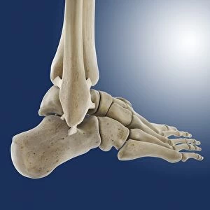
Metal Print : Inner ankle ligaments, artwork C013 / 4451


![]()
Metal Prints from Science Photo Library
Inner ankle ligaments, artwork C013 / 4451
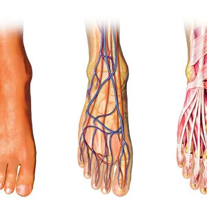
Inner ankle ligaments. Computer artwork of the bones and ligaments (white) of the right foot and ankle seen from the side. The two lower leg bones (upper right) are the tibia and fibula (behind tibia). In the foot, from right to left, are the heel bone (calcaneus), the tarsal bones, the metatarsals, and the phalanges (toes). The tibia and fibula articulate with the talus bone in the foot to form the ankle joint. Ligaments are tough bands of fibrous connective tissue that hold the bones of a joint together. These ligaments of the inner side of the ankle are the tibionavicular, tibiocalcaneal and posterior tibiotalar (collectively the deltoid ligament)
Science Photo Library features Science and Medical images including photos and illustrations
Media ID 9196075
© SPRINGER MEDIZIN/SCIENCE PHOTO LIBRARY
Ankle Arthrology Articulating Articulation Bones Calcaneus Connective Tissue Fibula Foot Heel Heel Bone Joint Joints Ligament Ligaments Lower Leg Metatarsal Metatarsals Metatarsus Muscular System Musculoskeletal System Osteology Outer Phalanges Phalanx Profile Shinbone Tarsals Tibia Tibial Toes Navicular Bone
20"x24" (61x51cm) Metal Print
Discover the intricacy of human anatomy with our Media Storehouse Metal Prints featuring the captivating image of Inner Ankle Ligaments (C013 / 4451) by Springer Medizin/Science Photo Library. This stunning computer artwork showcases the bones and ligaments of the right foot and ankle in meticulous detail, bringing the complexity of the human body to life. Our high-quality metal prints are not only a beautiful addition to any space, but also a testament to the wonders of science and anatomy. Order yours today and bring the marvels of the body into your home or office.
Made with durable metal and luxurious printing techniques, our metal photo prints go beyond traditional canvases, adding a cool, modern touch to your space. Wall mount on back. Eco-friendly 100% post-consumer recycled ChromaLuxe aluminum surface. The thickness of the print is 0.045". Featuring a Scratch-resistant surface and Rounded corners. Backing hangers are attached to the back of the print and float the print 1/2-inch off the wall when hung, the choice of hanger may vary depending on size and International orders will come with Float Mount hangers only. Finished with a brilliant white high gloss surface for unsurpassed detail and vibrance. Printed using Dye-Sublimation and for best care we recommend a non-ammonia glass cleaner, water, or isopropyl (rubbing) alcohol to prevent harming the print surface. We recommend using a clean, lint-free cloth to wipe off the print. The ultra-hard surface is scratch-resistant, waterproof and weatherproof. Avoid direct sunlight exposure.
Made with durable metal and luxurious printing techniques, metal prints bring images to life and add a modern touch to any space
Estimated Image Size (if not cropped) is 50.8cm x 60.9cm (20" x 24")
Estimated Product Size is 51.4cm x 61.5cm (20.2" x 24.2")
These are individually made so all sizes are approximate
Artwork printed orientated as per the preview above, with portrait (vertical) orientation to match the source image.
FEATURES IN THESE COLLECTIONS
> Animals
> Mammals
> Cricetidae
> White-footed Mouse
EDITORS COMMENTS
This print showcases the intricate inner ankle ligaments of the right foot and ankle, providing a detailed view from the side. The computer artwork beautifully highlights the bones and ligaments in white against a dark background, allowing for clear visibility and examination. Starting from right to left in the foot, we can observe the heel bone (calcaneus), followed by the tarsal bones, metatarsals, and finally, the phalanges (toes). Above these structures are two lower leg bones known as the tibia and fibula. These bones articulate with the talus bone in order to form our essential ankle joint. The ligaments depicted here play a crucial role in maintaining stability within this joint. They are tough bands of fibrous connective tissue that securely hold together these various bones. Specifically, on this inner side of the ankle, we find three important ligaments: tibionavicular, tibiocalcaneal, and posterior tibiotalar. Collectively referred to as deltoid ligament, they contribute significantly to overall joint integrity. This remarkable artwork not only provides an insight into human anatomy but also serves as a reminder of our body's complexity and resilience. It is through such meticulous illustrations that we gain a deeper understanding of our musculoskeletal system's intricate workings while appreciating its natural beauty.
MADE IN THE USA
Safe Shipping with 30 Day Money Back Guarantee
FREE PERSONALISATION*
We are proud to offer a range of customisation features including Personalised Captions, Color Filters and Picture Zoom Tools
SECURE PAYMENTS
We happily accept a wide range of payment options so you can pay for the things you need in the way that is most convenient for you
* Options may vary by product and licensing agreement. Zoomed Pictures can be adjusted in the Cart.