Photographic Print > Animals > Mammals > Cricetidae > White-footed Mouse
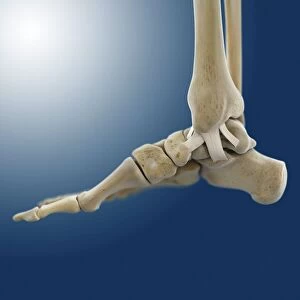
Photographic Print : Outer ankle ligaments, artwork C013 / 4452

![]()
Photo Prints from Science Photo Library
Outer ankle ligaments, artwork C013 / 4452
Outer ankle ligaments. Computer artwork of the bones and ligaments (white) of the right foot and ankle seen from the side. The two lower leg bones (upper left) are the fibula and tibia (behind fibula). In the foot, from left to right, are the heel bone (calcaneus), the tarsal bones, the metatarsals, and the phalanges (toes). The tibia and fibula articulate with the talus bone in the foot to form the ankle joint. Ligaments are tough bands of fibrous connective tissue that hold the bones of a joint together. These ligaments of the outer side of the ankle, named for the bones they join, are the tibiofibular, talofibular, and calcaneofibular
Science Photo Library features Science and Medical images including photos and illustrations
Media ID 9196087
© SPRINGER MEDIZIN/SCIENCE PHOTO LIBRARY
Anatomie Ankle Arthrology Articulating Articulation Band Calcaneus Connective Tissue Fibula Fibular Foot Heel Heel Bone Joint Joints Lateral Ligament Ligaments Metatarsal Bones Metatarsals Metatarsus Muscular System Musculoskeletal System Oblique Osteology Phalanges Phalanx Profile Shinbone Skelett Tarsals Tibia Toes Navicular Bone Rechts
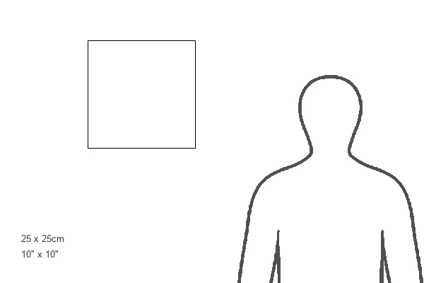
10"x10" Photo Print
Discover the intricacy of the human body with our latest addition to the Media Storehouse range of Photographic Prints. This captivating image, titled "Outer ankle ligaments, artwork C013 / 4452" by Springer Medizin/Science Photo Library, showcases the white-depicted bones and ligaments of the right foot and ankle from a side view. Delve into the world of anatomy and biology with this stunning, high-definition print, perfect for enhancing any space dedicated to science, education, or personal inspiration.
Photo prints are produced on Kodak professional photo paper resulting in timeless and breath-taking prints which are also ideal for framing. The colors produced are rich and vivid, with accurate blacks and pristine whites, resulting in prints that are truly timeless and magnificent. Whether you're looking to display your prints in your home, office, or gallery, our range of photographic prints are sure to impress. Dimensions refers to the size of the paper in inches.
Our Photo Prints are in a large range of sizes and are printed on Archival Quality Paper for excellent colour reproduction and longevity. They are ideal for framing (our Framed Prints use these) at a reasonable cost. Alternatives include cheaper Poster Prints and higher quality Fine Art Paper, the choice of which is largely dependant on your budget.
Estimated Product Size is 25.4cm x 25.4cm (10" x 10")
These are individually made so all sizes are approximate
Artwork printed orientated as per the preview above, with landscape (horizontal) or portrait (vertical) orientation to match the source image.
FEATURES IN THESE COLLECTIONS
> Animals
> Mammals
> Cricetidae
> White-footed Mouse
EDITORS COMMENTS
This print showcases the intricate network of outer ankle ligaments in exquisite detail. Crafted with precision and artistry, artwork C013 / 4452 from Springer Medizin/Science Photo Library offers a fascinating glimpse into the anatomy of the right foot and ankle seen from a side view. The image begins by highlighting the fibula and tibia, the two lower leg bones that form a sturdy foundation for this complex joint. Moving down to the foot, we encounter an array of essential structures: starting with the calcaneus or heel bone, followed by the tarsal bones, metatarsals, and phalanges (toes). These elements work harmoniously to support our weight while enabling fluid movement. However, what truly captures attention are the tough bands of fibrous connective tissue known as ligaments. In this artwork, we witness three specific ligaments on the outer side of the ankle: tibiofibular, talofibular, and calcaneofibular. These resilient connectors play a crucial role in maintaining stability within joints by securely holding bones together. By combining scientific accuracy with artistic flair, this print not only educates but also captivates viewers. It serves as a testament to human ingenuity in unraveling nature's mysteries while showcasing our body's remarkable design. Whether you're an anatomy enthusiast or simply appreciate beauty in science, this stunning illustration is sure to leave you awestruck at our wondrous musculoskeletal system.
MADE IN THE USA
Safe Shipping with 30 Day Money Back Guarantee
FREE PERSONALISATION*
We are proud to offer a range of customisation features including Personalised Captions, Color Filters and Picture Zoom Tools
SECURE PAYMENTS
We happily accept a wide range of payment options so you can pay for the things you need in the way that is most convenient for you
* Options may vary by product and licensing agreement. Zoomed Pictures can be adjusted in the Cart.