Canvas Print : Paprosky femur defect, type IIIA lateral


![]()
Canvas Prints from Science Photo Library
Paprosky femur defect, type IIIA lateral
Paprosky femur defect. Cutaway artwork of bone degradation in a type IV medial-lateral femur cortex defect (Paprosky classification system). This system is used for revision (replacement or repair) of a hip implant. The ball and socket part of the implant is at top. The rest of the implant consists of a shaft inside the femur (thigh bone). The amount of degradation in the cortical (outer) bone layer determines the amount of bone grafting needed, and whether a cementless implant can replace a cemented one. Type IV defects make the shaft unable to support weight (see C016/6620 for other types). Named for US surgeon Wayne G. Paprosky, the classification was developed in the 1980s and 1990s
Science Photo Library features Science and Medical images including photos and illustrations
Media ID 9214781
© D & L GRAPHICS / SCIENCE PHOTO LIBRARY
Arthrology Arthroplasty Bone Cement Cemented Cortex Cortical Cutaway Defect Defects Diagram Diaphysis Femur Grafting Hip Implant Hip Replacement Hip Revision Joint Medial Orthopaedic Orthopaedics Orthopedic Orthopedics Osteological Osteology Prostheses Prosthesis Prosthetic Prosthetics Repair Replacement Shaft Surgery Surgical Type Types Condition Cutouts Disorder Type 3
30"x20" (76x51cm) Canvas Print
Bring the intricacies of medical science into your home or office with Media Storehouse's Canvas Prints. This stunning, high-quality print features a captivating image of "Paprosky femur defect, type IIIA lateral" by D & L Graphics / Science Photo Library. This cutaway artwork offers a unique perspective into the complex world of bone degradation, showcasing the intricate details of the Paprosky classification system. Our Canvas Prints are not only a beautiful addition to any space, but also serve as a thought-provoking conversation starter. Order yours today and bring the wonders of science into your world.
Delivered stretched and ready to hang our premium quality canvas prints are made from a polyester/cotton blend canvas and stretched over a 1.25" (32mm) kiln dried knot free wood stretcher bar. Packaged in a plastic bag and secured to a cardboard insert for safe transit.
Canvas Prints add colour, depth and texture to any space. Professionally Stretched Canvas over a hidden Wooden Box Frame and Ready to Hang
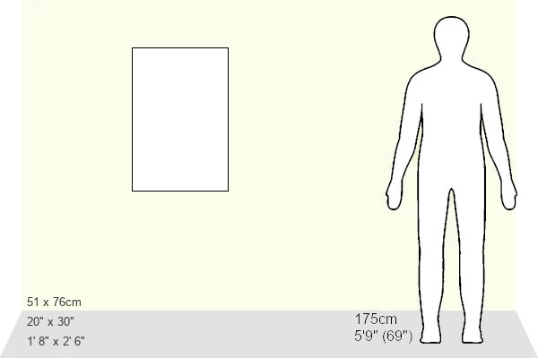
Estimated Image Size (if not cropped) is 40.6cm x 76.2cm (16" x 30")
Estimated Product Size is 50.8cm x 76.2cm (20" x 30")
These are individually made so all sizes are approximate
Artwork printed orientated as per the preview above, with portrait (vertical) orientation to match the source image.
EDITORS COMMENTS
This detailed print showcases a Paprosky femur defect, specifically a type IIIA lateral defect. The image is an intricate cutaway artwork that depicts the degradation of bone in the medial-lateral femur cortex, following the Paprosky classification system. This classification system is crucial for guiding revision procedures of hip implants. At the top of the illustration, we can observe the ball and socket component of the implant, while the rest consists of a shaft placed inside the femur (thigh bone). The severity of cortical bone degradation determines whether bone grafting is necessary and if a cementless implant can replace a cemented one. Type IV defects render the shaft incapable of supporting weight. Named after esteemed US surgeon Wayne G. Paprosky, this classification was developed during the 1980s and 1990s to aid orthopedic surgeons in determining appropriate treatment plans for patients requiring hip implant revisions. The image provides valuable insight into this medical condition by highlighting various anatomical structures such as metaphysis, metaphyseal region, diaphysis, and cortical layers affected by deterioration. It serves as an educational tool for healthcare professionals involved in arthroplasty surgeries or those studying osteology and arthrology. With its white background and precise detailing, this print from D & L GRAPHICS / SCIENCE PHOTO LIBRARY offers an informative visual representation that aids understanding of large bone defects like Paprosky femur defects within medical contexts.
MADE IN THE USA
Safe Shipping with 30 Day Money Back Guarantee
FREE PERSONALISATION*
We are proud to offer a range of customisation features including Personalised Captions, Color Filters and Picture Zoom Tools
SECURE PAYMENTS
We happily accept a wide range of payment options so you can pay for the things you need in the way that is most convenient for you
* Options may vary by product and licensing agreement. Zoomed Pictures can be adjusted in the Cart.