Beta Sheet Collection (page 3)

The beta sheet, a crucial element in the world of molecular biology and protein structure, plays a significant role in various biological processes

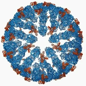
Background imageBeta Sheet Collection: Bluetongue virus capsid, molecular model F006 / 9687

Bluetongue virus capsid, molecular model F006 / 9687
Bluetongue virus capsid, molecular model. This virus, transmitted by midges, causes bluetongue disease in ruminants (commonly sheep, and less frequently cattle, goats and deer)

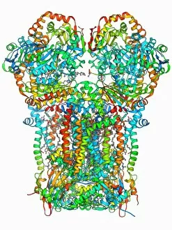
Background imageBeta Sheet Collection: Chaperonin protein F006 / 9690

Chaperonin protein F006 / 9690
Chaperonin protein, molecular mode. Chaperonins are proteins that provide favourable conditions for the correct folding of other proteins

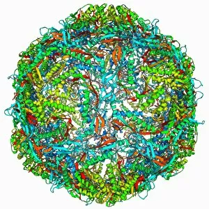
Background imageBeta Sheet Collection: Hepatitis E virus capsid, molecular model F006 / 9688

Hepatitis E virus capsid, molecular model F006 / 9688
Hepatitis E virus capsid, molecular model. This virus causes the disease hepatitis E in humans. Transmitted by faecal-oral contact

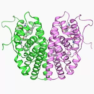
Background imageBeta Sheet Collection: Oestrogen receptor and tamoxifen drug F006 / 9674

Oestrogen receptor and tamoxifen drug F006 / 9674
Oestrogen receptor and tamoxifen drug, molecular model. Tamoxifen is a selective oestrogen receptor modulator (SERM) that blocks oestrogen production in breast tissue by inhibiting the oestrogen

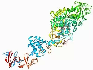
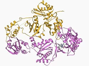
Background imageBeta Sheet Collection: Glucansucrase enzyme molecule F006 / 9685

Glucansucrase enzyme molecule F006 / 9685
Glucansucrase, molecular model. This enzyme catalyses the cleavage of sucrose into fructose and glucose

Background imageBeta Sheet Collection: Cytochrome BC1 molecule F006 / 9681

Cytochrome BC1 molecule F006 / 9681
Cytochrome BC1, Molecular model. Cytochrome molecules perform oxidation and reduction reactions for electron transport, a chain of reactions used to power cellular processes that require energy

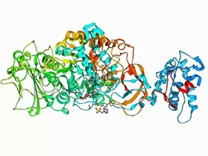
Background imageBeta Sheet Collection: HIV reverse transcription enzyme F006 / 9684

HIV reverse transcription enzyme F006 / 9684
HIV reverse transcription enzyme. Molecular model of the reverse transcriptase enzyme found in HIV (the human immunodeficiency virus) bound to the inhibitor nevirapine

Background imageBeta Sheet Collection: Human growth hormone molecule F006 / 9683

Human growth hormone molecule F006 / 9683
Human growth hormone. Molecular model of human growth hormone (hGH, orange) bound to the extracellular domain of the human growth hormone binding protein (hGHBP)

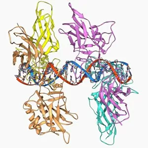
Background imageBeta Sheet Collection: HIV DNA and transcription factor F006 / 9680

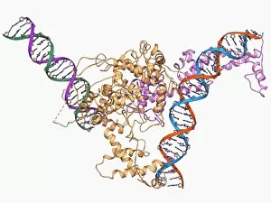
HIV DNA and transcription factor F006 / 9680
HIV DNA and transcription factor. Molecular model of DNA (deoxyribonucleic acid) from HIV-1 (human immunodeficiency virus type 1) complexed with the transcription factor kappa B

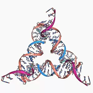
Background imageBeta Sheet Collection: Self-assembled DNA triangle F006 / 9679

Self-assembled DNA triangle F006 / 9679
Self-assembled DNA triangle. Molecular model of DNA (deoxyribonucleic acid) strands forming what is called a tensegrity triangle

Background imageBeta Sheet Collection: Human pathogen recognition molecule F006 / 9677

Human pathogen recognition molecule F006 / 9677
Human pathogen recognition molecule. Molecular model of a complex between human lymphocyte antigen 96 and toll-like receptor 4

Background imageBeta Sheet Collection: Antibodies and their antigen F006 / 9678

Antibodies and their antigen F006 / 9678
Antibodies and their antigen. Molecular model showing the molecular structure of two anti-P-glycoprotein antibodies (upper left and right) and the P-glycoprotein (v-shaped) to which they respond

Background imageBeta Sheet Collection: Bacterial nanocompartment F006 / 9672

Bacterial nanocompartment F006 / 9672
Bacterial nanocompartment. Molecular molecule of a nanocompartment from the bacterium Thermotoga maritima. This is a shell formed from encapsulin proteins that encloses enzymes that defend the cell

Background imageBeta Sheet Collection: E coli virulence factor molecule F006 / 9675

E coli virulence factor molecule F006 / 9675
E. coli virulence factor molecule. Molecular model of the enzyme arylsulfate sulfotransferase (ASST) from an Escherichia coli bacterium

Background imageBeta Sheet Collection: Fatty acid binding protein and inhibitor F006 / 9676

Fatty acid binding protein and inhibitor F006 / 9676
Fatty acid binding protein and inhibitor. Molecular model of adipocyte fatty-acid binding protein (A-FABP) bound to an inhibitor

Background imageBeta Sheet Collection: Fenna-Matthews-Olson complex molecule F006 / 9673

Fenna-Matthews-Olson complex molecule F006 / 9673
Fenna-Matthews-Olson complex. Molecular model of the Fenna-Matthews-Olson complex from the green sulphur bacterium Prosthecochloris aestuarii

Background imageBeta Sheet Collection: Nucleoside diphosphate kinase molecule F006 / 9671

Nucleoside diphosphate kinase molecule F006 / 9671
Nucleoside diphosphate kinase. Molecular model of the enzyme nucleoside diphosphate kinase from the Acanthamoeba polyphaga mimivirus

Background imageBeta Sheet Collection: Cytochrome C, molecular model F006 / 9670

Cytochrome C, molecular model F006 / 9670
Cytochrome C, molecular model. Cytochrome molecules perform oxidation and reduction reactions for electron transport, a chain of reactions used to power cellular processes that require energy

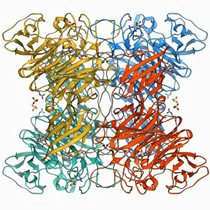
Background imageBeta Sheet Collection: Concanavalin A F006 / 9668

Concanavalin A F006 / 9668
Concanavalin A, molecular model. This lectin protein is found in legumes. Lectins are highly specific sugar-binding proteins that play a role in biological recognition

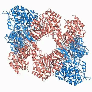
Background imageBeta Sheet Collection: Toll-like receptor 3 and RNA F006 / 9666

Toll-like receptor 3 and RNA F006 / 9666
Toll-like receptor 3 and RNA. Molecular model of the toll-like receptor 3 (TLR3) protein (pink and blue) bound to a strand of RNA (ribonucleic acid, green and yellow)

Background imageBeta Sheet Collection: Calcium-binding protein molecule F006 / 9667

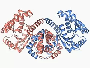
Calcium-binding protein molecule F006 / 9667
Calcium-binding protein. Molecule model of the calcium-binding protein calmodulin (CaM). This protein is found in all eukaryotic cells

Background imageBeta Sheet Collection: Isocitrate dehydrogenase molecule F006 / 9663

Isocitrate dehydrogenase molecule F006 / 9663
Isocitrate dehydrogenase, molecular model. This enzyme catalyses the third step in the citric acid (or Krebs) cycle, the process by which mitochondria convert glucose to energy
Background imageBeta Sheet Collection: Opsin molecule F006 / 9665

Opsin molecule F006 / 9665
Opsin. Molecular model of a ligand-free opsin molecule. Opsins are found in photoreceptor cells (rods and cones) in the retina of the eye

Background imageBeta Sheet Collection: Interferon antagonism by viral protein F006 / 9662

Interferon antagonism by viral protein F006 / 9662
Interferon (IFN) antagonism by viral protein. Molecular model of an orthopoxvirus IFN-gamma-binding protein bound to an IFN-gamma molecule

Background imageBeta Sheet Collection: DNA and MECP2 complex, molecular model F006 / 9664

DNA and MECP2 complex, molecular model F006 / 9664
DNA and MECP2 complex. Molecular model of MECP2 (methyl CpG binding protein 2 (Rett syndrome)) bound to the BDNF (brain-derived neurotrophic factor)

Background imageBeta Sheet Collection: Selenocysteine synthase enzyme molecule F006 / 9661

Selenocysteine synthase enzyme molecule F006 / 9661
Selenocysteine synthase enzyme molecule. Computer model showing the molecular structure of the enzyme selenocysteine synthase (SecS)

Background imageBeta Sheet Collection: Androgen receptor and modulator F006 / 9660

Androgen receptor and modulator F006 / 9660
Androgen receptor and modulator. Molecular model of a selective androgen receptor modulator (SARM) bound to an androgen receptor

Background imageBeta Sheet Collection: Bacterial biofilm enzyme F006 / 9658

Bacterial biofilm enzyme F006 / 9658
Bacterial biofilm enzyme. Molecular model of the enzyme glucansucrase from the bacterium Streptococcus mutans. This enzyme is used to form bacterial biofilms on teeth that cause dental caries

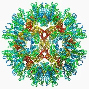
Background imageBeta Sheet Collection: Rous sarcoma virus capsid F006 / 9653

Rous sarcoma virus capsid F006 / 9653
Rous sarcoma virus capsid, molecular model. In viruses, the capsid is the protein shell that encloses the genetic material

Background imageBeta Sheet Collection: Rabbit eye lens protein molecule F006 / 9657

Rabbit eye lens protein molecule F006 / 9657
Rabbit eye lens protein. Molecular model of lambda-crystallin, a structural protein found in the eye lenses of rabbits (family Leporidae)

Background imageBeta Sheet Collection: Sodium-potassium ion pump protein F006 / 9656

Sodium-potassium ion pump protein F006 / 9656
Sodium-potassium ion pump protein, molecular model. Sodium-potassium ATPase (adenosine triphosphatase) is an ATP-powered ion pump found in all animal cells

Background imageBeta Sheet Collection: Chromatin remodelling factor and DNA F006 / 9655

Chromatin remodelling factor and DNA F006 / 9655
Chromatin remodelling factor and DNA, molecular model. The strands of DNA (deoxyribonucleic acid) are at left and right. This chromatin remodelling factor is ISW1a

Background imageBeta Sheet Collection: 70S ribosome, molecular model F006 / 9651

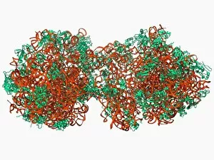
70S ribosome, molecular model F006 / 9651
70S ribosome, molecular model. Ribosomes are composed of protein and RNA (ribonucleic acid). In bacteria each ribosome consists of a small (30S) subunit and a large (50S) subunit

Background imageBeta Sheet Collection: MscS ion channel protein structure F006 / 9650

MscS ion channel protein structure F006 / 9650
MscS ion channel protein structure. Molecular model of a mechanosensitive channel of small conductance (MscS) from an Escherichia coli bacterium

Background imageBeta Sheet Collection: Streptavidin bacterial protein F006 / 9654

Streptavidin bacterial protein F006 / 9654
Streptavidin bacterial protein, molecular model. Streptavidin is a protein obtained from the bacterium Streptomyces avidinii

Background imageBeta Sheet Collection: Fatty acid synthase molecule F006 / 9647

Fatty acid synthase molecule F006 / 9647
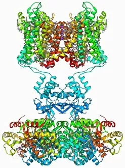
Fatty acid synthase (FAS), molecular model. FAS is a multi-enzyme that plays a key role in the synthesis of fatty acids (lipids) in the human body. It is not a single enzyme but a whole enzyme system

Background imageBeta Sheet Collection: Eye lens protein molecule F006 / 9652

Eye lens protein molecule F006 / 9652
Eye lens protein. Molecular model of alpha-crystallin, a protein found in the lens of the eye. The regular arrangement of the protein in the lens is thought to be responsible for its transparency

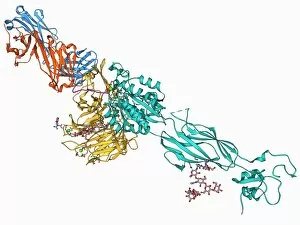
Background imageBeta Sheet Collection: Integrin and fibrinogen complex molecule F006 / 9649

Integrin and fibrinogen complex molecule F006 / 9649
Integrin and fibrinogen complex. Molecular model of integrin alpha-II beta-3 complexed with fibrinogen. Integrin alpha-II beta-3 is a transmembrane protein found on platelets

Background imageBeta Sheet Collection: SelB elongation factor bound to RNA F006 / 9648

SelB elongation factor bound to RNA F006 / 9648
SelB elongation factor bound to RNA. Molecular model of the SelB elongation factor bound to an mRNA (messenger ribonucleic acid) hairpin formed by the selenocysteine insertion sequence (SECIS)

Background imageBeta Sheet Collection: Voltage-gated potassium channel F006 / 9642

Voltage-gated potassium channel F006 / 9642
Voltage-gated potassium channel. Molecular model of a voltage-gated potassium (Kv) ion channel. Ion channels are membrane-spanning proteins that form pores in cell membranes

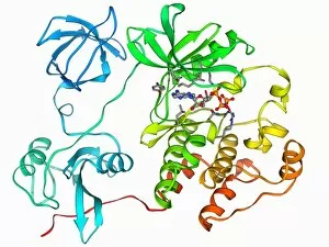
Background imageBeta Sheet Collection: Src protein molecule F006 / 9646

Src protein molecule F006 / 9646
Src protein, molecular model. Src is a tyrosine kinase, a signalling protein in cells that has the ability to turn on protein synthesis and cellular growth

Background imageBeta Sheet Collection: Copper, zinc superoxide dismutase enzyme F006 / 9645

Copper, zinc superoxide dismutase enzyme F006 / 9645
Copper, zinc superoxide dismutase enzyme

Background imageBeta Sheet Collection: Fungal prion protein F006 / 9644

Fungal prion protein F006 / 9644
Fungal prion protein. Molecular model of the amyloid form of the HET prion protein

Background imageBeta Sheet Collection: Beta-2 adrenergic receptor molecule F006 / 9643

Beta-2 adrenergic receptor molecule F006 / 9643
Beta-2 adrenergic receptor. Molecular model of a human beta-2 adrenergic receptor bound to an antibody. Beta receptors respond to adrenalin, causing a sympathetic (fight or flight) response

Background imageBeta Sheet Collection: Adenovirus protein and tumor suppressor F006 / 9641

Adenovirus protein and tumor suppressor F006 / 9641
Adenovirus protein and tumour suppressor. Molecular model of the E1A protein from human adenovirus bound to a retinoblastoma tumour suppressor

Background imageBeta Sheet Collection: Beta-2 adrenergic receptor molecule F006 / 9640

Beta-2 adrenergic receptor molecule F006 / 9640
Beta-2 adrenergic receptor. Molecular model of a human beta-2 adrenergic receptor bound to an antibody. Beta receptors respond to adrenalin, causing a sympathetic (fight or flight) response

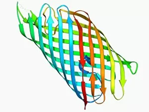
Background imageBeta Sheet Collection: E coli virulence factor F006 / 9639

E coli virulence factor F006 / 9639
E. coli virulence factor. Molecular model of the beta-domain of the EspP autotransporter protein from the bacterium Escherichia coli

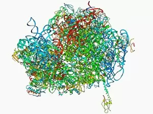
Background imageBeta Sheet Collection: 70S ribosome, molecular model F006 / 9638

70S ribosome, molecular model F006 / 9638
70S ribosome. Molecular model of a 70S ribosome complex containing a Shine-Dalgarno helix, the point of mRNA (messenger ribonucleic acid) binding


For sale as Licensed Images

Choose your image, Select your licence and Download the media

The beta sheet, a crucial element in the world of molecular biology and protein structure, plays a significant role in various biological processes. From DNA transcription to HIV reverse transcription enzyme, this secondary structure of proteins is an intricate masterpiece. In the realm of DNA transcription, the beta sheet takes center stage as it assists in unraveling genetic information. Its molecular model showcases its complexity and elegance, resembling an artistic creation that captivates scientists worldwide. Not only does the beta sheet contribute to DNA transcription, but it also plays a vital role in combating diseases. The Hepatitis C virus enzyme's molecular model reveals how this secondary structure aids in understanding and potentially treating this infectious disease. Another remarkable example lies within Manganese superoxide dismutase enzyme F006 / 9423. This intricate arrangement highlights how the beta sheet contributes to antioxidant defense mechanisms within our bodies. Moreover, Argonaute protein molecule F006 / 9526 demonstrates how the beta sheet collaborates with microRNA to regulate gene expression. This interaction holds immense potential for therapeutic interventions targeting various diseases. Immunoglobulin G antibody and egg white F006 / 9682 showcase yet another fascinating aspect of the beta sheet's versatility. Here we witness its involvement in immune responses against foreign substances present within our bodies. Furthermore, Cytochrome P450 complex F006 / 9669 illustrates how this secondary structure enables drug metabolism and detoxification processes essential for maintaining human health. Succinyl-CoA synthetase enzyme F006 / 9592 emphasizes another critical function of the beta sheet: energy production through cellular respiration pathways. Its presence ensures efficient conversion of succinyl-CoA into ATP molecules necessary for sustaining life processes. Additionally, RNA-induced silencing complex F006 / 9586 unveils how the beta sheet participates in gene regulation by suppressing specific mRNA molecules through small interfering RNAs (siRNAs).