Beta Sheet Collection (page 14)

The beta sheet, a crucial element in the world of molecular biology and protein structure, plays a significant role in various biological processes

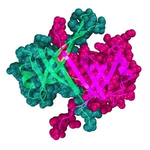
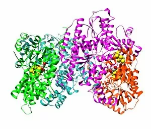
Background imageBeta Sheet Collection: Bacteriochlorophyll-containing protein

Bacteriochlorophyll-containing protein. Molecular models showing two views of the bacteriochlorophyll-containing protein found in the bacterium Prosthecochloris aestuarii

Background imageBeta Sheet Collection: Antithrombin molecule

Antithrombin molecule
Antithrombin, molecular model. Antithrombin is a glycoprotein produced in the liver that inhibits enzymes involved in blood clotting

Background imageBeta Sheet Collection: Interleukin-12 protein molecule

Interleukin-12 protein molecule. Molecular model of a molecule of the cytokine protein interleukin-12 (IL-12). The secondary structure of the molecule is seen here

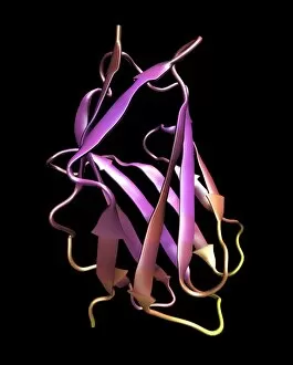
Background imageBeta Sheet Collection: Microglobulin protein, molecular model

Microglobulin protein, molecular model
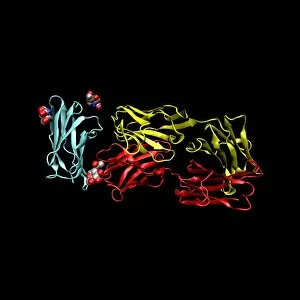
Microglobulin protein. Molecular models of two overlapping views of the protein beta-2 microglobulin. This protein, with a relatively small molecular mass

Background imageBeta Sheet Collection: Birch pollen allergen

Birch pollen allergen. Molecular model of the secondary structure of Bet v 1l, the molecule responsible for allergic reactions to birch pollen

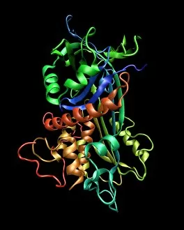
Background imageBeta Sheet Collection: Cytochrome P450 molecule

Cytochrome P450 molecule
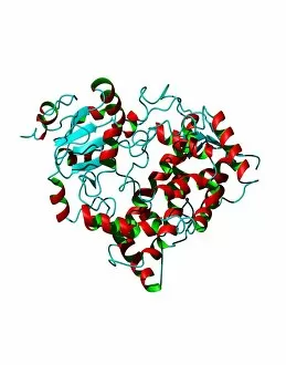
Cytochrome P450 protein. Molecular model showing the secondary structure of cytochrome P450. This protein plays a crucial role in metabolism in animals (including humans), fungi, plants and bacteria

Background imageBeta Sheet Collection: Cd28 antigen molecule

Cd28 antigen molecule. Computer model showing the secondary structure of cd28. Cd28 is an antigen found on the surface of T cells

Background imageBeta Sheet Collection: Mouse urinary protein, molecular model

Mouse urinary protein, molecular model
Mouse urinary protein (MUP), molecular model. MUP refers to a family of similar proteins found in mouse urine. The proteins act as pheromones, airborne chemical signals

Background imageBeta Sheet Collection: Bluetongue virus protein structure

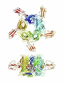
Bluetongue virus protein structure. Molecular model of some of the protein structures found in the protein coating of the bluetongue virus (BTV)

Background imageBeta Sheet Collection: Erythropoietin hormone

Erythropoietin hormone
Erythropoietin. Computer model of a molecule of the hormone erythropoietin. This is a glycoprotein that consists of a single-chain polypeptide of 165 amino acids

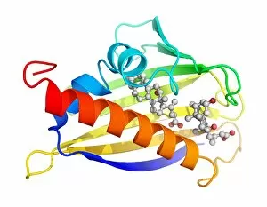
Background imageBeta Sheet Collection: Testosterone bound to receptor, artwork

Testosterone bound to receptor, artwork
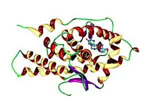
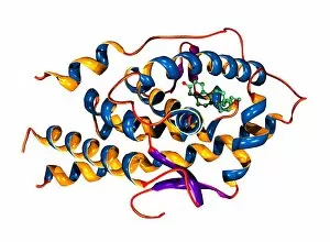
Testosterone bound to receptor. Molecular model of a molecule of the male sex hormone testosterone (ball-and-stick primary structure) bound to a human androgen receptor (secondary structure)

Background imageBeta Sheet Collection: Green fluorescent protein, computer model

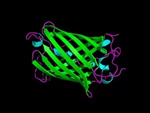
Green fluorescent protein, computer model. This protein is found in the jellyfish Aequorea victoria. When ultraviolet or blue light shines on the protein, it emits green light

Background imageBeta Sheet Collection: Cobra venom action, molecular model

Cobra venom action, molecular model
Cobra (Naja sp.) venom action. Molecular model showing top (upper centre) and side (lower centre) views of the secondary structure of an alpha-cobratoxin (snake venom protein)

Background imageBeta Sheet Collection: RNA polymerase from Norwalk virus

RNA polymerase from Norwalk virus
RNA polymerase enzyme from Norwalk virus, molecular model. This enzyme makes RNA (ribonucleic acid) from the RNA template within the virus

Background imageBeta Sheet Collection: TGF beta, molecular model

TGF beta, molecular model
TGF beta molecule. Molecular model showing the primary (rods) and secondary structure (arrows) of transforming growth factor beta (TGF beta)

Background imageBeta Sheet Collection: TGN1412 drug molecule

TGN1412 drug molecule. Computer model showing the secondary structure of the drug TGN1412. This drug was shown to have serious side effects during a clinical trial in the UK in 2006 when it caused

Background imageBeta Sheet Collection: THG anabolic steroid and receptor

THG anabolic steroid and receptor
THG bound to receptor. Molecular model of a molecule of the anabolic steroid drug tetrahydrogestrinone (THG, ball-and-stick primary structure) bound to a human androgen receptor (secondary structure)

Background imageBeta Sheet Collection: HIV antibody therapy

HIV antibody therapy. Molecular model of the interaction of the HIV surface protein gp120 (green, lower right) as it interacts with a human white blood cell surface protein (CD4, blue)

Background imageBeta Sheet Collection: Cholesterol enzyme affected by a drug

Cholesterol enzyme affected by a drug
Cholesterol enzyme being affected by a drug. Molecular model of the shape of the human enzyme Hmg-Coa reductase interacting (complexed) with the anti-cholesterol drug Fluvastatin (not seen)

Background imageBeta Sheet Collection: HIV enzyme being affected by a drug

HIV enzyme being affected by a drug. Molecular model of HIVs reverse transcriptase enzyme as it interacts with a drug (not seen)

Background imageBeta Sheet Collection: Insulin hormone, molecular model

Insulin hormone, molecular model
Insulin hormone. Molecular model of the bovine form of the hormone insulin, produced by the pancreas in mammals to aid the body in metabolising sugars

Background imageBeta Sheet Collection: Thrombin protein, secondary structure

Thrombin protein, secondary structure
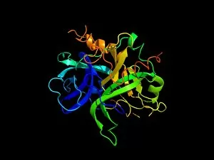
Thrombin protein, computer model. Thrombin is a protein involved in the blood coagulation (clotting) process. It converts fibrinogen (a soluble plasma glycoprotein synthesised in the liver)

Background imageBeta Sheet Collection: Cytochrome P450 protein, molecular model

Cytochrome P450 protein, molecular model. This protein plays a crucial role in metabolism in animals (including humans), fungi, plants and bacteria

Background imageBeta Sheet Collection: Src protein domain

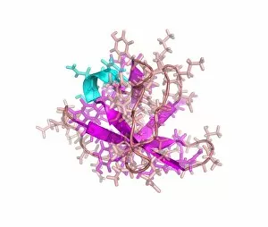
Src protein domain. Computer model showing the primary (rods) and secondary (alpha helix, blue and beta sheets, purple) structures of the Src homology domain 3 (SH3)

Background imageBeta Sheet Collection: Enzyme from a sulphur-reducing bacterium

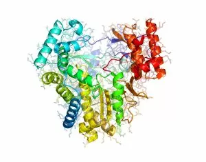
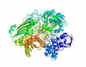
Enzyme from a sulphur-reducing bacterium. Molecular model of the enzyme aldehyde oxidoreductase from the Desulfovibrio gigas sulphur-reducing bacterium

Background imageBeta Sheet Collection: Yeast enzyme, molecular model

Yeast enzyme, molecular model
Yeast enzyme. Molecular model of an enzyme from Saccharomyces cerevisiae (Bakers yeast). This enzyme is 20S proteasome. A proteasome is a complex type of proteinase (protein-digesting enzyme)

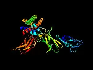
Background imageBeta Sheet Collection: Fibroblast growth factor receptor 2

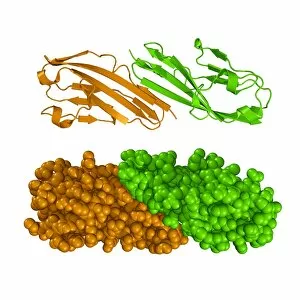
Fibroblast growth factor receptor 2 (FGFR2). Molecular models of the secondary structure (top) and the tertiary structure (bottom) of FGFR2

Background imageBeta Sheet Collection: Single stranded DNA-binding protein

Single stranded DNA-binding protein (SSBP). Molecular model showing the secondary and tertiary structures of a protein that binds to the single stranded DNA (deoxyribonucleic acid)

Background imageBeta Sheet Collection: Nerve growth factor, molecular model

Nerve growth factor, molecular model
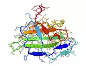
Nerve growth factor. Molecular model showing the secondary structure of nerve growth factor (NGF). NGF is a small protein, which is involved in the growth

Background imageBeta Sheet Collection: Nitrogenase protein, molecular model

Nitrogenase protein, molecular model
Nitrogenase protein. Molecular model of the MoFe protein, one of two proteins (MoFe and Fe) that combine to form the enzyme nitrogenase


For sale as Licensed Images

Choose your image, Select your licence and Download the media

The beta sheet, a crucial element in the world of molecular biology and protein structure, plays a significant role in various biological processes. From DNA transcription to HIV reverse transcription enzyme, this secondary structure of proteins is an intricate masterpiece. In the realm of DNA transcription, the beta sheet takes center stage as it assists in unraveling genetic information. Its molecular model showcases its complexity and elegance, resembling an artistic creation that captivates scientists worldwide. Not only does the beta sheet contribute to DNA transcription, but it also plays a vital role in combating diseases. The Hepatitis C virus enzyme's molecular model reveals how this secondary structure aids in understanding and potentially treating this infectious disease. Another remarkable example lies within Manganese superoxide dismutase enzyme F006 / 9423. This intricate arrangement highlights how the beta sheet contributes to antioxidant defense mechanisms within our bodies. Moreover, Argonaute protein molecule F006 / 9526 demonstrates how the beta sheet collaborates with microRNA to regulate gene expression. This interaction holds immense potential for therapeutic interventions targeting various diseases. Immunoglobulin G antibody and egg white F006 / 9682 showcase yet another fascinating aspect of the beta sheet's versatility. Here we witness its involvement in immune responses against foreign substances present within our bodies. Furthermore, Cytochrome P450 complex F006 / 9669 illustrates how this secondary structure enables drug metabolism and detoxification processes essential for maintaining human health. Succinyl-CoA synthetase enzyme F006 / 9592 emphasizes another critical function of the beta sheet: energy production through cellular respiration pathways. Its presence ensures efficient conversion of succinyl-CoA into ATP molecules necessary for sustaining life processes. Additionally, RNA-induced silencing complex F006 / 9586 unveils how the beta sheet participates in gene regulation by suppressing specific mRNA molecules through small interfering RNAs (siRNAs).