Beta Sheet Collection (#8)

The beta sheet, a crucial element in the world of molecular biology and protein structure, plays a significant role in various biological processes

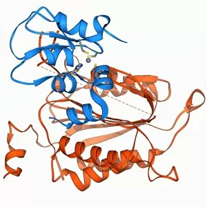
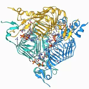
Background imageBeta Sheet Collection: Caspase-9 with inhibitor, molecular model F006 / 9442

Caspase-9 with inhibitor, molecular model F006 / 9442
Caspase-9 complexed with an inhibitor, molecular model. Caspase-9 is a protease, an enzyme that cleaves proteins, that plays a role in apoptosis (programmed cell death)

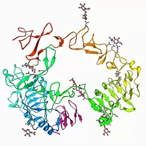
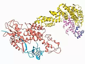
Background imageBeta Sheet Collection: Epidermal growth factor and receptor F006 / 9440

Epidermal growth factor and receptor F006 / 9440
Epidermal growth factor molecule. Molecular model of epidermal growth factor (EGF) bound to a receptor. EGF plays an important role in the regulation of cell growth, proliferation and differentiation

Background imageBeta Sheet Collection: Oxytocin and carrier protein F006 / 9439

Oxytocin and carrier protein F006 / 9439
Oxytocin and carrier protein. Molecular model of the hormone oxytocin bound to its carrier protein neurophysin I. Oxytocin is a neurotransmitter that plays a role in labour, sexual arousal, bonding

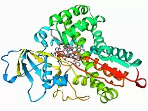
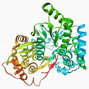
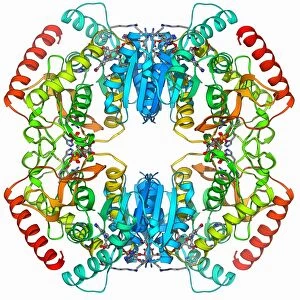
Background imageBeta Sheet Collection: Cytochrome P450 F006 / 9437

Cytochrome P450 F006 / 9437
Cytochrome P450. Molecular model of cytochrome P450 complexed with its catalytic product hydroxycamphor. This protein plays a crucial role in metabolism in animals (including humans), fungi

Background imageBeta Sheet Collection: DNA transcription, molecular model F006 / 9424

DNA transcription, molecular model F006 / 9424
DNA transcription. Molecular model of the enzyme RNA polymerase II synthesising a mRNA (messenger ribonucleic acid) strand from a DNA (deoxyribonucleic acid) template

Background imageBeta Sheet Collection: Nitric oxide synthase molecule F006 / 9436

Nitric oxide synthase molecule F006 / 9436
Nitric oxide synthase, molecular model. This enzyme catalyses the production of nitric oxide from L-arginine. Nitric oxide is involved in cellular signalling

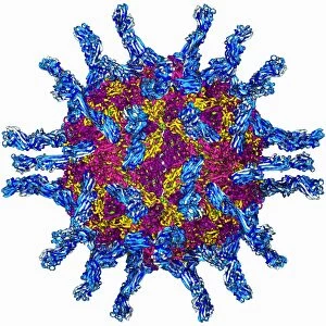
Background imageBeta Sheet Collection: Human poliovirus, molecular model F006 / 9434

Human poliovirus, molecular model F006 / 9434
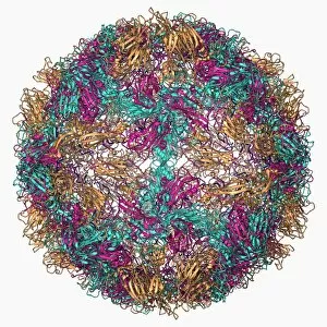
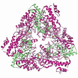
Human poliovirus particle. Molecular model of the capsid of the human poliovirus. The capsid is a protein coat that encloses the viruss genetic information (genome), stored as RNA (ribonucleic acid)

Background imageBeta Sheet Collection: Herpesvirus capsid protein F006 / 9435

Herpesvirus capsid protein F006 / 9435
Herpesvirus capsid protein. Molecular model of the major capsid protein VP5 from herpes simplex virus-1 (HSV-1). This virus infects epithelial cells around the mouth

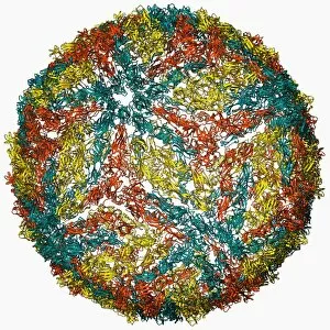
Background imageBeta Sheet Collection: Rhinovirus 14 capsid, molecular model F006 / 9430

Rhinovirus 14 capsid, molecular model F006 / 9430
Rhinovirus 14 capsid, molecular model. This is human rhinovirus 14. The rhinovirus infects the upper respiratory tract and is the cause of the common cold. It is spread by coughs and sneezes

Background imageBeta Sheet Collection: Flu virus surface protein molecule F006 / 9433

Flu virus surface protein molecule F006 / 9433
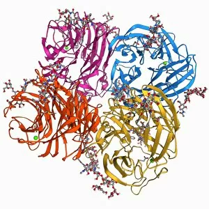
Flu virus surface protein molecule. Molecular model of the neuraminidase glycoprotein enzyme found on the surface of the influenza A (flu) virus

Background imageBeta Sheet Collection: Succinate dehydrogenase enzyme F006 / 9432

Succinate dehydrogenase enzyme F006 / 9432
Succinate dehydrogenase enzyme. Molecular model of the succinate dehydrogenase (complex II) enzyme from an Escherichia coli bacterium

Background imageBeta Sheet Collection: Flu virus surface protein and antibody F006 / 9428

Flu virus surface protein and antibody F006 / 9428
Flu virus surface protein and antibody. Molecular model of the neuraminidase glycoprotein enzyme found on the surface of the influenza (flu) virus

Background imageBeta Sheet Collection: Cell adhesion protein molecule F006 / 9429

Cell adhesion protein molecule F006 / 9429
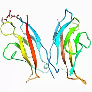
Cell adhesion protein. Molecular model of the cell adhesion protein n-cadherin (neural cadherin). This transmembrane protein facilitates adhesion between cells

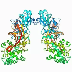
Background imageBeta Sheet Collection: Transposase enzyme and DNA complex F006 / 9425

Transposase enzyme and DNA complex F006 / 9425
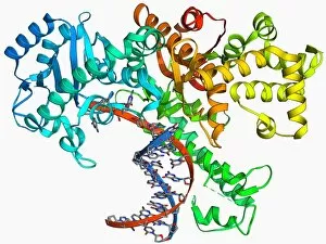
Transposase enzyme and DNA complex. Molecular model of a TN5 transposase enzyme bound to the transposon end of a DNA molecule (red and blue)

Background imageBeta Sheet Collection: DNA helicase molecule F006 / 9426

DNA helicase molecule F006 / 9426
DNA helicase. Molecular model of a helicase molecule from the SV40 virus. Helicases are enzymes that separate the two strands of the DNA double helix

Background imageBeta Sheet Collection: Hepatitis C polymerase enzyme F006 / 9427

Hepatitis C polymerase enzyme F006 / 9427
Hepatitis C polymerase enzyme, molecular model. This protein is the NS5b RNA polymerase found in the virus hepatitis C (genotype-1b, strain J4)

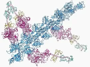
Background imageBeta Sheet Collection: Bacteriophage alpha 3 capsid F006 / 9414

Bacteriophage alpha 3 capsid F006 / 9414
Bacteriophage alpha 3 capsid, molecular model. Bacteriophages are viruses that infect bacteria. Bacteriophage alpha 3 has a capsid consisting of 60 coat proteins (blue) and 12 pentameric (5-subunit)

Background imageBeta Sheet Collection: Hammerhead ribozyme molecule F006 / 9422

Hammerhead ribozyme molecule F006 / 9422
Hammerhead ribozyme, molecular model. Ribozymes are RNA (ribonucleic acid) molecules that catalyse certain biochemical reactions

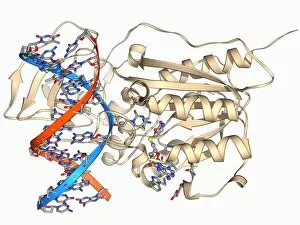
Background imageBeta Sheet Collection: Methyltransferase and DNA F006 / 9421

Methyltransferase and DNA F006 / 9421
Methyltransferase and DNA. Molecular model of the enzyme HhaI methyltransferase (beige) complexed with a molecule of DNA (deoxyribonucleic acid, red and blue)

Background imageBeta Sheet Collection: Insect flight muscle, molecular model F006 / 9416

Insect flight muscle, molecular model F006 / 9416
Insect flight muscle. Molecular model of myosin crossbridges in insect flight muscles in the nucleotide-free (rigor) state. This conformation is thought to represent the end of the power stroke

Background imageBeta Sheet Collection: Diphtheria toxin, molecular model F006 / 9419

Diphtheria toxin, molecular model F006 / 9419
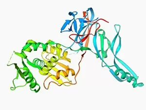
Diphtheria toxin, molecular model. Diphtheria is caused by the bacterium Corynebacterium diphtheriae. Symptoms include sore throat, fever and breathing difficulties

Background imageBeta Sheet Collection: Myoglobin molecule F006 / 9418

Myoglobin molecule F006 / 9418
Myoglobin, molecular model. Myoglobin is a protein found in muscle tissue, where it binds to and stores oxygen to be used during strenuous exercise

Background imageBeta Sheet Collection: Myoglobin molecule F006 / 9417

Myoglobin molecule F006 / 9417
Myoglobin, molecular model. Myoglobin is a protein found in muscle tissue, where it binds to and stores oxygen to be used during strenuous exercise

Background imageBeta Sheet Collection: Endonuclease and DNA, molecular model F006 / 9413

Endonuclease and DNA, molecular model F006 / 9413
Endonuclease and DNA. Molecular model of an endonuclease restriction enzyme (yellow) bound to a molecule of DNA (deoxyribonucleic acid)

Background imageBeta Sheet Collection: Lactate dehydrogenase enzyme molecule F006 / 9411

Lactate dehydrogenase enzyme molecule F006 / 9411
Lactate dehydrogenase enzyme, molecular model. This enzyme converts pyruvate to lactate in the final steps of glycolysis, and lactate to pyruvate during the lactic acid cycle

Background imageBeta Sheet Collection: Escherichia coli heat-labile enterotoxin F006 / 9410

Escherichia coli heat-labile enterotoxin F006 / 9410
Escherichia coli heat-labile enterotoxin, molecular model. This is one of several proteins produced by pathogenic E. coli bacteria in the intestines

Background imageBeta Sheet Collection: Lipase molecule F006 / 9409

Lipase molecule F006 / 9409
Lipase molecule, molecular model. Lipase is an enzyme that breaks lipids (fats) into fatty acids and glycerol. Human pancreatic lipase is the main enzyme responsible for breaking down fat in

Background imageBeta Sheet Collection: Ubiquitin ligase molecule F006 / 9407

Ubiquitin ligase molecule F006 / 9407
Ubiquitin ligase, molecular model. This enzyme tags proteins for degradation by attaching a ubiquitin molecule to them

Background imageBeta Sheet Collection: Gene activator protein F006 / 9406

Gene activator protein F006 / 9406
Gene activator protein. Molecular model of catabolite gene activator protein (CAP, yellow) complexed with deoxyribonucleic acid (DNA, red and blue) and RNA polymerase (green and pink)

Background imageBeta Sheet Collection: Microglobulin protein, molecular model F006 / 9408

Microglobulin protein, molecular model F006 / 9408
Microglobulin protein. Molecular model of the protein beta-2 microglobulin. This protein, with a relatively small molecular mass, is part of the immunoglobulin protein family

Background imageBeta Sheet Collection: Antibiotic resistance enzyme molecule F006 / 9405

Antibiotic resistance enzyme molecule F006 / 9405
Antibiotic resistance enzyme. Molecular model of an aminoglycoside phosphotransferase enzyme. This enzyme confers resistance to aminoglycoside antibiotics

Background imageBeta Sheet Collection: Cell adhesion protein molecule F006 / 9404

Cell adhesion protein molecule F006 / 9404
Cell adhesion protein. Molecular model of the ectodomain of the cell adhesion protein c-cadherin. This transmembrane protein facilitates adhesion between cells in solid tissues

Background imageBeta Sheet Collection: Cytochrome complex molecule F006 / 9401

Cytochrome complex molecule F006 / 9401
Cytochrome complex. Molecular model of cytochrome BC1 complex with bound cytochrome C. Cytochrome molecules perform oxidation and reduction reactions for electron transport

Background imageBeta Sheet Collection: Dengue virus capsid, molecular model F006 / 9392

Dengue virus capsid, molecular model F006 / 9392
Dengue virus capsid, molecular model. This virus, transmitted by mosquito bites, causes the tropical disease dengue fever in humans

Background imageBeta Sheet Collection: Galactoside acetyltransferase molecule F006 / 9400

Galactoside acetyltransferase molecule F006 / 9400
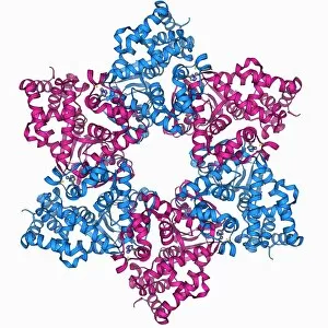
Galactoside acetyltransferase, molecular model. This enzyme from the bacterium Escherichia coli is involved in the lac operon

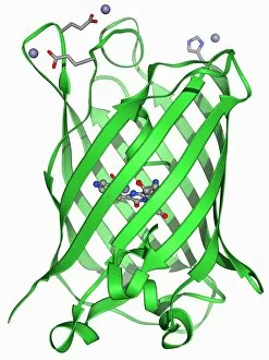
Background imageBeta Sheet Collection: Green fluorescent protein molecule F006 / 9402

Green fluorescent protein molecule F006 / 9402
Green fluorescent protein (GFP), molecular model. The molecule has a cylindrical structure formed from beta sheets (ribbons). GFP is found in the Pacific jellyfish Aequorea victoria
Background imageBeta Sheet Collection: Outer membrane receptor protein molecule F006 / 9398

Outer membrane receptor protein molecule F006 / 9398
Outer membrane receptor protein. Molecular model of FecA an outer membrane receptor protein

Background imageBeta Sheet Collection: Antibiotic resistance enzyme molecule F006 / 9399

Antibiotic resistance enzyme molecule F006 / 9399
Antibiotic resistance enzyme. Molecular model of kanamycin nucleotidyltransferase (KNTase). This bacterial enzyme is responsible for resistance to a number of antibiotics

Background imageBeta Sheet Collection: Enzyme catalysing DNA recombination F006 / 9395

Enzyme catalysing DNA recombination F006 / 9395
Enzyme catalysing DNA recombination. Molecular model of the enzyme CRE (cyclization recombination) recombinase (green and purple) mediating the recombination of strands of DNA (deoxyribonucleic acid)

Background imageBeta Sheet Collection: DNA polymerase Klenow fragment F006 / 9397

DNA polymerase Klenow fragment F006 / 9397
Klenow fragment of DNA polymerase I. Molecule model of the Klenow, or large, fragment from DNA polymerase I complexed with DNA (deoxyribonucleic acid, red and blue)

Background imageBeta Sheet Collection: Anthrax oedema factor molecule F006 / 9393

Anthrax oedema factor molecule F006 / 9393
Anthrax oedema factor. Molecular model of oedema factor (EF) from the anthrax bacterium Bacillus antracis complexed with a calmodulin protein molecule

Background imageBeta Sheet Collection: Fish antifreeze protein F006 / 9396

Fish antifreeze protein F006 / 9396
Fish antifreeze protein. Molecular model of a fish antifreeze protein (AFP) from the ocean pout (Zoarces americanus)

Background imageBeta Sheet Collection: Proteinase inhibitor molecule F006 / 9394

Proteinase inhibitor molecule F006 / 9394
Proteinase inhibitor. Molecular model of a proteinase inhibitor, or antitrypsin, molecule bound to a tryspin protease. The proteinase inhibitor is a type of serine protease inhibitor (serpin)

Background imageBeta Sheet Collection: Voltage-gated potassium channel F006 / 9391

Voltage-gated potassium channel F006 / 9391
Voltage-gated potassium channel. Molecular model of a voltage-gated potassium (Kv) ion channel complexed with the antigen-binding fragment (Fab) of a monoclonal antibody

Background imageBeta Sheet Collection: DNA polymerase IV with DNA F006 / 9390

DNA polymerase IV with DNA F006 / 9390
polymerase IV with DNA. Molecular model of DNA polymerase IV (beige) complexed with a molecule of DNA (deoxyribonucleic acid, red and blue)

Background imageBeta Sheet Collection: Coxsackie B3 virus particle F006 / 9381

Coxsackie B3 virus particle F006 / 9381
Coxsackie B3 virus. Computer model of the capsid of the Coxsackie B3 virus

Background imageBeta Sheet Collection: Integrin, molecular model F006 / 9388

Integrin, molecular model F006 / 9388
Integrin. Molecular model of the integrin protein alpha-v beta-3. This is a transmembrane protein that is found on platelets. It is composed of two subunits; integrin alpha-V and integrin beta 3

Background imageBeta Sheet Collection: VMA-1 derived endonuclease molecule F006 / 9389

VMA-1 derived endonuclease molecule F006 / 9389
VMA-1 derived endonuclease. Molecular model of the VMA-1 derived endonuclease restriction enzyme. Restriction enzymes, also known as restriction endonucleases


For sale as Licensed Images

Choose your image, Select your licence and Download the media

The beta sheet, a crucial element in the world of molecular biology and protein structure, plays a significant role in various biological processes. From DNA transcription to HIV reverse transcription enzyme, this secondary structure of proteins is an intricate masterpiece. In the realm of DNA transcription, the beta sheet takes center stage as it assists in unraveling genetic information. Its molecular model showcases its complexity and elegance, resembling an artistic creation that captivates scientists worldwide. Not only does the beta sheet contribute to DNA transcription, but it also plays a vital role in combating diseases. The Hepatitis C virus enzyme's molecular model reveals how this secondary structure aids in understanding and potentially treating this infectious disease. Another remarkable example lies within Manganese superoxide dismutase enzyme F006 / 9423. This intricate arrangement highlights how the beta sheet contributes to antioxidant defense mechanisms within our bodies. Moreover, Argonaute protein molecule F006 / 9526 demonstrates how the beta sheet collaborates with microRNA to regulate gene expression. This interaction holds immense potential for therapeutic interventions targeting various diseases. Immunoglobulin G antibody and egg white F006 / 9682 showcase yet another fascinating aspect of the beta sheet's versatility. Here we witness its involvement in immune responses against foreign substances present within our bodies. Furthermore, Cytochrome P450 complex F006 / 9669 illustrates how this secondary structure enables drug metabolism and detoxification processes essential for maintaining human health. Succinyl-CoA synthetase enzyme F006 / 9592 emphasizes another critical function of the beta sheet: energy production through cellular respiration pathways. Its presence ensures efficient conversion of succinyl-CoA into ATP molecules necessary for sustaining life processes. Additionally, RNA-induced silencing complex F006 / 9586 unveils how the beta sheet participates in gene regulation by suppressing specific mRNA molecules through small interfering RNAs (siRNAs).