Light Microscope Collection (page 9)

Exploring the Intricacies of Life: A Glimpse through the Light Microscope Unveiling the Wonders of Cerebellum Tissue

Background imageLight Microscope Collection: Beelouse, light micrograph C014 / 4841

Beelouse, light micrograph C014 / 4841
Beelouse (Braula coeca), dark-field illuminated light micrograph. This wingless fly is a parasite of the honeybee (Apis mellifera)

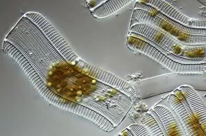
Background imageLight Microscope Collection: Diatom, light micrograph C014 / 4669

Diatom, light micrograph C014 / 4669
Diatom. Differential interference contrast micrograph of an Achnantes longipes diatom. Diatoms are a group of photosynthetic, single-celled algae containing about 10, 000 species

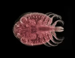
Background imageLight Microscope Collection: Fish louse C014 / 4837

Fish louse C014 / 4837
Fish louse. Dark-field illuminated light micrograph of a fish louse (Argulus sp.). This freshwater crustacean is a serious pest in commercial fish farms and fisheries

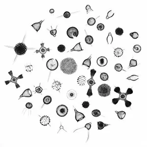
Background imageLight Microscope Collection: Radiolaria, light micrograph C014 / 4672

Radiolaria, light micrograph C014 / 4672
Radiolaria. Collection of light micrographs showing various different species of radiolaria. Radiolaria (or radiolarians) are amoeboid protozoa that produce intricate mineral skeletons

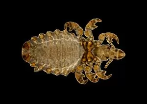
Background imageLight Microscope Collection: Parasitic louse, light micrograph C014 / 4850

Parasitic louse, light micrograph C014 / 4850
Parasitic louse (Haematopinus asini), light micrograph. This louse commonly infects horses. It causes irritation that can cause damage by the horse rubbing itself raw

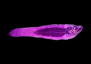
Background imageLight Microscope Collection: Paraneplocephala tapeworm, micrograph C014 / 4859

Paraneplocephala tapeworm, micrograph C014 / 4859
Paraneplocephala tapeworm. Light micrograph of a Paraneplocephala sp. tapeworm. This short parasitic tapeworm (order Cestoidea) is found in horses and other herbivores

Background imageLight Microscope Collection: Plant seedling, light micrograph C014 / 4741

Plant seedling, light micrograph C014 / 4741
Plant seedling, light micrograph. Magnification: x5 when printed 10 cm wide

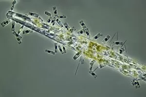
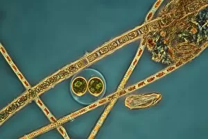
Background imageLight Microscope Collection: Diatoms, light micrograph C014 / 4668

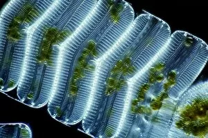
Diatoms, light micrograph C014 / 4668
Diatoms. Differential interference contrast micrograph of a group of Achnantes longipes diatoms. Diatoms are a group of photosynthetic, single-celled algae containing about 10, 000 species
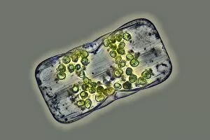
Background imageLight Microscope Collection: Diatoms, light micrograph C014 / 4674

Diatoms, light micrograph C014 / 4674
Diatoms. Differential interference contrast micrograph of a group of Achnantes longipes diatoms. Diatoms are a group of photosynthetic, single-celled algae containing about 10, 000 species

Background imageLight Microscope Collection: Green hydra, light micrograph C014 / 4678

Green hydra, light micrograph C014 / 4678
Green hydra. Light micrograph of the tentacled head of a green hydra (Hydra viridis). Hydra are small simple predatory fresh-water animals, belonging to the phylum Cnidaria

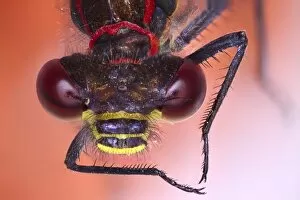
Background imageLight Microscope Collection: Damselfly head, light micrograph C014 / 4665

Damselfly head, light micrograph C014 / 4665
Damselfly head. Light micrograph of the head of a damselfly (order Odonata), showing its large compound eyes. Each compound eye is made up of numerous simple eyes called ommatidia

Background imageLight Microscope Collection: Damselfly eye, light micrograph C014 / 4663

Damselfly eye, light micrograph C014 / 4663
Damselfly eye. Light micrograph showing one of the compound eyes of a damselfly (order Odonata). Each compound eye is made up of numerous simple eyes called ommatidia

Background imageLight Microscope Collection: Poppy seed germinating, light micrograph C014 / 4660

Poppy seed germinating, light micrograph C014 / 4660
Poppy (Papaver sp.) seed germinating, light micrograph. Magnification: x34, when printed 10 centimetres wide

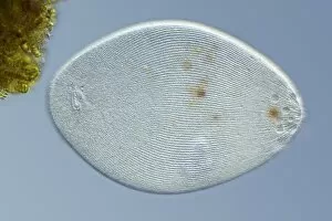
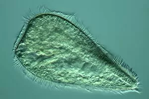
Background imageLight Microscope Collection: Ciliate protozoan, light micrograph C014 / 4676

Ciliate protozoan, light micrograph C014 / 4676
Ciliate protozoan. Differential interference contrast micrograph of a ciliate protozoan, showing the symbiotic green algae (Zoochlorellae, green) contained in vacuoles within

Background imageLight Microscope Collection: Dragonfly wing, light micrograph C014 / 4655

Dragonfly wing, light micrograph C014 / 4655
Dragonfly wing. Light micrograph of the wing of a dragonfly (order Odonata), showing its numerous veins. Magnification: x6 when printed 10 centimetres wide

Background imageLight Microscope Collection: Rotifer, light micrograph C014 / 4666

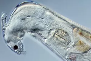
Rotifer, light micrograph C014 / 4666
Rotifer, differential interference contrast micrograph. These animals are related to roundworms. They are usually freshwater organisms though a few marine sp

Background imageLight Microscope Collection: Green hydra, light micrograph C014 / 4679

Green hydra, light micrograph C014 / 4679
Green hydra. Darkfield illuminated light micrograph of a green hydra (Hydra viridis), showing its tentacled head. Hydra are small simple predatory fresh-water animals

Background imageLight Microscope Collection: Damselfly head, light micrograph C014 / 4662

Damselfly head, light micrograph C014 / 4662
Damselfly head. Light micrograph of the head of a damselfly (order Odonata), showing its large compound eyes. Each compound eye is made up of numerous simple eyes called ommatidia

Background imageLight Microscope Collection: Ceropegia sp. flower, light micrograph C014 / 4729

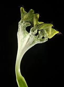
Ceropegia sp. flower, light micrograph C014 / 4729
Lantern flower (Ceropegia sp.), light micrograph. Magnification: x2.5 when printed 10 cm wide

Background imageLight Microscope Collection: Paraneplocephala tapeworm, micrograph C014 / 4860

Paraneplocephala tapeworm, micrograph C014 / 4860
Paraneplocephala tapeworm. Light micrograph showing the internal anatomy of a Paraneplocephala sp. tapeworm. This short parasitic tapeworm (order Cestoidea) is found in horses and other herbivores

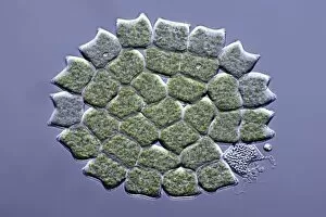
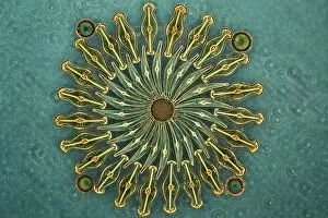
Background imageLight Microscope Collection: Pediastrum green algae, micrograph

Pediastrum green algae, micrograph
Pediastrum green algae. Differential interference contrast light micrograph of a circular colony of Pediastrum sp. green algae (Chlorophyte). Pediastrum is a type of freshwater colonial algae

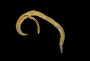
Background imageLight Microscope Collection: Schistosome flukes mating, micrograph C014 / 4867

Schistosome flukes mating, micrograph C014 / 4867
Schistosome flukes mating. Light micrograph of Schistosoma japonicum fluke worms mating. The male is the smaller animal at centre

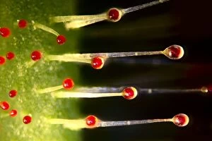
Background imageLight Microscope Collection: Sundew (Drosera sp. ) light micrograph C014 / 4657

Sundew (Drosera sp. ) light micrograph C014 / 4657
Sundew (Drosera sp.). Light micrograph of a sundew carnivorous plant, showing the glandular hairs. The hairs secrete a sticky liquid with a fragrance that attracts insects

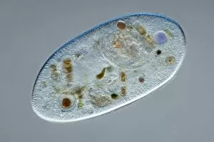
Background imageLight Microscope Collection: Ciliate protozoan, light micrograph C014 / 4667

Ciliate protozoan, light micrograph C014 / 4667
Ciliate protozoan. Differential interference contrast micrograph of a ciliate protozoan, showing the symbiotic green algae (Zoochlorellae, green) contained in vacuoles within

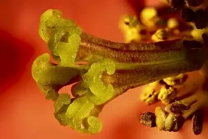
Background imageLight Microscope Collection: Cydonia oblonga flower, light micrograph C014 / 4656

Cydonia oblonga flower, light micrograph C014 / 4656
Quince (Cydonia oblonga) flower. Light micrograph of a quince flower, showing the female stigma (centre). Magnification: x17 when printed 10 centimetres wide

Background imageLight Microscope Collection: Damselfly head, light micrograph C014 / 4661

Damselfly head, light micrograph C014 / 4661
Damselfly head. Light micrograph of the head of a damselfly (order Odonata), showing its large compound eyes. Each compound eye is made up of numerous simple eyes called ommatidia

Background imageLight Microscope Collection: Foraminiferan

Foraminiferan, darkfield light micrograph. Foraminifera are marine single-celled protozoa that construct and inhabit shells composed of several chambers

Background imageLight Microscope Collection: Common liver fluke C014 / 4847

Common liver fluke C014 / 4847
Common liver fluke (Fasciola hepatica). This parasitic trematode (flatworm) infects the livers of various mammals, including humans

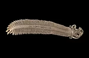
Background imageLight Microscope Collection: LM of the red algae, Plumaria elegans C014 / 8486

LM of the red algae, Plumaria elegans C014 / 8486
Plumaria algae. Light micrograph of part of the finely branched thallus of the red algae Plumaria elegans (family: Florideophycidae)

Background imageLight Microscope Collection: LM of the red algae, Plumaria elegans C014 / 8488

LM of the red algae, Plumaria elegans C014 / 8488
Plumaria algae. Light micrograph of part of the finely branched thallus of the red algae Plumaria elegans (family: Florideophycidae)

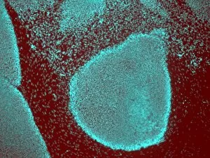
Background imageLight Microscope Collection: Induced stem cells, light micrograph

Induced stem cells, light micrograph
Induced stem cells. Light micrograph of a cultured colony of induced human pluripotent stem (IPS) cells. Oct-4 (octamer-binding transcription factor 4) is cyan

Background imageLight Microscope Collection: Induced nerve stem cells, micrograph

Induced nerve stem cells, micrograph
Induced nerve stem cells. Fluorescence light micrograph of neural (nerve) stem cells that have been created (induced) from human adult skin fibroblast cells by gene manipulation

Background imageLight Microscope Collection: LM of the red algae, Plumaria elegans C014 / 8487

LM of the red algae, Plumaria elegans C014 / 8487
Plumaria algae. Light micrograph of part of the finely branched thallus of the red algae Plumaria elegans (family: Florideophycidae)

Background imageLight Microscope Collection: Stem cell-derived nerve cells

Stem cell-derived nerve cells. Fluorescence light micrograph of neural (nerve) stem cells that have been derived from human embryonic stem cells (HESC)

Background imageLight Microscope Collection: Closterium desmid, light micrograph C016 / 8608

Closterium desmid, light micrograph C016 / 8608
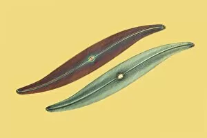
Closterium desmid. Interphase contrast micrograph of a Closterium sp. desmid green alga. Desmids are a common group of freshwater single-celled algae that have intricate cell walls

Background imageLight Microscope Collection: Amoeba protozoa, light micrograph C016 / 8607

Amoeba protozoa, light micrograph C016 / 8607
Amoeba protozoa. Interphase contrast light micrograph of two Amoeba sp. protozoa eating each other, showing its numerous pseudopodia (cytoplasmic extensions, leg-like)

Background imageLight Microscope Collection: Diatom, light micrograph C016 / 8606

Diatom, light micrograph C016 / 8606
Diatom. Phase-contrast light micrograph of a diatom from the Mediterranean. Diatoms are a group of photosynthetic, single-celled algae containing about 10, 000 species

Background imageLight Microscope Collection: Diatoms, light micrograph C016 / 8605

Diatoms, light micrograph C016 / 8605
Diatoms, Shearing contrast light micrograph. Diatoms are a group of photosynthetic, single-celled algae containing about 10, 000 species

Background imageLight Microscope Collection: Diatoms, light micrograph C016 / 8598

Diatoms, light micrograph C016 / 8598
Diatoms. Interphase contrast light micrograph of a selection of differently shaped diatoms. Diatoms are a group of photosynthetic, single-celled algae containing about 10, 000 species

Background imageLight Microscope Collection: Diatoms, light micrograph C016 / 8602

Diatoms, light micrograph C016 / 8602
Diatoms. Phase-contrast light micrograph of diatoms from the Mediterranean. Diatoms are a group of photosynthetic, single-celled algae containing about 10, 000 species

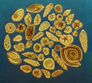
Background imageLight Microscope Collection: Foraminifera, light micrograph C016 / 8597

Foraminifera, light micrograph C016 / 8597
Foraminifera. Interphase contrast light micrograph of a selection of different foraminifera. Foraminifera are marine single-celled protozoa that construct

Background imageLight Microscope Collection: Diatoms, light micrograph C016 / 8600

Diatoms, light micrograph C016 / 8600
Diatoms. Differential interference contrast (DIC) light micrograph of diatoms from the Mediterranean. Diatoms are a group of photosynthetic, single-celled algae containing about 10, 000 species

Background imageLight Microscope Collection: Green algae, light micrograph C016 / 8595

Green algae, light micrograph C016 / 8595
Green algae, Interphase contrast light micrograph. Magnification: x280, when printed 10 centimetres wide

Background imageLight Microscope Collection: Ciliate protozoan, light micrograph C016 / 8593

Ciliate protozoan, light micrograph C016 / 8593
Ciliate protozoan. Differential interference contrast (DIC) light micrograph of a ciliate protozoan. Magnification: x280 when printed 10 centimetres wide

Background imageLight Microscope Collection: Hypotrich protozoan, light micrograph C016 / 8592

Hypotrich protozoan, light micrograph C016 / 8592
Hypotrich protozoan. Differential interference contrast (DIC) light micrograph of a Euplotes sp. hypotrich ciliate protozoan. Magnification: x280 when printed 10 centimetres wide

Background imageLight Microscope Collection: Fossil diatom, light micrograph C016 / 8586

Fossil diatom, light micrograph C016 / 8586
Fossil diatom. Interphase contrast light micrograph of a fossilised diatom. Diatoms are a group of photosynthetic, single-celled algae containing about 10, 000 species

Background imageLight Microscope Collection: Hypotrich protozoan, light micrograph C016 / 8590

Hypotrich protozoan, light micrograph C016 / 8590
Hypotrich protozoan. Differential interference contrast (DIC) light micrograph of a Euplotes sp. hypotrich ciliate protozoan. Magnification: x280 when printed 10 centimetres wide

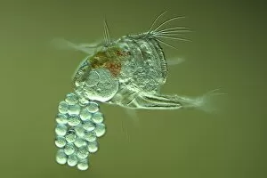
Background imageLight Microscope Collection: Rotifer with eggs, light micrograph C016 / 8588

Rotifer with eggs, light micrograph C016 / 8588
Rotifer with eggs. Differential interference contrast (DIC) light micrograph of a rotifer (phylum Rotifera) carrying eggs (small, round)


For sale as Licensed Images

Choose your image, Select your licence and Download the media

Exploring the Intricacies of Life: A Glimpse through the Light Microscope Unveiling the Wonders of Cerebellum Tissue: Witness the intricate network of cells in cerebellum tissue as seen through a captivating light micrograph. Nerve and Glial Cells Revealed: Dive into the mesmerizing world of nerve and glial cells, captured in stunning detail by a powerful light microscope. Unraveling Chemical Structures: Behold the ethereal beauty of copper and magnesium sulphate crystals, magnified under a light micrograph to expose their delicate arrangements. The Hidden Beauty Within Caffeine Crystals: Marvel at the crystalline structures of caffeine, brought to life through an enchanting light micrograph that unveils its hidden allure. Journey into Hippocampus Brain Tissue: Embark on an extraordinary expedition deep within hippocampus brain tissue, where mysteries unfold with every glance through our remarkable light microscope lens. Illuminating Glial Cells' Secrets: Delve into the enigmatic realm of glial cells as they reveal their intricate patterns and connections in a mesmerizing confocal light micrograph. HeLa Cells Exposed - A Window to Immortality? Explore HeLa cells like never before with our cutting-edge technology, providing unprecedented insight into these immortalized wonders under brightfield illumination (C017 / 8299). Peering Into Purkinje Nerve Cells in Cerebellum: Discover the elegance and complexity displayed by Purkinje nerve cells residing within cerebellar tissue when observed using a high-resolution light microscope. Dicotyledon Plant Stem's Inner Workings Revealed. Get lost amidst nature's marvels as we uncover the intricacies present within dicotyledon plant stems using our advanced techniques for capturing breathtakingly detailed images under brightfield illumination. Unveiling the Secrets of Kidney Tubules.