Light Microscope Collection (#3)

Exploring the Intricacies of Life: A Glimpse through the Light Microscope Unveiling the Wonders of Cerebellum Tissue

Background imageLight Microscope Collection: Light Micrograph : Flattened Mayfly Nymph, Magnification x 10 (A4 size: 29. 7 cm width)

Light Micrograph : Flattened Mayfly Nymph, Magnification x 10 (A4 size: 29. 7 cm width)
LRDS-80 Flattened Mayfly Nymph Light Micrograph Ecdyonurus venosus Magnification x 10 (A4 size: 29.7 cm width) Credit: David Spears (last refuge)

Background imageLight Microscope Collection: Light Micrograph (LM): Transverse section of Dehiscence Lilium Anthers with Pollen; Magnification

Light Micrograph (LM): Transverse section of Dehiscence Lilium Anthers with Pollen; Magnification x300 (on 10)
LRDS-200 Light Micrograph (LM): Transverse section of Dehiscence Lilium Anthers with Pollen Magnification x300 (on 10.5 cm width print) David Spears (Last Refuge)

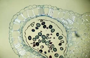
Background imageLight Microscope Collection: Apple stem, light micrograph

Apple stem, light micrograph
Apple stem. Polarised light micrograph of a section through the stem of a young apple (Malus domestica) tree plant. Magnification: x100 when printed 10 centimetres wide

Background imageLight Microscope Collection: Light microscope F006 / 7165

Light microscope F006 / 7165
Light microscope on a laboratory bench

Background imageLight Microscope Collection: Light microscope F006 / 7167

Light microscope F006 / 7167
Light microscope on a laboratory bench

Background imageLight Microscope Collection: Light microscope F006 / 7186

Light microscope F006 / 7186
Light microscope on a laboratory bench

Background imageLight Microscope Collection: Light microscope F006 / 7190

Light microscope F006 / 7190
Light microscope

Background imageLight Microscope Collection: Light microscope F006 / 7166

Light microscope F006 / 7166
Light microscope on a laboratory bench

Background imageLight Microscope Collection: Light microscope eyepieces F006 / 7188

Light microscope eyepieces F006 / 7188
Light microscope eyepieces

Background imageLight Microscope Collection: Light microscope F006 / 7184

Light microscope F006 / 7184
Light microscope on a laboratory bench

Background imageLight Microscope Collection: Human skin section, light micrograph

Human skin section, light micrograph
Human skin. Light micrograph of a section through healthy human skin. The outer surface of the skin is at top. The uppermost surface is the epidermis

Background imageLight Microscope Collection: Chilodonella ciliate protozoa, LM

Chilodonella ciliate protozoa, LM
Chilodonella protozoa. Light micrograph of several Chilodonella sp. ciliate protozoa. Chilodonella is a single-celled animal that is found in freshwater

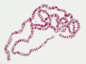
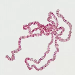
Background imageLight Microscope Collection: Giant chromosomes, light micrograph P657 / 0038

Giant chromosomes, light micrograph P657 / 0038
Giant chromosomes. Light micrograph of giant (polytene) chromosomes from a fruit fly (Drosophila busckii). These chromosomes are formed from repeated rounds of DNA (deoxyribonucleic acid)

Background imageLight Microscope Collection: Human skin section, light micrograph P710 / 0472

Human skin section, light micrograph P710 / 0472
Human skin. Light micrograph of a section through healthy human skin. The outer surface of the skin is at top. The uppermost surface is the epidermis

Background imageLight Microscope Collection: Giant chromosomes, light micrograph P657 / 0037

Giant chromosomes, light micrograph P657 / 0037
Giant chromosomes. Light micrograph of giant (polytene) chromosomes from a fruit fly (Drosophila busckii). These chromosomes are formed from repeated rounds of DNA (deoxyribonucleic acid)

Background imageLight Microscope Collection: Optical light microscope with camera

Optical light microscope with camera
Microscope. Computer artwork of a typical optical microscope with a digital camera attached for photography. Below the camera these are the two eyepieces (binocular) for viewing the specimen

Background imageLight Microscope Collection: Optical light microscope, artwork

Optical light microscope, artwork
Microscope. Computer artwork of a typical optical microscope. Below the camera these are the two eyepieces (binocular) for viewing the specimen

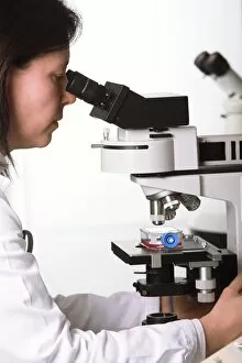
Background imageLight Microscope Collection: Biological research F005 / 0936

Biological research F005 / 0936
MODEL RELEASED. Biological research. Researcher using a light microscope

Background imageLight Microscope Collection: Biological research F005 / 0937

Biological research F005 / 0937
MODEL RELEASED. Biological research. Researcher using a light microscope

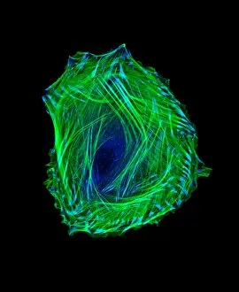
Background imageLight Microscope Collection: Embryonic smooth muscle cell C018 / 8595

Embryonic smooth muscle cell C018 / 8595
Embryonic smooth muscle cell, immunofluorescence micrograph. Actin filaments are green, the protein vinculin is light blue. Actin is part of the cytoskeleton

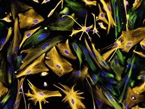
Background imageLight Microscope Collection: Neural progenitor cell differentiation C018 / 8758

Neural progenitor cell differentiation C018 / 8758
Neural progenitor cell differentiation. Fluorescence light micrograph of neural progenitor cells that have been grown in a medium that is selective for astrocytes for three weeks

Background imageLight Microscope Collection: Mouse embryo, light micrograph C018 / 8600

Mouse embryo, light micrograph C018 / 8600
Mouse embryo head. Light micrograph of the head of a mouse (Mus sp.) embryo 13 days after fertilisation

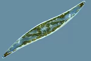
Background imageLight Microscope Collection: Diatom, light micrograph C014 / 4673

Diatom, light micrograph C014 / 4673
Diatom. Differential interference contrast micrograph of a Gyrosigma sp. diatom. Diatoms are a group of photosynthetic, single-celled algae containing about 10, 000 species

Background imageLight Microscope Collection: Stem cell-derived retinal cells

Stem cell-derived retinal cells. Fluorescence light micrograph of retinal pigment epithelium (RPE) cells that have been derived from human embryonic stem cells (HESC)

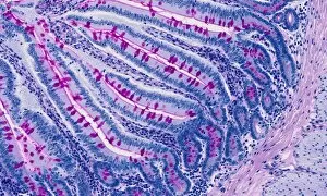
Background imageLight Microscope Collection: Small intestine, light micrograph

Small intestine, light micrograph
Small intestine. Light micrograph of a section through the small intestine. The finger-like projections at left are the villi. These increase the surface area for the absorption of food

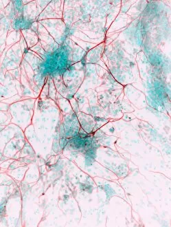
Background imageLight Microscope Collection: Stem cell-derived neurons, micrograph

Stem cell-derived neurons, micrograph
Stem cell-derived neurons. Light micrograph of human nerve cells (neurons) that have been derived from induced pluripotent stem cells (IPS). Tuj1 proteins are cyan, and cell nuclei are red

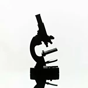
Background imageLight Microscope Collection: Light microscope C014 / 1407

Light microscope C014 / 1407
Light microscope, seen in silhouetted profile. This microscope has a binocular eyepiece (an eyepiece for each eye). The sample is placed on a glass slide on the mount below centre

Background imageLight Microscope Collection: MDCK cells, inverted light micrograph C017 / 7647

MDCK cells, inverted light micrograph C017 / 7647
MDCK cells. Differential interference contrast inverted light micrograph of Madin-Darby canine kidney (MDCK) cells. This is a kidney epithelial cell line from a cocker spaniel

Background imageLight Microscope Collection: Biological research F006 / 8581

Biological research F006 / 8581
Biological research

Background imageLight Microscope Collection: Light microscope F006 / 8553

Light microscope F006 / 8553
Light microscope

Background imageLight Microscope Collection: Light microscope F006 / 8554

Light microscope F006 / 8554
Light microscope

Background imageLight Microscope Collection: Light microscope F006 / 8551

Light microscope F006 / 8551
Light microscope

Background imageLight Microscope Collection: Light microscope F006 / 8545

Light microscope F006 / 8545
Light microscope

Background imageLight Microscope Collection: Light microscope F006 / 9033

Light microscope F006 / 9033
Light microscope. Close up of a slide on a microscope stage

Background imageLight Microscope Collection: Light microscope F006 / 9034

Light microscope F006 / 9034
Light microscope. Close up of a slide on a microscope stage

Background imageLight Microscope Collection: Light microscope F006 / 9032

Light microscope F006 / 9032
Light microscope. Close up of a slide on a microscope stage

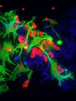
Background imageLight Microscope Collection: Stem cell-derived astrocyte brain cells

Stem cell-derived astrocyte brain cells
Stem cell-derived nerve cells. Fluorescence light micrograph of astrocyte brain cells that have been derived from neural (nerve) stem cells from a mouse

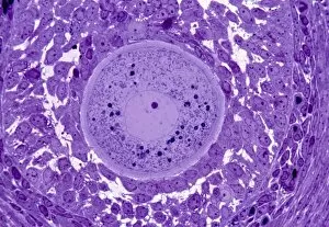
Background imageLight Microscope Collection: Oocyte, light micrograph

Oocyte, light micrograph
Oocyte. Light micrograph of a section through an oocyte within a early antral follicle in an ovary. Oocytes are immature ova, or egg cells

Background imageLight Microscope Collection: Cardiac muscle, light micrograph

Cardiac muscle, light micrograph
Cardiac muscle. Light micrograph of a section through muscle tissue from a heart, showing the cardiomyocyte cells. Heart muscle cells show central nuclei but the dominant feature is the abundance of

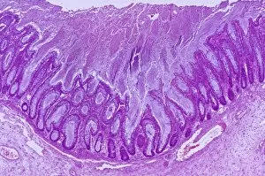
Background imageLight Microscope Collection: Colitis, light micrograph

Colitis, light micrograph
Colitis. Light micrograph of a section through colon (large intestine) tissue affected by bacterial infection, causing injury to the mucosal lining, which has resulted in inflammation (colitis)

Background imageLight Microscope Collection: Hairy scalp skin, light micrograph

Hairy scalp skin, light micrograph
Hairy scalp skin. Light micrograph of a section through healthy skin from a human scalp, showing the numerous hair follicles. The scalp is an example of hairy skin

Background imageLight Microscope Collection: Oxalosis, light micrograph

Oxalosis, light micrograph
Oxalosis. Light micrograph of a section through adult bone tissue affected by oxalosis (primary hyperoxaluria), showing mature bone (green)
Background imageLight Microscope Collection: Peripheral nerve, light micrograph

Peripheral nerve, light micrograph
Peripheral nerve. Light micrograph of a section through a peripheral nerve. This is a mixed nerve with myelinated axons (dark blue circles)
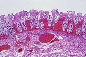
Background imageLight Microscope Collection: Ischaemic bowel, light micrograph

Ischaemic bowel, light micrograph
Ischaemic bowel. Light micrograph of a section through a portion of intestine that has been damaged by an interruption of the blood supply, or ischaemia

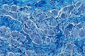
Background imageLight Microscope Collection: Nerve ganglion, light micrograph

Nerve ganglion, light micrograph
Nerve ganglion. Light micrograph of a section through a dorsal (sensory) spinal root ganglion associated with a sensory nerve root of the spinal cord. Sensory information from peripheral sites e.g

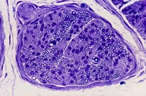
Background imageLight Microscope Collection: Myelinated nerve, light micrograph

Myelinated nerve, light micrograph
Myelinated nerve. Light micrograph of a section through a peripheral myelinated nerve, showing many individual axons each covered with deep blue-staining myelin sheaths

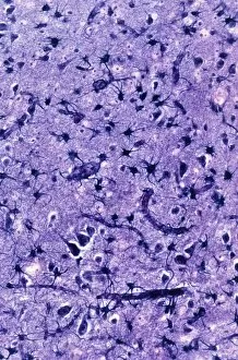
Background imageLight Microscope Collection: Glial cells, light micrograph

Glial cells, light micrograph
Glial cells in the brain. Light micrograph of a section through glial cells (dark) in the grey matter of the brain. Due to their star shape these glial cells are called astrocytes

Background imageLight Microscope Collection: Copper sulphate crystals light micrograph

Copper sulphate crystals light micrograph
Copper sulphate crystals, polarised light micrograph. Magnification: x2.5, when printed 10 centimetres wide


For sale as Licensed Images

Choose your image, Select your licence and Download the media

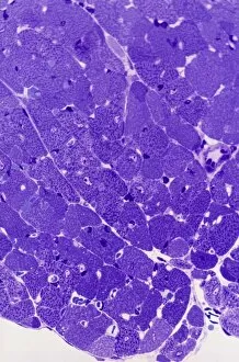
Exploring the Intricacies of Life: A Glimpse through the Light Microscope Unveiling the Wonders of Cerebellum Tissue: Witness the intricate network of cells in cerebellum tissue as seen through a captivating light micrograph. Nerve and Glial Cells Revealed: Dive into the mesmerizing world of nerve and glial cells, captured in stunning detail by a powerful light microscope. Unraveling Chemical Structures: Behold the ethereal beauty of copper and magnesium sulphate crystals, magnified under a light micrograph to expose their delicate arrangements. The Hidden Beauty Within Caffeine Crystals: Marvel at the crystalline structures of caffeine, brought to life through an enchanting light micrograph that unveils its hidden allure. Journey into Hippocampus Brain Tissue: Embark on an extraordinary expedition deep within hippocampus brain tissue, where mysteries unfold with every glance through our remarkable light microscope lens. Illuminating Glial Cells' Secrets: Delve into the enigmatic realm of glial cells as they reveal their intricate patterns and connections in a mesmerizing confocal light micrograph. HeLa Cells Exposed - A Window to Immortality? Explore HeLa cells like never before with our cutting-edge technology, providing unprecedented insight into these immortalized wonders under brightfield illumination (C017 / 8299). Peering Into Purkinje Nerve Cells in Cerebellum: Discover the elegance and complexity displayed by Purkinje nerve cells residing within cerebellar tissue when observed using a high-resolution light microscope. Dicotyledon Plant Stem's Inner Workings Revealed. Get lost amidst nature's marvels as we uncover the intricacies present within dicotyledon plant stems using our advanced techniques for capturing breathtakingly detailed images under brightfield illumination. Unveiling the Secrets of Kidney Tubules.