Light Microscope Collection (#4)

Exploring the Intricacies of Life: A Glimpse through the Light Microscope Unveiling the Wonders of Cerebellum Tissue

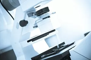
Background imageLight Microscope Collection: Light microscope F006 / 8551

Light microscope F006 / 8551
Light microscope

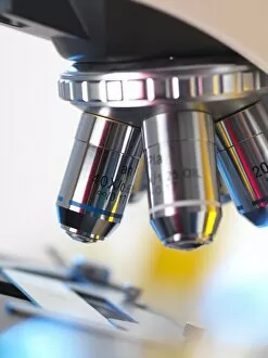
Background imageLight Microscope Collection: Light microscope F006 / 8545

Light microscope F006 / 8545
Light microscope

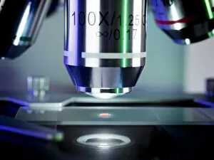
Background imageLight Microscope Collection: Light microscope F006 / 9033

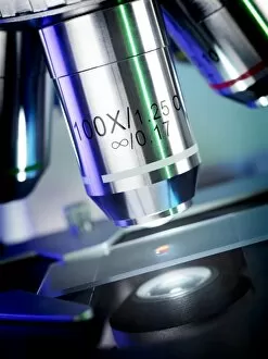
Light microscope F006 / 9033
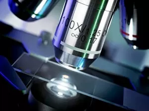
Light microscope. Close up of a slide on a microscope stage

Background imageLight Microscope Collection: Light microscope F006 / 9034

Light microscope F006 / 9034
Light microscope. Close up of a slide on a microscope stage

Background imageLight Microscope Collection: Light microscope F006 / 9032

Light microscope F006 / 9032
Light microscope. Close up of a slide on a microscope stage

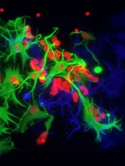
Background imageLight Microscope Collection: Stem cell-derived astrocyte brain cells

Stem cell-derived astrocyte brain cells
Stem cell-derived nerve cells. Fluorescence light micrograph of astrocyte brain cells that have been derived from neural (nerve) stem cells from a mouse

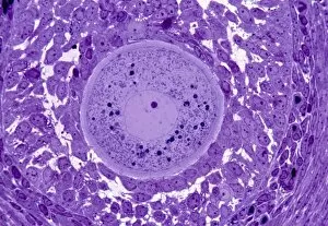
Background imageLight Microscope Collection: Oocyte, light micrograph

Oocyte, light micrograph
Oocyte. Light micrograph of a section through an oocyte within a early antral follicle in an ovary. Oocytes are immature ova, or egg cells

Background imageLight Microscope Collection: Cardiac muscle, light micrograph

Cardiac muscle, light micrograph
Cardiac muscle. Light micrograph of a section through muscle tissue from a heart, showing the cardiomyocyte cells. Heart muscle cells show central nuclei but the dominant feature is the abundance of

Background imageLight Microscope Collection: Colitis, light micrograph

Colitis, light micrograph
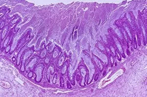
Colitis. Light micrograph of a section through colon (large intestine) tissue affected by bacterial infection, causing injury to the mucosal lining, which has resulted in inflammation (colitis)

Background imageLight Microscope Collection: Hairy scalp skin, light micrograph

Hairy scalp skin, light micrograph
Hairy scalp skin. Light micrograph of a section through healthy skin from a human scalp, showing the numerous hair follicles. The scalp is an example of hairy skin
Background imageLight Microscope Collection: Oxalosis, light micrograph

Oxalosis, light micrograph
Oxalosis. Light micrograph of a section through adult bone tissue affected by oxalosis (primary hyperoxaluria), showing mature bone (green)
Background imageLight Microscope Collection: Peripheral nerve, light micrograph

Peripheral nerve, light micrograph
Peripheral nerve. Light micrograph of a section through a peripheral nerve. This is a mixed nerve with myelinated axons (dark blue circles)
Background imageLight Microscope Collection: Ischaemic bowel, light micrograph

Ischaemic bowel, light micrograph
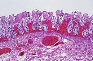
Ischaemic bowel. Light micrograph of a section through a portion of intestine that has been damaged by an interruption of the blood supply, or ischaemia

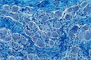
Background imageLight Microscope Collection: Nerve ganglion, light micrograph

Nerve ganglion, light micrograph
Nerve ganglion. Light micrograph of a section through a dorsal (sensory) spinal root ganglion associated with a sensory nerve root of the spinal cord. Sensory information from peripheral sites e.g

Background imageLight Microscope Collection: Myelinated nerve, light micrograph

Myelinated nerve, light micrograph
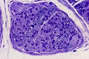
Myelinated nerve. Light micrograph of a section through a peripheral myelinated nerve, showing many individual axons each covered with deep blue-staining myelin sheaths

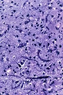
Background imageLight Microscope Collection: Glial cells, light micrograph

Glial cells, light micrograph
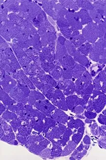
Glial cells in the brain. Light micrograph of a section through glial cells (dark) in the grey matter of the brain. Due to their star shape these glial cells are called astrocytes

Background imageLight Microscope Collection: Copper sulphate crystals light micrograph

Copper sulphate crystals light micrograph
Copper sulphate crystals, polarised light micrograph. Magnification: x2.5, when printed 10 centimetres wide

Background imageLight Microscope Collection: Cirrhosis of the liver, light micrograph

Cirrhosis of the liver, light micrograph
Cirrhosis of the liver. Light micrograph of a section through liver tissue affected by cirrhosis, showing a cluster of hepatocyte cells (orange)

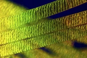
Background imageLight Microscope Collection: Spirogyra algae, light micrograph C016 / 9592

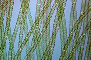
Spirogyra algae, light micrograph C016 / 9592
Spirogyra algae. Polarised light micrograph of Spirogyra sp. algae. This filamentous green algae is named for the spiral arrangement of its chloroplasts (green)

Background imageLight Microscope Collection: Spirogyra algae, light micrograph C016 / 9594

Spirogyra algae, light micrograph C016 / 9594
Spirogyra algae. Rheinberg illuminated light micrograph of Spirogyra sp. algae. This filamentous green algae is named for the spiral arrangement of its chloroplasts (green)

Background imageLight Microscope Collection: Light microscopy C016 / 9700

Light microscopy C016 / 9700
Light microscopy. Light microscope being used to magnify a blood sample

Background imageLight Microscope Collection: Desmids and spirogyra, light micrograph C016 / 9595

Desmids and spirogyra, light micrograph C016 / 9595
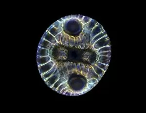
Desmids and spirogyra. Polarised light micrograph of two Micrasterias rotata desmids (round) and a single filament of Spirogyra sp. green alga (lower right)

Background imageLight Microscope Collection: Light microscopy C016 / 9703

Light microscopy C016 / 9703
Light microscopy. Light microscope being used to magnify a blood sample

Background imageLight Microscope Collection: Fossil diatom, light micrograph C016 / 8603

Fossil diatom, light micrograph C016 / 8603
Fossil diatom. Darkfield illuminated light micrograph of a fossilised diatom. Diatoms are a group of photosynthetic, single-celled algae containing about 10, 000 species

Background imageLight Microscope Collection: IVF treatment, light microscope C016 / 6536

IVF treatment, light microscope C016 / 6536
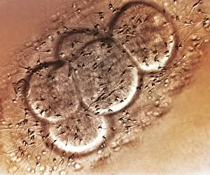
IVF treatment. Light microscope of human egg cells (large, round) and sperm (small dark ovals with tails) in a petri dish during in vitro fertilisation (IVF)

Background imageLight Microscope Collection: IVF treatment, light microscope C016 / 6535

IVF treatment, light microscope C016 / 6535
IVF treatment. Light microscope of human egg cells (large, round) and sperm (small dark ovals with tails) in a petri dish during in vitro fertilisation (IVF)

Background imageLight Microscope Collection: Netrium desmid, light micrograph C016 / 9591

Netrium desmid, light micrograph C016 / 9591
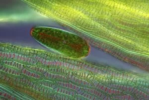
Netrium desmid. Polarised light micrograph of a Netrium sp. desmid (oval, centre) among peat moss (Sphagnum sp.) leaves (green)

Background imageLight Microscope Collection: IVF treatment, light microscope C016 / 6512

IVF treatment, light microscope C016 / 6512
IVF treatment. Light microscope of human egg cells (large, round) and sperm (small dark ovals with tails) in a petri dish during in vitro fertilisation (IVF)

Background imageLight Microscope Collection: Light microscopy C016 / 9702

Light microscopy C016 / 9702
Light microscopy. Light microscope being used to magnify a blood sample

Background imageLight Microscope Collection: Spirogyra algae, light micrograph C016 / 9593

Spirogyra algae, light micrograph C016 / 9593
Spirogyra algae. Polarised light micrograph of Spirogyra sp. algae. This filamentous green algae is named for the spiral arrangement of its chloroplasts (green)

Background imageLight Microscope Collection: Micrasterias desmids, light micrograph C016 / 9596

Micrasterias desmids, light micrograph C016 / 9596
Micrasterias desmids. Rheinberg illuminated light micrograph of Micrasterias rotata desmid green algae. Desmids are a common group of freshwater single-celled algae that have intricate cell walls

Background imageLight Microscope Collection: Rotifer, light micrograph C016 / 9550

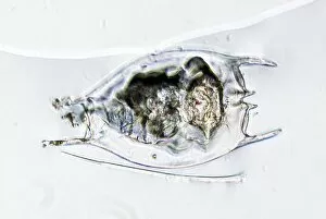
Rotifer, light micrograph C016 / 9550
Rotifer. Light micrograph of a freshwater rotifer (Brachionus calyciflorus). Rotifers are microscopic aquatic animals that are related to roundworms

Background imageLight Microscope Collection: Rotifer with eggs, light micrograph C016 / 8587

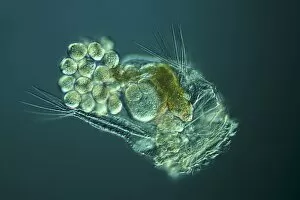
Rotifer with eggs, light micrograph C016 / 8587
Rotifer with eggs. Differential interference contrast (DIC) light micrograph of a rotifer (phylum Rotifera) carrying eggs (small, round)

Background imageLight Microscope Collection: Light microscopy C016 / 9701

Light microscopy C016 / 9701
Light microscopy. Light microscope being used to magnify a blood sample

Background imageLight Microscope Collection: Acute myeloid leukaemia, micrograph

Acute myeloid leukaemia, micrograph
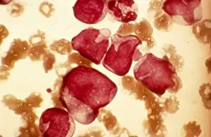
Acute myeloid leukaemia. Light micrograph of blood cells from bone marrow in a case of acute myeloid leukaemia. These cells include a monocytoid myeloblast stem cell

Background imageLight Microscope Collection: Elliptocytosis, light micrograph

Elliptocytosis, light micrograph
Elliptocytosis. Light micrograph of red blood cells in a case of elliptocytosis. Red blood cells (erythrocytes) carry oxygen and carbon dioxide to and from body tissues

Background imageLight Microscope Collection: Cystine in bone marrow, light micrograph

Cystine in bone marrow, light micrograph
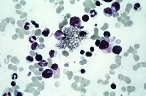
Cystine in bone marrow. Light micrograph of crystals of cystine among blood cells in a sample of bone marrow. Cystine is an amino acid that can form crystals in urine

Background imageLight Microscope Collection: Bone marrow blood cells, light micrograph

Bone marrow blood cells, light micrograph

Background imageLight Microscope Collection: Ciliate protozoan ingesting algae

Ciliate protozoan ingesting algae. Darkfield illuminated light micrograph of a ciliate protozoan ingesting green algae (green)

Background imageLight Microscope Collection: Parkinsons disease, light micrograph

Parkinsons disease, light micrograph
Parkinsons disease. Light micrograph of a section through the sabstantia nigra of the brain of a patient with Parkinsons disease showing a neuron (nerve cell)

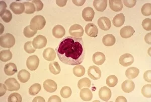
Background imageLight Microscope Collection: Monocyte blood cell, light micrograph

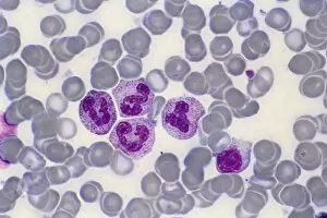
Monocyte blood cell, light micrograph. Monocytes are a type of white blood cell, part of the bodys immune system. Monocytes are the largest white blood cell

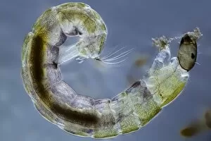
Background imageLight Microscope Collection: Midge larva, light micrograph

Midge larva, light micrograph
Midge larva. Differential interference contrast (DIC) micrograph of the larva of a non-biting midge (family Chironomidae). Midge refers to many kinds of very small two-winged flies found world-wide

Background imageLight Microscope Collection: Chebarkul meteorite, light micrograph C015 / 2863

Chebarkul meteorite, light micrograph C015 / 2863
Chebarkul meteorite fragment seen through a microscope in polarised light. This research is being carried out at the Vernadsky Institute, Moscow, Russia

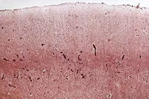
Background imageLight Microscope Collection: Dementia, light micrograph

Dementia, light micrograph
Dementia. Light micrograph of a section through the brain of a patient with dementia. Dementia is mainly a disease of old age

Background imageLight Microscope Collection: Desmid, light micrograph

Desmid, light micrograph
Desmid. Darkfield illuminated light micrograph of a desmid green alga. Desmids are a common group of freshwater single-celled algae that have intricate cell walls

Background imageLight Microscope Collection: Pernicious anaemia, light micrograph

Pernicious anaemia, light micrograph
Pernicious anaemia. Light micrograph of megaloblast blood cells from bone marrow in a case of pernicious anaemia, also known as Biermers anaemia

Background imageLight Microscope Collection: Blood cell cancer, light micrograph

Blood cell cancer, light micrograph
Blood cell cancer. Light micrograph of blood cells from a lymphatic ganglion in a case of a blood cancer of a mixed cell type

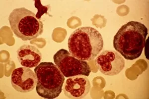
Background imageLight Microscope Collection: Erythroblast blood cell, light micrograph

Erythroblast blood cell, light micrograph
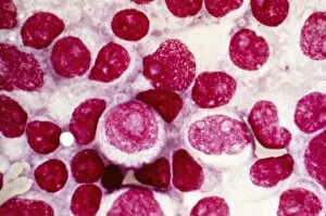
Erythroblast blood cell. Light micrograph of cells from a sample of bone marrow, including a polychromatic erythroblast (upper left). This one has multiple cell nuclei (dark red, four in total)


For sale as Licensed Images

Choose your image, Select your licence and Download the media

Exploring the Intricacies of Life: A Glimpse through the Light Microscope Unveiling the Wonders of Cerebellum Tissue: Witness the intricate network of cells in cerebellum tissue as seen through a captivating light micrograph. Nerve and Glial Cells Revealed: Dive into the mesmerizing world of nerve and glial cells, captured in stunning detail by a powerful light microscope. Unraveling Chemical Structures: Behold the ethereal beauty of copper and magnesium sulphate crystals, magnified under a light micrograph to expose their delicate arrangements. The Hidden Beauty Within Caffeine Crystals: Marvel at the crystalline structures of caffeine, brought to life through an enchanting light micrograph that unveils its hidden allure. Journey into Hippocampus Brain Tissue: Embark on an extraordinary expedition deep within hippocampus brain tissue, where mysteries unfold with every glance through our remarkable light microscope lens. Illuminating Glial Cells' Secrets: Delve into the enigmatic realm of glial cells as they reveal their intricate patterns and connections in a mesmerizing confocal light micrograph. HeLa Cells Exposed - A Window to Immortality? Explore HeLa cells like never before with our cutting-edge technology, providing unprecedented insight into these immortalized wonders under brightfield illumination (C017 / 8299). Peering Into Purkinje Nerve Cells in Cerebellum: Discover the elegance and complexity displayed by Purkinje nerve cells residing within cerebellar tissue when observed using a high-resolution light microscope. Dicotyledon Plant Stem's Inner Workings Revealed. Get lost amidst nature's marvels as we uncover the intricacies present within dicotyledon plant stems using our advanced techniques for capturing breathtakingly detailed images under brightfield illumination. Unveiling the Secrets of Kidney Tubules.